Biochemistry - Chemical Reactions in Living Cells Volume 3 - D. Metzler 1980
Cell Growth, Differentiation, and Chemical Communication
Tissue Differentiation and Developmental Biology
Changes in DNA Content
There are many known cases where Cell Differentiation leads to a temporary or stable Modification of the genome. For instance, the Amplification of rRNA genes (ch. 15, sec. I, 1, d) in oocytes is accompanied by a temporary increase in the overall DNA content of The Cell. Some highly specialized Cells, such as Purkinje cells in the Cerebellum or numerous cells in dipteran larvae (ch. 15, sec. G, 9, c), are characterized by polyploidy. As a rule, these are cells that have reached the limit of their specialization and lost the capacity for Cell Division. Each copy of DNA in polyploidy cells typically contains a complete set of genes (most of which remain phenotypically silent).
On the other hand, in certain cells The process of irreversible differentiation is coupled with the loss of a portion of The Genome. An extreme manifestation of this is found in human erythrocytes, which completely lose their nucleus. In other cells, individual Chromosomes undergo degradation. There are also instances where a chromosome or part of it is irreversibly inactivated and remains in The Cell as a compact Structure known as heterochromatin—a term used to designate intensely staining Regions of the Cell Nucleus. While some heterochromatin contains highly repetitive sequences (ch. 15, sec. I, 1, b), specific heterochromatic regions harbor clusters of inactivated genes. A case of profound interest is the complete inactivation of one of the two X chromosomes in female mammalian cells [181]. The entire chromosome in this state appears as heterochromatin. This inactivation occurs early in embryonic development and affects either the maternal or paternal X chromosome at random: the maternal X is inactivated in some cells, the paternal in others. However, through subsequent cell divisions, the exact same chromosome remains inactive throughout the entire cell clone. As a result, female organisms exhibit mosaicism for heterozygous X-linked genes.
The mechanism underlying chromosome inactivation and selective degradation remains elusive; it has been suggested to share a chemical basis with the modification and restriction processes characteristic of Bacteria (ch. 15, sec. E) [182]. Via a methylation system, one chromosome might be "tagged" and preserved, whereas the other becomes subject to successive assaults by a cleaving endonuclease. Alternatively, it is conceivable that a different enzyme initiates the transition of chromosomes into heterochromatin.
While certain genes are selectively inactivated or alternately switched on and off, others are in some instances irreversibly lost during cellular differentiation. Genetic recombination apparently takes place within the chromosomes of individual cells during mitosis, and sister chromatid exchange has been detected. However, if such an exchange involves equal amounts of genetic material, no genetic alteration occurs in the daughter cells. Conversely, if two or more identical base sequences reside within a single DNA molecule, unequal Crossing-over (ch. 16, sec. J, 3) can take place, resulting in the loss of genetic material in one of the daughter cells. Essentially, this mechanism may constitute a predetermined differentiation program for specific cell types.
The loss of genes from chromosomes can also proceed via another mechanism, namely the looping-out mechanism [183].
By analogy with the excision of phage λ (ch. 15, sec. G, 8) from the E. coli chromosome, such Gene loss must occur at specific DNA sites. Permanent loss of genetic material presumably takes place during the differentiation of pluripotent stem cells that give rise to Blood Cells. From these pluripotent precursors, three distinct stem cell lineages are initially formed—namely, myeloid, erythroid, and lymphoid lines—which undergo further differentiation as illustrated in the scheme.
Class="center">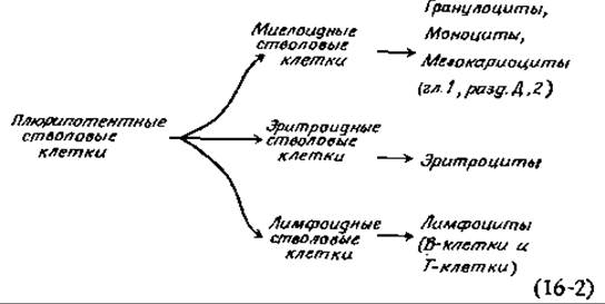
Erythroid stem cells serve as precursors to Hemoglobin-containing erythrocytes. Recall (ch. 4, sec. D, 7) that mammalian Hemoglobins consist of two a-chains and two additional chains that are either ß, y, δ, or ε. Adult hemoglobin predominantly possesses the a2ß2 structure, though small amounts of a2δ2 hemoglobin are also present. Early embryonic development is characterized by a2ε2 hemoglobin, but at later stages the ε-chains are replaced by two other embryonic hemoglobin chains, specifically Gy and Ay. Genetic studies have demonstrated that the e-, y-, ß-, and δ-globin genes are tightly linked [188]. Why, then, does an individual erythrocyte contain only a single type of hemoglobin? The reason apparently lies in the presence of only one promoter for this given gene cluster. If a terminator signal follows each gene, METABOLISM/31.html">Transcription will obviously proceed only for the gene located closest to the promoter. Should this gene be lost at some developmental stage, the Transcription of the next gene will initiate, and so forth; in this manner, progressive, stepwise changes in Gene Expression can occur within erythrocytes. Another notable feature of erythrocyte differentiation is its sensitivity to Erythropoietin, a glycoprotein hormone produced in the Kidneys [184–186]. Under The Influence of erythropoietin, differentiating stem cells initiate intensive hemoglobin synthesis and ultimately transform into mature erythrocytes [186a].

Last update: 06/08/2026
Editorial and Educational Adaptation: This material has been compiled based on the primary/original source text. The project team performed an editorial review, corrected technical inaccuracies, structured sections, and adapted the content for an educational format.
What was processed:
- elimination of formatting defects (OCR errors, structural breaks, corrupted characters);
- editorial organization of content;
- standardization of terminology in accordance with academic sources;
- verification of factual statements against the original source text.
All mentions of the author, publication year, and origin of the primary text have been preserved in accordance with the source.