BIOCHEMISTRY - L. Stryer - 1984
VOLUME 3
Part IV INFORMATION
CHAPTER 31. GENE REARRANGEMENTS: RECOMBINATION, TRANSPOSITION, AND CLONING
31.2. Genetic Recombination Involves the Pairing of Homologous DNA Strands to Form a Double-Stranded Intermediate
Intermediates in genetic recombination have been visualized by Electron Cell/15.html">Microscopy. These studies utilized plasmid DNA from E. coli. As we shall soon see (Section 31.4), these small, circular, double-stranded DNAs replicate autonomously within the bacterial cell. In the presence of chloramphenicol, the number of Plasmids per bacterium increases from about 20 to 1,000. This antibiotic, an inhibitor of Protein Synthesis, halts the Replication of the bacterial chromosome while leaving plasmid replication unaffected. Consequently, the bacterial cell becomes packed with plasmid molecules capable of undergoing recombination. Electron Microscopy of plasmids isolated from these Cells reveals that approximately a quarter of them exist as dimers in the shape of the figure eight (Fig. 31.4A). These dimers were subsequently cleaved with the restriction endonuclease EcoRI, which cuts a plasmid monomer at a single, highly specific site. Had the dimers been interlocked monomers or double-length circles, Treatment with the restriction endonuclease would have yielded linear rods of uniform length. On the otherخرى hand, if two plasmid circles are covalently joined at a region of Homology, a four-branched Structure resembling the Greek letter ӽ should be visible. In reality, nearly all of the figure-eights are converted into the ӽ-form (Fig. 31.4B). This provides compelling evidence that the figure-eights are indeed replication intermediates. This Conclusion is further supported by the observation that certain E. coli mutants defective in recombination fail to form figure-eights.
Class="center">Fig. 31.4. Electron micrograph of DNA molecules undergoing recombination: (A) A figure-eight intermediate composed of two DNA molecules; (B) Cleavage of this intermediate with a restriction endonuclease yields the ӽ-form.
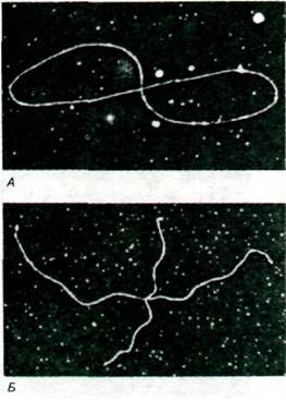
Fig. 31.5. Proposed cleavage scheme for two DNA molecules joined at nonhomologous (A) and homologous (B) regions. The observed Symmetry of the ӽ-forms (as in Fig. 31.4B) indicates that the figure-eight DNA molecules are joined at homologous regions.
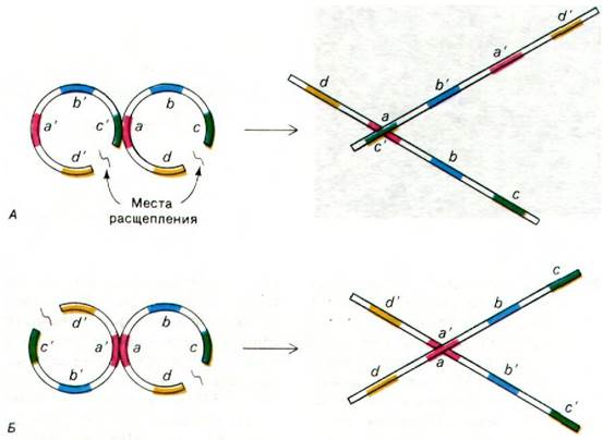
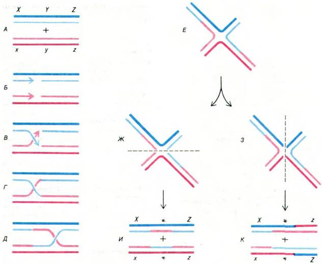
What is The structure of the crossover region in the figure-eights? The contact point (intersection) of the ӽ-forms always divides the entire structure into two pairs of arms of equal length. This implies that the genomes are joined within a region of homology (Fig. 31.5). If the plasmids were joined at nonhomologous sequences, the lengths of all four arms would be distributed at random. Furthermore, the contact point is located with roughly equal probability along the entire length of the plasmid, indicating that pairing can occur at many positions. The manner in which the strands are connected at the crossover region was investigated using selective Denaturation. Electron micrographs reveal that four double-stranded molecules diverge from a single-stranded ring at the junctions of the two genomes (Fig. 31.6). This intermediate can subsequently be cleaved and ligated to produce two pairs of distinct recombinant molecules (Fig. 31.7).
Fig. 31.6. (A) Electron micrograph of the ӽ-form; (B) Schematic diagram of the molecule shown in the micrograph. The region of homology (rich in A-T Base Pairs) was selectively denatured with formamide to clearly reveal the strand connection at the crossover region.
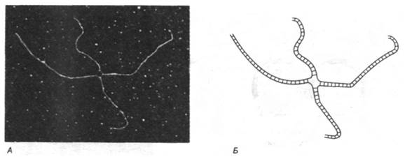
Fig. 31.7. A model of genetic recombination proposed by Robin Holliday. One parental double-stranded molecule is shown in blue, the other in red. The darker strand in each duplex represents the (+) chain. The letters X, Y, and Z designate three genes, and x, y, and z represent their alleles. (B and C) Covalent joining of the parental DNA molecules. (E) An alternative representation of the molecular complex shown in (D). Note that this structure in (E) can be cleaved along either a horizontal or a vertical axis. Rejoining of the strands in (G) and (H) yields two different sets of recombinants (I and J). Regions containing a single strand from each parental duplex are marked with asterisks.
31.3. The recA Protein Catalyzes ATP-Dependent DNA Strand Exchange in Genetic Recombination
The process discussed thus far is termed general genetic recombination because exchanges can occur between any pair of homologous sequences within the parental DNA molecules. In E. coli, general recombination is dependent on rec genes. In rec- cells, Bacterial DNA is unable to recombine with exogenous DNA molecules. Three rec genes have been identified: recA, recB, and recC. The recB protein (molecular mass 140 kDa) and recC protein (128 kDa) are two subunits of a single nuclease that unwinds double-helical DNA and degrades one of the unwound strands, followed by the other. These fragments, several hundred NUCLEOTIDES in length, undergo further degradation by the recBC enzyme, which exhibits exonuclease activity. The unwinding and nuclease activities of this enzyme complex are dependent on ATP Hydrolysis. Most likely, The Role of the recBC protein in recombination is to generate single-stranded DNA capable of invading a double-stranded DNA molecule.
How does single-stranded DNA locate a homologous sequence within a double-stranded molecule, pair with the complementary strand, and displace the second strand? It has recently been demonstrated that this reaction is catalyzed by the recA protein, which has a mass of 40 kDa and utilizes the energy derived from ATP hydrolysis. The product of this reaction consists of a double-stranded region and a displaced single-stranded loop, forming a D-shaped structure known as a D-loop (Fig. 31.8). D-loop formation is facilitated by a protein that specifically binds to single-stranded DNA. This protein also plays a crucial role in METABOLISM/36.html">DNA replication (Section 24.21); it stabilizes the single-stranded DNA generated by the recBC nuclease and stimulates the strand invasion of the homologous double helix, a process catalyzed by the recA protein.
Fig. 31.8. Pairing of a single-stranded DNA molecule (shown in red) with the complementary strand (yellow) of a duplex, catalyzed by the recA protein. This results in The formation of a structure termed a D-loop.

31.4. Bacteria Contain Plasmids and Other Mobile Genetic Elements
General genetic recombination gives rise to new combinations of specific alleles but does not alter the arrangement of loci. In other words, Homologous Recombination between ABCDE and A'B'C'D'E' readily produces ABC'D'E', but cannot produce ABXYZCDE or ABE. Such major genetic rearrangements
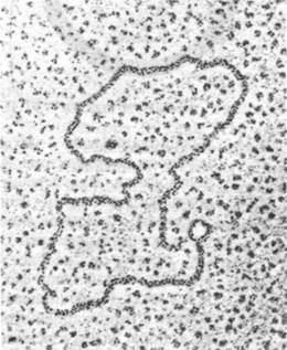
occur through the action of Mobile Genetic Elements (Table 31.1). An important class of mobile genetic elements is plasmids. These are circular, double-stranded DNA molecules (Fig. 31.9) ranging in size from two to several hundred thousand base pairs (kb). Plasmids carry genes responsible for Antibiotic Resistance, the metabolism of natural compounds, and toxin production. In essence, plasmids function as accessory Chromosomes. They differ from the bacterial chromosome in that The Cell can survive without them under certain conditions. Furthermore, plasmids possess The ability to replicate independently of the cellular chromosome. An E. coli cell typically contains about 20 copies of small plasmids and 1 to 2 large ones.
Table 31.1. Mobile genetic elements
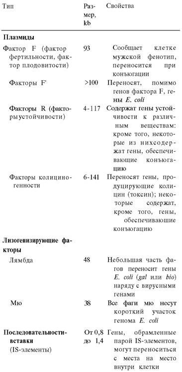
Fig. 31.9. Electron micrograph of a small R-factor plasmid
Last update: 06/08/2026
Editorial and Educational Adaptation: This material has been compiled based on the primary/original source text. The project team performed an editorial review, corrected technical inaccuracies, structured sections, and adapted the content for an educational format.
What was processed:
- elimination of formatting defects (OCR errors, structural breaks, corrupted characters);
- editorial organization of content;
- standardization of terminology in accordance with academic sources;
- verification of factual statements against the original source text.
All mentions of the author, publication year, and origin of the primary text have been preserved in accordance with the source.