Principles of Biochemistry Volume 3 - A. Lehninger 1985
Molecular mechanisms of genetic information transfer
DNA: structure of chromosomes and genes
Bacterial DNA is protected by restriction-modification systems
It has long been known that bacterial DNA contains bases bearing additional methyl groups, interspersed among millions of ordinary bases (A, T, G, and C). The Biological Significance of these methylated bases became clear As a result of several major discoveries that profoundly influenced the Selection/4.html">Development of Genetics, particularly biochemical genetics. Each bacterial species is characterized by a specific pattern of methyl-base distribution along its DNA, distinguishing it from the DNA of other species. If foreign DNA from another species somehow penetrates a living bacterial Cell, it is recognized as "foreign" precisely because it lacks the species-specific distribution pattern of methylated bases inherent to the host cell's DNA. In such cases, the foreign DNA is degraded by a specific nuclease that cleaves both DNA strands right at—or very near—the sites lacking the methylated bases characteristic of the host cell. Thus, foreign DNA undergoes restriction: it is degraded by specific Nucleases produced by each bacterial species.
The DNA of a given bacterial species is protected by two closely related enzymatic systems: 1) a modifying methylase and 2) a restricting endonuclease. The modifying methylase is responsible for establishing the species-specific methylation pattern at specific short sequences within The Cell's own DNA. The methyl groups at these sites remain unchanged throughout the cell's life. Conversely, the corresponding restricting endonuclease cleaves both strands of any other DNA in which these specific base sequences are unmethylated. A classic example is the restriction endonuclease HindII from Haemophilus influenzae (each restriction endonuclease is designated by a specific abbreviation). This enzyme cleaves both strands of any DNA containing a specific base sequence at the sites indicated by the arrows:
Class="center">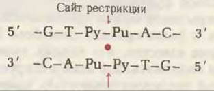
but fails to cleave the same sequence if the bases marked with a red asterisk are methylated:
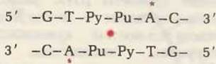
Interestingly, this short stretch of DNA, whether methylated or unmethylated, possesses internal Symmetry with respect to a central point marked in red. Rotating this segment by 180° within the plane of the drawing around the central point leaves it reading identically to its pre-rotation state. This type of symmetry is characterized by a two-fold axis of symmetry. Most of the modification-restriction sequences examined to date exhibit two-fold symmetry. The restriction endonuclease HindII cleaves both strands at the midpoint of this segment in any DNA where the sequence is unmethylated. Once cleaved in this manner, the foreign DNA cannot be repaired and therefore cannot replicate.
Subsequently, other cellular nucleases degrade such damaged DNA down to mononucleotides.
Table 27-7 lists the specific sequences attacked by typical Restriction Endonucleases (indicated by symbols) from various bacterial species. Each sequence recognized by such an enzyme possesses a two-fold axis of symmetry. Depending on the type of restriction endonuclease, Cleavage of double-stranded DNA produces either "blunt" (i.e., flush) ends (as in the case of HindII described above) or protruding, sticky ends (exemplified by the endonuclease EcoRI from E. coli). In the latter case, the two overlapping ends are called sticky because they are capable of forming complementary Base Pairs with each other.
Table 21-1. Specificity of some restriction endonucleases1)
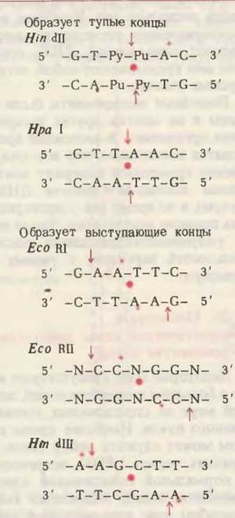
1) The bold red dot indicates the two-fold axis of symmetry, and the red arrows show the cleavage sites. Red asterisks denote methylation sites (where known) in the Organism from which the enzyme was isolated: Haemophilus influenzae for HindII and HindIII, E. coli for EcoRI and EcoRII, and Haemophilus parainfluenzae for HpaII. Pu denotes purine, Py denotes pyrimidine, and N denotes A or T.
It has been calculated that hexanucleotide sequences with two-fold rotational symmetry can occur in any DNA, regardless of species, with a probability of 1 in 4,000. Because bacterial DNA molecules consist of millions of nucleotide pairs, the likelihood that any given bacterial DNA will be cleaved at least once by any specific restriction endonuclease is extremely high. However, the host cell protects its own DNA by methylating one or more bases within the restriction-susceptible sequence; the methylated sequence fails to bind the restriction endonuclease and thus escapes cleavage.
More than 150 different restriction endonucleases have been discovered in various bacterial species to date. Some Bacteria contain more than one set of Modifying Methylases and restricting endonucleases. Nevertheless, bacterial Viruses (phages) have evolved various ways to bypass the restriction defenses of their host Cells. Certain viral DNAs contain modified bases of various kinds that allow them to evade cleavage by the restriction endonucleases of the invaded host cell. The modifying groups in such viral DNAs include methyl, hydroxymethyl, and glucosyl groups. Other viruses have evolved sequences in their DNA that lack the sites recognized by certain restriction endonucleases.
Restriction nucleases have proved to be exceptionally valuable tools in genetic research because they allow reproducible cleavage of both DNA strands at strictly defined sites. The discovery of Enzymes with such properties inaugurated a new era in Gene biochemistry. Thanks to restriction endonucleases—many of which are now commercially produced and available on the market—targeted chromosome cleavage and mapping have become possible, and these enzymes have also become indispensable tools for determining DNA nucleotide sequences. Restriction endonucleases paved the way for splicing genes from one organism into The Genome of another (Chapter 30). The 1978 Nobel Prize in Physiology and Medicine was awarded to the scientists who discovered The phenomenon of DNA Restriction, elucidated The Nature of restriction endonuclease action, and demonstrated their utility in gene excision: Werner Arber of Switzerland, and Hamilton Smith and Daniel Nathans (both of the USA).
Last update: 06/08/2026
Editorial and Educational Adaptation: This material has been compiled based on the primary/original source text. The project team performed an editorial review, corrected technical inaccuracies, structured sections, and adapted the content for an educational format.
What was processed:
- elimination of formatting defects (OCR errors, structural breaks, corrupted characters);
- editorial organization of content;
- standardization of terminology in accordance with academic sources;
- verification of factual statements against the original source text.
All mentions of the author, publication year, and origin of the primary text have been preserved in accordance with the source.