BIOCHEMISTRY - L. Stryer - 1984
VOLUME 3
Part IV INFORMATION
CHAPTER 31. GENE REARRANGEMENTS: RECOMBINATION, TRANSPOSITION, AND CLONING
31.5. The F Factor Enables Bacteria to Transfer Genes to Recipients via Conjugation
Some Plasmids mediate The transfer of bacterial genetic material to other Bacteria through The formation of direct intercellular contacts. This process, termed conjugation, was discovered in 1946 by Joshua Lederberg and Edward Tatum. During E. coli Cell conjugation, one partner (male) acts as the donor of genetic material, while the other (female) acts as the recipient. Male bacteria feature specialized surface appendages known as sex pili, whereas female Cells possess receptor sites that bind these pili. A pilus connects the male and female cells (Fig. 31.1). It then retracts, bringing the cells into close contact for DNA transfer (Fig. 31.10). The male bacterium harbors a plasmid called the F factor (from fertility), which carries the genes determining the formation of sex pili and other components involved in conjugation. During conjugation, one strand of the F factor plasmid is nicked at a specific site, and the double-stranded molecule unwinds (Fig. 31.11). The 5' end of the nicked strand enters the recipient cell, where a complementary strand is synthesized, resulting in a closed, circular, double-stranded molecule. The presence of the F factor plasmid in a recipient cell (initially F-) converts it into a male cell (F+). A male cell can spontaneously lose its F factor and thereby revert to the F- genotype.
Class="center">Fig. 31.10. Electron micrograph of two E. coli cells during conjugation
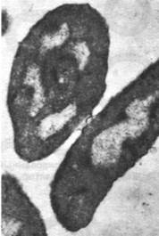
Fig. 31.11. Proposed mechanism of R factor strand transfer during conjugation. A single-strand break is introduced into the supercoiled R factor molecule within the F+ donor cell, after which it unwinds. The transfer of a single R factor strand to the F- recipient cell is coupled with the Replication of this donor strand, followed by the Synthesis of the complementary strand in the recipient cell

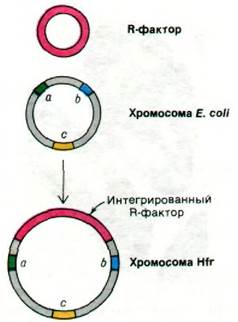
The F factor plasmid can integrate into the bacterial chromosome. Integration occurs via Crossing-over at one of multiple sites on the bacterial chromosome, with an integration frequency of approximately 10-5 per generation. Bacteria carrying the F factor in their Chromosomes are designated as Hfr cells (high frequency of recombination). Like F+ cells, Hfr cells participate in conjugation as Donors (Fig. 31.12). The distinction between them is that an Hfr cell transfers the entire bacterial chromosome (including the integrated F factor), whereas an F+ cell transmits only the F factor to the recipient. In an Hfr x F- mating, the entire chromosome is transferred in approximately 90 minutes. The order of entry of transferred genes into the recipient cell depends on the site of F factor integration and its polarity. Consequently, the Gene order on the donor chromosome can be readily mapped by interrupting conjugation at various time intervals and determining which markers have successfully been transferred. The transferred donor chromosome can recombine with the recipient chromosome. Recombination frequency is highest for genes that enter the recipient cell first, as they reside there the longest. Thus, genetic maps can be constructed by determining the entry times and recombination frequencies of markers transferred by the donor.
Fig. 31.12. Scheme of Hfr Cell Formation via Integration of the F factor with the E. coli chromosome
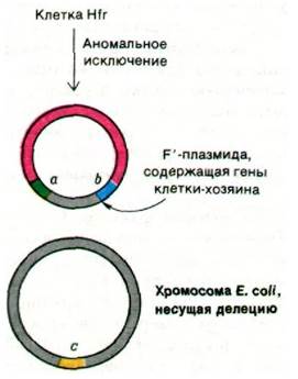
Upon excision of the F factor from the Hfr cell chromosome, it reverts to the F+ state. This process, the reverse of F factor integration, also occurs at a frequency of about 10-5 per generation. In a small fraction of revertants, crossing-over occurs at a site distinct from the integration site, generating a plasmid that carries chromosomal genes In addition to F factor genes (Fig. 31.13). Such a plasmid is termed an F' factor; the prime symbol indicates the presence of chromosomal genes. Conjugation of an F' cell with an F- cell results in the transfer of these chromosomal genes from the donor to the recipient cell, which consequently becomes diploid for those genes.
Fig. 31.13. Aberrant excision of the F factor leads to the formation of an F' plasmid carrying a portion of the E. coli chromosome
Thus, bacteria possess a mechanism for transferring entire chromosomes or multiple genes from one cell to another. The F factor can be viewed as a specialized vector that evolved specifically for the EXCHANGE OF GENETIC material. Interestingly, lysogenic phages can also participate in host gene exchange. For example, the DNA of phage  can integrate between the gal and bio GENES OF THE E. coli chromosome (Section 30.16). Prophage excision from the chromosome is normally precise, but not always. Approximately one in 105 virions of phage
  contains the gal Operon or the bio gene. Upon infection by such phages, designated as gal and bio, these genes are introduced into the E. coli cell along with phage
  genes. Another related phage, designated ɸ80, integrates near the trp operon and can transfer trp genes from one infected cell to another. Bacteriophage Mu integrates almost anywhere in the E. coli chromosome and invariably packages a piece of the bacterial chromosome upon excision. These transducing phages, much like the F factor, represent Mobile Genetic Elements that facilitate the mutual exchange of bacterial genes. It is plausible that Transduction accelerates bacterial evolution.
31.6. R Factor Plasmids Confer Antibiotic Resistance in Bacteria
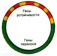
A striking example of remarkably rapid bacterial evolution is the 1955 epidemic of bacterial dysentery. A strain of Shigella dysenteriae acquired simultaneous resistance to chloramphenicol, streptomycin, sulfonamides, and tetracycline. Such multiple drug resistance is now widespread among many pathogenic microorganisms. Genes conferring resistance to numerous Antibiotics are clustered within plasmid R factors (resistance factors), also referred to as resistance plasmids. The largest of these plasmids contain several r genes along with a resistance transfer factor (RTF) region (Fig. 31.14). The RTF region enables the plasmid to be transferred to other bacteria via conjugation. In fact, The genes of the RTF region bear a close resemblance to the corresponding F factor genes. The r genes encode Enzymes that inactivate specific drugs. R factors possessing the RTF region can be transferred among different bacterial species during co-cultivation; consequently, multiple Antibiotic Resistance can be transmissible.
Fig. 31.14. Schematic representation of an R factor. RTF genes (responsible for conjugation and replication) are shown in green, r genes (responsible for resistance to various drugs) are shown in red, and IS elements are shown in yellow
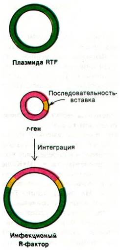
Smaller R factor plasmids lack the RTF region and typically confer resistance to only a single antibiotic. For example, the 8.2 kb plasmid pSC101 carries a tetracycline resistance gene but cannot be transferred by conjugation. However, this r gene can associate with another plasmid carrying a different resistance gene (Fig. 31.15). If these r genes integrate with a plasmid containing the RTF region, a transmissible R plasmid is generated. Therefore, complex R factor plasmids are assembled from highly mobile genetic elements that confer resistance to individual chemical compounds. Such transferable genetic elements are now termed Transposons.
Fig. 31.15. An infectious R factor is formed when an r gene associates with an RTF plasmid
31.7. IS Elements Can Integrate into Unrelated Genes
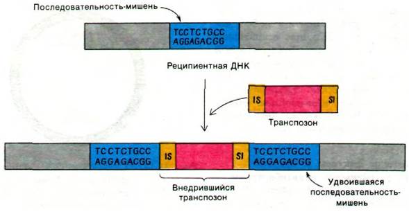
What is the basis for the high mobility of transposons? Electron Microscopy studies and DNA nucleotide sequencing have demonstrated that the sequence located at one end of a transposon is repeated at the other end. For example, the ends of the Tn3 transposon, which encodes ampicillin resistance, consist of 38-base-pair inverted repeats. The nucleotide sequences in the recipient DNA flanking the transposon on both sides form a direct repeat of a 5-9 base pair sequence that was present prior to transposon insertion (Fig. 31.16). There is no Homology whatsoever between these flanking sequences of the recipient DNA and the terminal sequences of the transposon. Furthermore, the rec genes of E. coli are not involved in transposon integration, which fundamentally distinguishes it from general genetic recombination. The ends of the transposon likely function as IS elements, directing the action of Nucleases and other Proteins involved in integration. Most importantly, the transposon does not need to be homologous to the recipient DNA, since integration Specificity is determined primarily by DNA-Structure/156.html">Protein Interactions rather than base pairing.
Fig. 31.16. Transfer of a gene (shown in red) flanked on both sides by IS elements (shown in yellow). During transposition, the recipient site (shown in blue) is duplicated. One end of the transposon represents an inverted repeat of the other end.
The smallest mobile genetic elements are insertion sequences (IS elements), which are approximately 1 kb in length. Unlike transposons, IS elements do not carry any genes. However, they exert a significant effect on the expression of neighboring genes. IS elements typically block the METABOLISM/31.html">Transcription of distal genes within a transcriptional unit. In addition, they can act as novel promoters. Furthermore, IS elements promote chromosomal rearrangements such as deletions and inversions. Multiple copies of four different IS elements (IS1, IS2, IS3, and IS4) have been identified in the E. coli chromosome. Moreover, the terminal sequences of certain transposons are identical to one of these IS elements. In all likelihood, a transposon is formed when a gene becomes flanked by a pair of IS elements.
We have already seen that plasmids and phages can exchange blocks of genes with bacterial chromosomes. In addition, plasmids and phages are capable of recombining with each other. It has been shown that the genetic element responsible for tetracycline resistance moves from an R-factor plasmid into a phage multiplying in Salmonella cells, and from there into the Salmonella chromosome, then into phage , and from phage into the E. coli trp operon, and back into phage . This remarkable journey demonstrates just how mobile prokaryotic genes truly are. It would be fascinating to discover whether eukaryotic genes possess a similarly high degree of mobility.
Last update: 06/08/2026
Editorial and Educational Adaptation: This material has been compiled based on the primary/original source text. The project team performed an editorial review, corrected technical inaccuracies, structured sections, and adapted the content for an educational format.
What was processed:
- elimination of formatting defects (OCR errors, structural breaks, corrupted characters);
- editorial organization of content;
- standardization of terminology in accordance with academic sources;
- verification of factual statements against the original source text.
All mentions of the author, publication year, and origin of the primary text have been preserved in accordance with the source.