BIOCHEMISTRY - L. Stryer - 1984
VOLUME 3
Part IV INFORMATION
CHAPTER 31. GENE REARRANGEMENTS: RECOMBINATION, TRANSPOSITION, AND CLONING
31.8. New Genomes Can Be Constructed in the Laboratory and Cloned in Host Cells
The technology of recombinant DNA, developed in recent years, represents a major milestone in molecular biology. It has made it possible to create novel combinations of unrelated genes under laboratory conditions. These new genomes can then be introduced into suitable host Cells and amplified manifold using the host Cell's DNA Synthesis machinery. Furthermore, some of these introduced genes can be transcribed and translated within their new environment. The fundamental steps of DNA Cloning are outlined below (Fig. 31.17).
Class="center">Fig. 31.17. Synthesis and Cloning of recombinant DNA molecules

1. Formation of a recombinant molecule. The DNA fragment of interest is covalently attached to a vector DNA. The essential property of a vector is its ability to replicate autonomously within a suitable host. For example, Plasmids and phage λ are the most convenient vectors for Gene cloning in E. coli cells. As will be described below, DNA molecules can be joined by ligating fragments (i.e., through the action of a ligase) that have cohesive single-stranded ends or blunt ends.
Chimeric DNA is a recombinant DNA molecule containing unrelated genes. The term originates from the chimera, a mythical creature with the HEAD of a lion, the body of a goat, and the tail of a serpent. "...A creature of divine, not mortal birth: a lion in the fore part, a dragon behind, and a goat in the middle, breathing forth a fierce blast of bright fire." Homer, *Iliad*, trans. A.T. Murray, Book VI, line 180.
2. Introduction into a host cell. Most bacterial and Eukaryotic cells take up naked DNA molecules from the medium. The uptake efficiency is low (approximately 1 per 106 DNA molecules), but under specially optimized experimental conditions, a significant fraction of the cells can be transformed. Another approach involves infecting cells with reconstituted virions containing recombinant DNA molecules. In such a synthetic viral genome, the gene of interest replaces a segment of the viral DNA that is non-essential for Replication.
3. Selection. The next step is to identify which cells carry the recombinant DNA molecule containing the desired gene. Such clones can be selected based on the presence of the vector or the inserted gene itself. For instance, certain Plasmid Vectors confer resistance to a specific antibiotic upon the host cell. Another approach is to determine which cells bind RNA complementary to the target gene or synthesize the protein it encodes. Clones containing recombinant DNA are stable, at least for several hundred generations.
Recombinant DNA cloning has already made a profound contribution to our understanding of Chromosome Structure and Gene Expression. Many inserted genes have been successfully amplified through cloning, yielding large quantities of DNA for base sequencing and Electron Microscopy studies. In addition, these clones have enabled the synthesis of large amounts of Proteins that are normally produced in minute quantities. Recombinant DNA Methods are also widely used to study complex genomes and The regulation of their expression.
31.9. Restriction Enzymes and DNA Ligase Are Essential Tools for Generating Recombinant DNA Molecules
DNA molecules can be readily joined in vitro using Restriction Endonucleases (Section 24.27), DNA ligases (Section 24.15), and other highly specific Enzymes acting on DNA. In a recombinant DNA experiment, the vector is prepared for joining with the cloned fragment by Cleavage at a single specific site using a restriction endonuclease. For example, the small plasmid pSC101 can be cleaved at a unique site by the restriction enzyme EcoR1. If this enzyme cuts the two DNA strands in a staggered fashion, complementary single-stranded ends (sticky ends) are generated. Suppose now that the DNA fragment to be inserted into this plasmid is produced by cleaving a large DNA molecule with the endonuclease EcoR1. The single-stranded ends of this fragment will then be complementary to the ends of the cleaved plasmid. The DNA fragment and the plasmid can now be annealed and joined by DNA ligase (Fig. 31.19). Bacteria are subsequently incubated in the presence of this mixture of DNA molecules. A small fraction of the bacteria carrying the plasmid is selected based on the fact that pSC101 confers tetracycline resistance to the cells. Another selection method is then used to identify clones containing the plasmid with the inserted foreign DNA.
Fig. 31.18. Electron micrograph of pSC101, a plasmid vector used for DNA cloning

Fig. 31.19. Joining of DNA molecules by the sticky-end method. One parental DNA molecule (shown in green) carries genes P and Q separated by a restriction site, whereas the other (red) carries genes X and Y. One recombinant molecule carries genes P and Y, while the other carries Q and X

A second method for joining two unrelated DNA molecules relies on The addition of poly(dA) tails to both 3' ends of one molecule and poly(dT) tails to both 3' ends of the other molecule (Fig. 31.20). These homopolymer sequences are synthesized by terminal deoxynucleotidyl transferase (terminal transferase), an enzyme that adds NUCLEOTIDES to the 3'-hydroxyl group of a DNA chain. Unlike DNA polymerase, this transferase is template-independent, and thus can append sequences consisting of only a single type of nucleotide (typically about 100 residues long). The poly(dA) ends of one DNA molecule are annealed with the poly(dT) ends of the other. Because the lengths of these terminal sequences may vary somewhat, the gaps are filled in using DNA polymerase I before sealing the strands with DNA ligase. Similarly, poly(dG) and poly(dC) terminal sequences can be used to join different DNA molecules.
Fig. 31.20. Joining of DNA molecules by the poly(dA) and poly(dT) tailing (homopolymer tailing) method

A third approach to joining DNA molecules combines the advantages of the sticky-end method with the universal applicability of the (dA–dT) tailing method. A chemically synthesized linker, i.e., a connecting duplex (six to ten Base Pairs long) that is susceptible to cleavage by a specific restriction enzyme, is covalently attached to the ends of the DNA fragment or vector. The 5' ends of the ten-nucleotide linker and the DNA molecule are phosphorylated using polynucleotide kinase and joined by phage T4 ligase, which can form a covalent bond between blunt-ended DNA molecules. Subsequent Treatment of these terminal regions with the appropriate restriction enzyme generates sticky ends (Fig. 31.21). Thus, sticky ends corresponding to the Specificity of a given restriction enzyme can be generated on virtually any DNA molecule.
Fig. 31.21. Generation of sticky ends by the attachment and cleavage of a chemically synthesized linker

31.10. Plasmids and Lambda Phage as the Most Suitable Vectors for DNA Cloning in Bacteria
To increase the efficiency of recombinant DNA uptake by cells and to facilitate the Selection of bacteria containing such molecules, new vectors are being developed. For instance, the plasmid pBR322 carries genes conferring resistance to tetracycline and ampicillin (an antibiotic related to penicillin). This plasmid is cleaved at a single unique site by each of five different restriction enzymes (Fig. 31.22). Insertion of DNA into the EcoRI restriction site does not affect the Antibiotic Resistance genes. Conversely, inserting DNA into the HindIII, SalI, or BamHI restriction sites inactivates the tetracycline resistance gene—a phenomenon known as insertional inactivation. Cells containing pBR322 with an inserted DNA fragment are resistant to ampicillin but sensitive to tetracycline, making them easy to select. Cells that fail to take up vector DNA are sensitive to both Antibiotics, whereas cells containing pBR322 without a DNA insert are resistant to both.
Fig. 31.22. Genetic Map of plasmid pBR322 carrying two antibiotic resistance genes

Another group of vectors has been derived from plasmid ColE1, which encodes colicin E, a protein toxin that kills certain strains of E. coli. The advantage of using these colicinogenic plasmids as vectors is that their replication is not under stringent control. A cell harboring ColE1 typically contains about 25 copies of the plasmid. Treating cells with chloramphenicol blocks Protein Synthesis AND chromosomal replication; however, ColE1 replication continues under these conditions. As a result, chloramphenicol-treated cells can accumulate up to 1,000 copies of ColE1, accounting for approximately half of the total cellular DNA1.
1 Plasmid pBR322 is derived from ColE1 and therefore retains The ability to similarly amplify in cells in the presence of chloramphenicol. — Transl. note.
Fig. 31.23. Electron micrograph of ColE1 plasmids

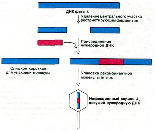
Phage  is another convenient vector. Large segments of its 48 kb DNA are non-essential for lytic infection or integration and can be replaced by foreign DNA. Mutant
  phages designed for DNA cloning have been engineered. One such mutant, designated gt = , contains two EcoRI cleavage sites instead of the five found in the wild-type phage (Fig. 31.24). Following Digestion, the central segment of this DNA molecule can be removed. The two remaining fragments together comprise 72% of the total genome length. This amount of DNA is insufficient for packaging into the phage head. The DNA length that can be efficiently packaged ranges from 75% to 105% of the wild-type genome. However, insertion of a sufficiently long DNA fragment (e.g., 10 kb) between the two DNA ends enables this recombinant DNA molecule (93% of The Genome) to be packaged (encapsidated) into the head. Nearly all infectious particles produced in this manner will contain an inserted piece of foreign DNA. An additional advantage of using these virions as vectors is that they infect bacteria with much higher efficiency than plasmids. Methods have now been developed for in vitro DNA packaging to generate infectious virions. A wide variety of phage mutants have been constructed for use as vectors, some of which can accommodate DNA inserts up to 40 kb in size.
Fig. 31.24. A phage mutant can be used as a cloning vector. The phage packaging reaction selectively selects for DNA molecules containing an insert
Last update: 06/08/2026
Editorial and Educational Adaptation: This material has been compiled based on the primary/original source text. The project team performed an editorial review, corrected technical inaccuracies, structured sections, and adapted the content for an educational format.
What was processed:
- elimination of formatting defects (OCR errors, structural breaks, corrupted characters);
- editorial organization of content;
- standardization of terminology in accordance with academic sources;
- verification of factual statements against the original source text.
All mentions of the author, publication year, and origin of the primary text have been preserved in accordance with the source.