MEDICAL BIOLOGY, HUMAN ANATOMY, PHYSIOLOGY AND PATHOLOGY - Y.I. Fedoniuk 2010
BIOLOGY
SECTION 1. BIOLOGICAL FOUNDATIONS OF HUMAN VITAL ACTIVITY
1.3. MOLECULAR-GENETIC AND CELLULAR LEVELS OF LIFE ORGANIZATION
1.3.1. The cell as the elementary genetic, structural, and functional unit of living matter
Cellular life forms: prokaryotic and Eukaryotic Cells
Cellular life forms are extremely diverse, yet evolutionary history has given rise to only two MAIN TYPES OF Cellular Organization: prokaryotic and eukaryotic. Accordingly, two groups of organisms are distinguished: prokaryotes (prenuclear) and eukaryotes (nuclear). Prokaryotes include Bacteria and blue-green Algae, while eukaryotes encompass Fungi, plants, animals, and humans. It is believed that prokaryotes were the first organisms to appear on Earth over 3.5 billion years ago As a result of chemical evolution.
The most essential feature of a Introduction/4.html">Prokaryotic Cell is the absence of a morphologically distinct Nucleus. Its role is performed by the nucleoid—nuclear material that is diffusely distributed within the Cytoplasm and not separated from it by a nuclear envelope (Fig. 1.28). The nucleoid consists of a single DNA molecule covalently closed into a ring and devoid of histone Proteins. The Molecular Weight of the nucleoid is (1-3) x 109 daltons, and it contains up to 8x10⁶ nucleotide pairs. Cytoplasmic DNA is localized in Plasmids. Prokaryotes lack a nucleolus, a system of internal Cytoplasmic membranes, and such Organelles as Mitochondria, centrosome, Golgi apparatus, Endoplasmic reticulum, Plastids, and Microbodies. Prokaryotic Ribosomes are smaller (sedimentation coefficient 70S) than eukaryotic ribosomes (sedimentation coefficient 80S). The bacterial envelope consists of a Plasma Membrane, Cell wall, and capsule. The Structure of The Plasma Membrane in PROKARYOTES AND EUKARYOTES is similar, with the difference that in prokaryotes it lacks sterols.
Class="center">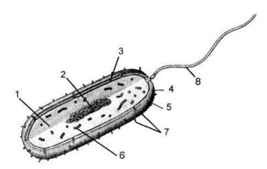
Fig. 1.28. Diagram of the organization of a prokaryotic cell:
1 - cytoplasm; 2 - nucleoid (DNA); 3 - Plasmalemma; 4 - cell wall; 5 - capsule; 6 - ribosomes; 7 - pili (fimbriae); 8 - flagellum.
The Cell wall contains peptidoglycans.
Respiratory Enzymes are concentrated in mesosomes, which are invaginations of the plasma membrane. Cytoplasmic streaming is not observed, and vacuoles are rare. Mitosis, the mitotic apparatus of division, and Meiosis are absent.
Prokaryotes are small in size (0.5-3.0 µm in diameter). They are characterized by immense biochemical diversity and rapid generation turnover (dividing every 20-30 minutes), making them convenient models for genetic research.
Eukaryotic cells possess a morphologically distinct nucleus (Figs. 1.2, 1.29). Genetic material (DNA) is almost exclusively localized within the nuclear Chromosomes and is segregated from the cytoplasm by a double-membrane nuclear envelope. Eukaryotic cells are characterized by the presence of a system of internal cytoplasmic membranes. They contain both membranous and non-membranous organelles. Eukaryotes exhibit mitosis, a mitotic division apparatus, and meiosis. Cytoplasmic DNA is concentrated in Mitochondria and Plastids. Respiratory enzymes are located on the mitochondrial cristae. In many cases, cytoplasmic streaming (cyclosis) is observed, and vacuoles are present. Unlike Prokaryotic Cells, eukaryotes are capable of coexisting with other cells and can consequently form Tissues. Among eukaryotes, unicellular, colonial, and Multicellular Organisms are distinguished. In multicellular organisms, cells are specialized and form tissues. In addition to cells, multicellular organisms contain non-cellular structures (symplasts, syncytia), but these are always cell derivatives.
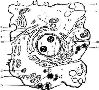
Fig. 1.29. Diagram of the structure of an animal cell.
Last update: 08/08/2026
Editorial and Educational Adaptation: This material has been compiled based on the primary/original source text. The project team performed an editorial review, corrected technical inaccuracies, structured sections, and adapted the content for an educational format.
What was processed:
- elimination of formatting defects (OCR errors, structural breaks, corrupted characters);
- editorial organization of content;
- standardization of terminology in accordance with academic sources;
- verification of factual statements against the original source text.
All mentions of the author, publication year, and origin of the primary text have been preserved in accordance with the source.