Human Anatomy Part 1 - K. A. Dubenko, A. K. Kolomiysev, Yu. B. Chaykovsky 2002
Special Part
Joints of the bones of the lower limb, juncturae membri inferioris - Joints of the bones of the leg
The foot as a whole
The FOOT, pes, is the distal segment of the lower extremity, playing a crucial role in supporting body weight while performing weight-bearing, Shock-absorbing, and locomotory Functions. This is reflected in its distinctive arched shape, configured as vaults firmly interconnected by ligaments and tendons. The arched Structure OF THE foot is unique to humans, associated with our upright bipedal posture.
The foot is formed by seven Tarsal Bones: the talus, talus; calcaneus, calcaneus; navicular bone, os naviculare; cuboid bone, os cuboideum; and three cuneiform bones — medial, intermediate, and lateral, ossa cuneiformia mediale, intermedia et laterale, as well as five (I-V) Metatarsal Bones, ossa metatarsi, and the BONES OF THE digits — Phalanges, ossa digitorum [phalanges]. Ten Bones of the foot (os naviculare, ossa cuneiformia mediale, intermedium, laterale, and ossa metatarsi I-V) form the rigid foundation of the foot (V. M. Tonkov).
The following regions and parts are distinguished on the foot: the heel, calx [reg. calcanea]; dorsum of the foot, dorsum pedis; SOLE OF THE foot, planta pedis; lateral margin, margo lateralis; medial margin, margo medialis; tarsus, tarsus; metatarsus, metatarsus; and digits, digiti; great toe, hallux; digits II–IV, digiti secundus, tertius, quartus; and digit V — the little toe, digitus minimus.
The bones of the foot articulate via joints: the talocrural (ankle) joint, intertarsal joints, tarsometatarsal joints, intermetatarsal joints, metatarsophalangeal joints, and interphalangeal joints. The talonavicular and calcaneocuboid joints are collectively known as the transverse tarsal joint (Chopart's joint), art. tarsi transversa. These joints do not communicate with each other, but their articular surfaces lie along a single S-shaped line. The tarsometatarsal joints, art. tarsometatarsale, consist of three joints collectively referred to as Lisfranc's joint.
The joints are reinforced by a series of ligaments — dorsal, plantar, and interosseous (see Figs. 107, 108). Among them, of primary importance are the interosseous talocalcaneal ligament, lig. talocalcaneum interosseum; the bifurcate ligament, lig. bifurcatum, which serves as the key of the transverse tarsal joint; and the long plantar ligament of the foot, lig. plantare longum. This is one of the strongest ligaments in The Human Body, second only to the iliofemoral ligament (according to P. F. Lesgaft, it withstands a load of 200 kg).
In addition to ligaments and bony elements, the foot is reinforced by Muscles extending from the leg to the foot and intrinsic foot muscles, which act as specific "slings" (P. F. Lesgaft) that maintain the arched shape of the foot. Mechanically, longitudinal and transverse slings are distinguished in the foot. Because the muscular component of these slings acts as an active factor, contraction of the longitudinal sling shortens the foot, while contraction of the transverse sling narrows it, thereby elevating its arches.
Class="center">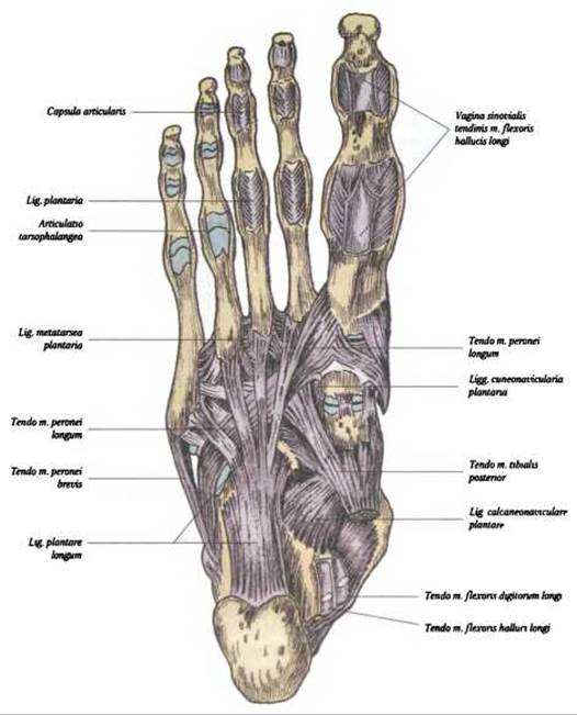
Fig. 108. Ligaments of the plantar surface of the foot
The longitudinal and transverse, active and passive slings of the arches are located on the sole and comprise the following layers: lig. calcaneocuboideum and lig. plantare longum. Furthermore, the arches are under the active influence of those muscles that insert onto them (Muscles of the great toe and the small toe).
2. The sling formed by the tendon of m. tibialis posterior, by its position and pull, predominantly suspends and activates the medial arch. The tendon of this Muscle inserts to a greater or lesser extent on all bones of the rearfoot except the talus. Additionally, this tendon serves as an insertion site for certain muscle bundles directed toward the digits. The tendon of m. flexor digitorum brevis also plays an important role in the longitudinal sling of the foot.
3. The most superficial sling is represented by the dense, resilient, and robust plantar aponeurosis, aponeurosis plantaris, and the flexor digitorum brevis muscle located deep to it.
4. The transverse sling is formed by the tendon of the fibularis (peroneus) longus muscle, the adductor hallucis muscle, and the transverse tarsal ligaments, which constitute the passive component of the slings.
In newborns and infants During the first two years of life, the foot is in a semi-supinated position. When walking, the child bears weight primarily on the medial side of the foot. The arches of the foot develop during the first two years of life, a factor that must be considered in orthopedic practice.
Due to their arrangement, ligaments, and muscles, the tarsal and metatarsal bones form a series of resilient arches of the foot (Fig. 109) that provide elasticity during walking and standing. The foot has five longitudinal arches corresponding to the five metatarsal bones, and one transverse arch formed by the stabilization of the bones. The longitudinal arches converge through the tarsus toward the weight-bearing calcaneus, while anteriorly the arches rest upon the heads of the metatarsal bones. The apex of the arch (the instep) is located between the navicular and talus bones.

Fig. 109. Longitudinal arch of the foot (diagram); the red line indicates the direction of the longitudinal arch
The arches of the foot are maintained by ligaments, muscles, and their tendons. The primary ligament reinforcing the arches of the foot is the long plantar ligament, lig. plantare longum (see Fig. 108), which originates from the Inferior surface of the calcaneus, runs anteriorly, and inserts into the tuberosity of the cuboid bone and the bases of the metatarsal bones. The three medial arches are classified as shock-absorbing (resilient), while the two lateral arches are weight-bearing. Flattening of the longitudinal and transverse arches of the foot results in a deformity known as Flatfoot, pes planus. In flatfoot, the entire surface of the sole acts as a support. This impairs the function of the extremity, alters gait, and results in a loss of elasticity. The footprint acquires a characteristic shape (Fig. 110). The shape and magnitude of the arches can change under METABOLISM/18.html">The Influence of various factors, depending on the displacement of bones relative to one another. Therefore, for prophylactic purposes, strengthening the musculoligamentous apparatus of the foot and leg through The Use of rational footwear is recommended.

Fig. 110. Footprints:
A — normal foot; B — flat foot
X-ray Anatomy of the JOINTS OF THE foot. Radiographic anatomy of the joints of the foot is evaluated in three standard projections: straight (anteroposterior), lateral, and oblique (Fig. 111 A, B). The anteroposterior radiograph of the distal leg and proximal foot demonstrates the region of the talocrural (ankle) joint. The joint space resembles the letter "U" (or "Pi"). Laterally, the radiograph reveals the sagittal curvature of the talar trochlea; the trabecular structure of the calcaneus, talus, and cuboid bones is clearly visible, and an overlay of the lateral malleolus onto the joint space is noted. The dorsoplantar radiograph of the foot visualizes all the tarsal, metatarsal, and phalangeal bones, excluding the talus and calcaneus. The joint spaces of the foot are clearly delineated. On the posterior radiograph, the region of the fibular notch of the Tibia appears as a prominence designated as the third malleolus, malleolus tertius.

Fig. 111. Overview radiograph of the ankle joint (after Ya. I. Kryzhanovsky):
A — anteroposterior PROJECTION OF THE ankle joint (joint space shaped like the letter "U" / "Pi"); 1 — tibia; 2 — Fibula; 3 — lateral malleolus; 4 — medial malleolus; 5 — talus; the dashed line indicates the joint space

Fig. 111. Overview radiograph of the foot joints (according to Ya. I. Kryzhanovsky) (Continuation):
B — lateral projection of the foot joints (the foot joints, the Bone Structure of the tarsus, and the distal PARTS OF THE leg bones are clearly visible): 1 - tibia; 2 - fibula; 3 - os calcaneus; 4 - os talus; 5 - os naviculare; 6 - os cuboideum
Last update: 08/08/2026
Editorial and Educational Adaptation: This material has been compiled based on the primary/original source text. The project team performed an editorial review, corrected technical inaccuracies, structured sections, and adapted the content for an educational format.
What was processed:
- elimination of formatting defects (OCR errors, structural breaks, corrupted characters);
- editorial organization of content;
- standardization of terminology in accordance with academic sources;
- verification of factual statements against the original source text.
All mentions of the author, publication year, and origin of the primary text have been preserved in accordance with the source.