Human Anatomy Part 1 - K. A. Dyubenko, A. K. Kolomiytsev, Yu. B. Chaykovsky 2002
Special Part
Joints of the lower limb bones, juncturae membri inferioris - Joints of the bones of the leg
Joints of the foot
The JOINTS OF THE FOOT, articulationes pedis, include:
- talocrural joint, art. talocruralis
- intertarsal joints, art. intertarseae
- tarsometatarsal joints, art. tarsometatarsales
- intermetatarsal joints, art. intermetatarsales
- metatarsophalangeal joints, art. metatarsophalangea
- interphalangeal joints of the foot, art. interphalangea pedis
The talocrural joint, art. talocruralis, is formed by the inferior articular surface of the Tibia, facies articularis inferior, flanked by the articular surfaces of the lateral and medial malleoli (of the tibia and Fibula), and by the superior surface of the body of the talus (trochlea tali) (Fig. 106). Mechanically, this is a ginglymus (hinge) joint, allowing movements around the frontal axis (flexion and extension). The Joint Capsule is thin and attaches to the margins of the articular surfaces of the connected bones, except for the anterior surface of the talus, where it attaches to the neck. Anteriorly and posteriorly, the joint capsule is roomy and thin, while laterally and medially it is reinforced by ligaments. Extending downwards from the medial malleolus is the medial ligament, lig. mediale [deltoideum], which has a triangular shape and attaches to the talus, calcaneus, and navicular bones (Fig. 107).
Class="center">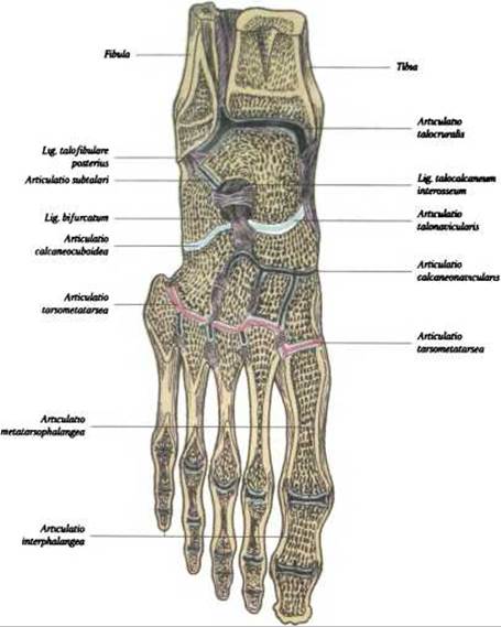
Fig. 106. Joints and ligaments of the foot - section of the talocrural joint and joints of the foot (after R. D. Sinelnikov)
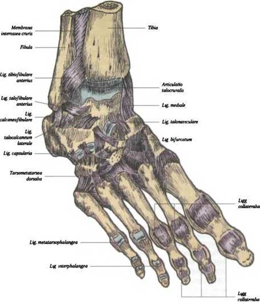
Fig. 107. Ligaments and joints of the dorsal aspect of the foot
Three independent ligaments extend from the lateral malleolus: the anterior and posterior talofibular ligaments, ligg. talofibulare, anteriores et posteriores, and the calcaneofibular ligament, lig. calcaneofibulare.
The exposed position of the talocrural joint and the weight-bearing demands placed upon it account for its frequent injuries, such as sprains, tears, dislocations, and malleolar fractures. When the talocrural joint is injured, the lateral ligaments are torn most frequently, whereas the medial deltoid ligament is rarely ruptured.
Blood supply. The talocrural joint receives its blood supply from the medial and lateral malleolar networks, rete malleolare mediale et laterale, formed by malleolar branches from the anterior and posterior tibial Arteries (a. tibialis anterior et posterior) and the fibular artery (a. peronea). Venous drainage is directed into the deep Veins OF THE leg - the anterior and posterior tibial veins (vv. tibiales anteriores et posteriores) and the fibular vein (v. peronea).
Lymphatic drainage is carried out via deep Lymphatic vessels into the popliteal Lymph Nodes (nodi lymphatici poplitei).
Innervation of the joint capsule is provided by the tibial nerve and deep fibular nerve (n. tibialis et n. peroneus profundus).
Intertarsal joints
The intertarsal joints, artt. intertarseae, are formed by the Tarsal Bones and are divided into four joints.
1. Talocalcaneonavicular joint, art. talocalcaneonavicularis - formed by three bones: the HEAD of the talus, the anterior superior surface of the calcaneus, and the posterior surface of the navicular bone; in form, it approaches a ball-and-socket (spheroid) joint. Movements occur in the sagittal plane between the talus and the calcaneus of the foot (pronation and supination). The articular capsule forms a single cavity.
The joint is reinforced by the following ligaments:
- interosseous talocalcaneal ligament, lig. talocalcaneum interosseum, which connects the talus and the calcaneus;
- plantar calcaneonavicular ligament, lig. calcaneonaviculare plantare, which runs between the sustentaculum tali and the margin of the navicular bone.
2. Subtalar joint, art. subtalaris - formed by the posterior surfaces of the talus and calcaneus; According to the shape of its articular surfaces, it belongs to the spiral joints (L. P. Afanasyev). Movements are possible around the sagittal axis (adduction and abduction). The joint has a thin articular capsule that attaches along the margin of the articular Cartilage. The joint is reinforced by the medial and lateral talocalcaneal ligaments, ligg. talocalcaneum mediale et laterale. The subtalar and talocalcaneonavicular joints are functionally combined into a single lower intertarsal (subtalar) complex, but anatomically they consist of two separate joints.
3. Calcaneocuboid joint, art. calcaneocuboidеa, - formed by the articular surfaces of the calcaneus and cuboid bones; it is classified as a saddle joint, but Functions as a uniaxial joint. Small rotational movements in the sagittal plane are possible within it. The joint has a strong, tightly stretched articular capsule reinforced by the long plantar ligament, lig. plantare longum, which extends from the calcaneus to the II–V Metatarsal Bones. The calcaneocuboid joint, together with the talonavicular joint, forms the transverse tarsal joint, art. tarsi transversa, also known as Chopart's joint1 (see Fig. 106). For disarticulation of the distal part of the foot, surgeons treat these two joints as a single functional unit. The joint line runs across both joints and resembles an "S" shape, with the medial part of this articulation convex forward and the lateral part convex backward. On the dorsal side, the joint is secured by the bifurcated ligament, lig. bifurcatum, which connects the calcaneus to the cuboid and navicular bones. This ligament serves as the "key" to the transverse tarsal joint and is of critical importance in surgery when disarticulating the distal part of the foot.
4. The cuneonavicular joint, art. cuneonavicularis, is a complex joint formed by the articular surfaces of the navicular and cuneiform bones. The articular surfaces are flat, and movements are minimal and gliding. The joint is stabilized on its dorsal and plantar aspects by strong interosseous ligaments extending from one bone to the other. The most prominent of these is the long plantar ligament of the foot, lig. plantare longum, which originates from the Inferior surface of the calcaneus and the tuberosity of the cuboid bone, and inserts into the bases of the I-V metatarsal bones.
The tarsometatarsal joints, art. tarsometatarsales (Lisfranc's joint)2, are formed by the distal row of tarsal bones and the metatarsal bones (see Fig. 106). They are plane joints and include the following:
- between the medial cuneiform and the I metatarsal bone;
- between the intermediate and lateral cuneiforms and the II-III metatarsal bones;
- between the cuboid and the IV-V metatarsal bones.
The tarsometatarsal joints are reinforced by strong plantar and dorsal ligaments of the same name, as well as the interosseous cuneometatarsal ligament, lig. cuneometatarsalia interossea, located between the I cuneiform and II metatarsal bones. This ligament is clinically significant when disarticulating the foot at this joint.
The intermetatarsal joints, art. intermetatarsales, lie between the adjacent lateral surfaces of the four lateral metatarsal bones. Based on the shape of their articular surfaces, they are plane joints, reinforced by interosseous, plantar, and dorsal metatarsal ligaments, ligg. metatarsalia interossea plantaria et dorsalia.
The metatarsophalangeales joints, art. metatarsophalangeales, are formed by the heads of the metatarsal bones and the bases of the proximal Phalanges of the I-V digits (see Fig. 106). According to the shape of their articular surfaces, these joints are classified as spheroidal (ball-and-socket), although movements within them are limited. Movements in the metatarsophalangeales joints occur around a frontal axis (flexion and extension); in addition, minor side-to-side movements are possible.
The interphalangeal joints of the foot, art. interphalangeales pedis, are located between the phalanges of the toes and are ginglymus (hinge) in shape (see Fig. 106). They are reinforced by collateral ligaments, ligg. collateralia, and plantar ligaments, ligg. plantaria, and allow movements around a frontal axis (flexion and extension).
1 François Chopart (1743-1795) was a professor of surgery, physiology, and pathology in Paris. He proposed a method of foot disarticulation, and the transverse tarsal joint is named after him.
2 Jacques Lisfranc (1790-1847) was a French surgeon who proposed the method of foot disarticulation at the tarsometatarsal joints.
The Blood supply to the joints of the foot is provided by Branches of the plantar arch, arcus plantaris, and the deep plantar branch, r. plantaris profundus, of the dorsal artery of the foot, a. dorsalis pedis.
The innervation of the joint capsules is supplied by branches of the medial and lateral plantar nerves, nn. plantares medialis et lateralis, and the superficial and deep fibular (peroneal) nerves, nn. peronei superficiales et profundis.
Last update: 08/08/2026
Editorial and Educational Adaptation: This material has been compiled based on the primary/original source text. The project team performed an editorial review, corrected technical inaccuracies, structured sections, and adapted the content for an educational format.
What was processed:
- elimination of formatting defects (OCR errors, structural breaks, corrupted characters);
- editorial organization of content;
- standardization of terminology in accordance with academic sources;
- verification of factual statements against the original source text.
All mentions of the author, publication year, and origin of the primary text have been preserved in accordance with the source.