MEDICAL BIOLOGY, HUMAN ANATOMY, PHYSIOLOGY AND PATHOLOGY - Ya.I. Fedoniuk 2010
ANATOMY, PHYSIOLOGY, PATHOLOGY
CHAPTER 5. THE PROCESS OF BLOOD AND LYMPH CIRCULATION. ITS PATHOLOGY
VESSELS OF THE SYSTEMIC CIRCULATION
1. AORTA
1.1. Branches of the aortic arch
The convex side of the arch gives rise to: 1) the brachiocephalic trunk; 2) the left common carotid artery; 3) the left Subclavian Artery (Fig. 5.15).
Class="center">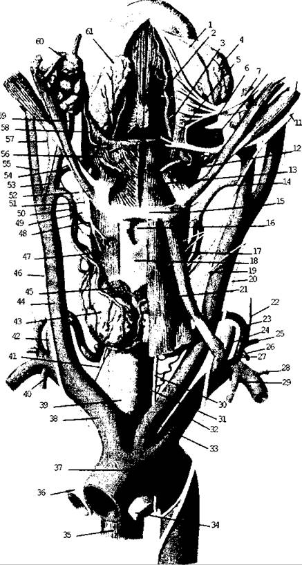
Fig. 5.15. Aortic arch and its branches
The concave side of the arch gives off small branches to the Trachea and Bronchi, as well as the ligamentum arteriosum. The latter is a remnant of the fetal ductus arteriosus, which connects the aorta to the pulmonary trunk or the left pulmonary artery.
1. The brachiocephalic trunk (truncus brachiocephalicus) bifurcates at the level of the right sternoclavicular joint into two branches: the right common carotid artery and the right subclavian artery.
2. The common carotid artery (arteria carotis communis) is a paired vessel that arises directly from the aortic arch on the left and from the brachiocephalic trunk on the right. Both Arteries ascend along the sides of the trachea and Esophagus, dividing at the level of the upper border of the thyroid Cartilage into the internal and external carotid arteries.
3. The External Carotid Artery (arteria carotis externa) supplies Blood to the external structures of the HEAD and Neck. It gives off anterior, posterior, medial, and Terminal Groups of branches.
Anterior group (Fig. 5.16):
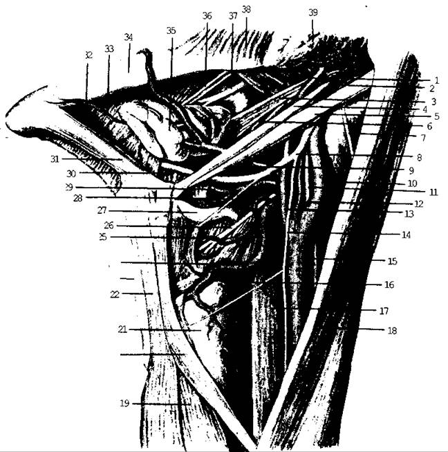
Fig. 5.16. External carotid artery
- superior thyroid artery – supplies The Thyroid Gland and Larynx;
- lingual artery – supplies the Tongue and sublingual salivary gland;
- facial artery – courses over the inferior border of the Mandible and supplies the palate with Tonsils, Cytology/practical/98.html">Submandibular salivary gland, chin, upper and lower Lips, and the ROOT of the Nose.
Posterior group (Fig. 5.17):
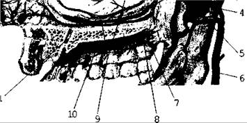
Fig. 5.17. Branches of the carotid artery
- sternocleidomastoid artery – supplies the sternocleidomastoid Muscle;
- occipital artery – supplies the occipital region;
- posterior auricular artery – supplies the external and Middle ear.
Medial group:
- ascending pharyngeal artery supplies the Pharynx, middle ear, and Meninges.
Terminal branches:
- superficial temporal artery
extends to the auricle, Parotid salivary gland, and the temporal, facial, and parietal regions;
- maxillary artery - supplies the maxillary Teeth, Muscles of Mastication, auricle, Hard and Soft 24 palate, dura 23 mater, and Nasal cavity (Fig. 5.18);
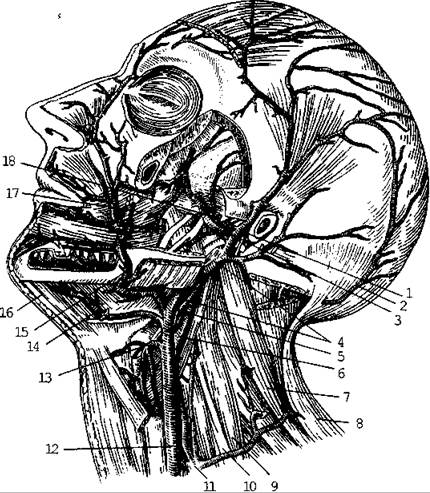
Fig. 5.18. Terminal branches of the maxillary artery
The Internal Carotid Artery (a. carotis interna) ascends to the base of The Skull and enters the cranial cavity through the carotid canal 20, lying lateral to the sella turcica.
It supplies blood to the Brain and the visual organ. It gives off no branches in the neck region, but within the cranial cavity, it gives rise to the following vessels:
- ophthalmic artery - passes through the optic canal into the Orbit along with the Optic nerve, supplying the orbital contents as well as the dura mater and the nasal mucosa;
- anterior cerebral artery (paired) - supplies the medial surfaces of the cerebral hemispheres. The left and right arteries are connected by the anterior communicating artery;
- middle cerebral artery (paired) - supplies the dorsolateral surface of the cerebral hemispheres;
- posterior communicating artery anastomoses with the posterior cerebral artery (a branch of the Basilar artery). The anterior, middle, and posterior cerebral arteries, along with the anterior and posterior communicating arteries, form a closed arterial ring at the Base of the brain known as the cerebral arterial circle (circle of Willis) (Fig. 5.19).
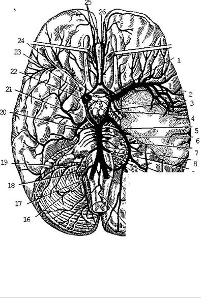
Fig. 5.19. Arteries of the brain, inferior view
The subclavian artery (a. subclavia) — the right arises from the brachiocephalic trunk, and the left from the aortic arch — exits the thoracic cavity through the superior thoracic aperture, rests in the corresponding groove on the first rib within the scalene interval, where it is accompanied by the Brachial Plexus. Below the lateral margin of the first rib, the subclavian artery enters the axillary cavity, continuing as the axillary artery.
Branches of the subclavian artery (Fig. 5.20):
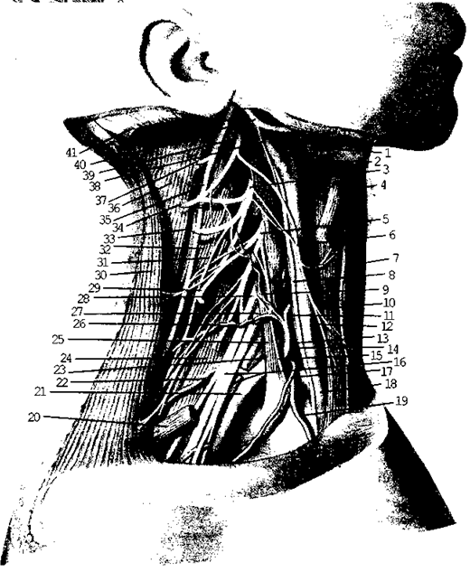
Fig. 5.20. Subclavian artery and its branches
- vertebral artery - ascends through the foramina in the transverse processes of the cervical vertebrae, enters the cranial cavity through the foramen magnum, and joins its counterpart from the opposite side to form the basilar artery. The terminal branches of the vertebral artery are the posterior cerebral arteries (right and left), which contribute to The formation of the circle of Willis and supply the occipital and temporal lobes of the cerebral hemispheres. Throughout its course, the vertebral artery gives off branches supplying the Spinal Cord, Medulla Oblongata, Cerebellum, and Inner ear;
- thyrocervical trunk - supplies the thyroid gland, larynx, and the Muscles of the neck and back;
- internal thoracic artery - descends along the posterior surface of the anterior thoracic wall, supplying the Thymus, mammary gland, intercostal muscles, Pericardium, and Diaphragm;
- costocervical trunk - supplies the muscles of the neck and the upper two intercostal spaces;
- transverse cervical artery - supplies the muscles and Skin of the back.
The subclavian artery continues as the axillary artery.
The axillary artery (a. axillaris) lies within the axillary cavity and extends into the arm, where it becomes the brachial artery. It gives off branches to the muscles of the shoulder girdle, the capsule of the shoulder joint, the muscles of the I and II intercostal spaces, and the superficial back muscles.
The brachial artery (a. brachialis) runs in the sulcus along the medial side of the biceps brachii, accompanied by the Long branches of the brachial plexus and the brachial Veins. Along its course, it gives off several branches that supply the humerus, the muscles and skin of the arm, and the elbow joint (Fig. 5.21). In the region of the cubital fossa, it divides into the ulnar and radial arteries.
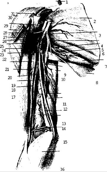
Fig. 5.21. The brachial artery and its branches
In the forearm, the ulnar and radial arteries run in their respective sulci, supplying the bones, muscles, and skin of the forearm. In the distal third of the forearm, the radial artery lies superficially, making it easily palpable for checking the pulse (Fig. 5.22).
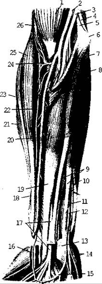
Fig. 5.22. The ulnar and radial arteries
Entering the hand, both arteries and their branches anastomose to form the superficial and deep palmar arterial arches, which supply blood to the bones and soft Tissues of the hand.
Last update: 08/08/2026
Editorial and Educational Adaptation: This material has been compiled based on the primary/original source text. The project team performed an editorial review, corrected technical inaccuracies, structured sections, and adapted the content for an educational format.
What was processed:
- elimination of formatting defects (OCR errors, structural breaks, corrupted characters);
- editorial organization of content;
- standardization of terminology in accordance with academic sources;
- verification of factual statements against the original source text.
All mentions of the author, publication year, and origin of the primary text have been preserved in accordance with the source.