Human Anatomy - Kotsan I. Ya. 2009
Vessels of the Lesser (Pulmonary) Circulation
Branches of the Abdominal Aorta
Parietal Branches of the Abdominal Aorta
The Abdominal Aorta is a direct continuation of the Thoracic Aorta (Fig. 208). The Arteries branching from the abdominal aorta are likewise divided into parietal and visceral groups.
1. The inferior phrenic artery (a. phrenica inferior) is the first branch of the abdominal aorta. It is a paired vessel that originates at the level of the 12th thoracic vertebra and extends toward the Inferior surface of the Diaphragm. On its path to the diaphragm, it gives off 1 to 24 superior suprarenal arteries (aa. suprarenales superiores). The inferior phrenic arteries supply Blood to the diaphragm and the Adrenal Glands.
Class="center">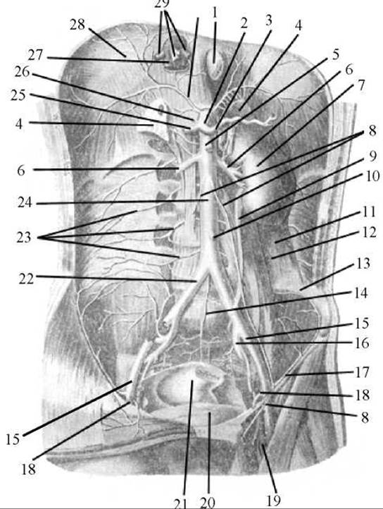
Fig. 208. Abdominal aorta (anterior view)
1 — Esophagus; 2 — celiac trunk; 3 — splenic artery; 4 — Adrenal gland; 5 — superior mesenteric artery; 6 — renal artery; 7 — left Kidney; 8 — testicular (in females, ovarian) arteries; 9 — left Ureter; 10 — inferior mesenteric artery; 11 — quadratus lumborum Muscle; 12 — psoas major muscle; 13 — iliac crest; 14 — median sacral artery; 15 — external iliac artery; 16 — left internal iliac artery; 17 — left inguinal ligament; 18 — external iliac vein; 19 — femoral vein; 20 — Urinary Bladder; 21 — rectum; 22 — right common iliac artery; 23 — lumbar arteries; 24 — abdominal aorta; 25 — common hepatic artery; 26 — left gastric artery; 27 — INFERIOR VENA CAVA; 28 — diaphragm; 29 — hepatic Veins
2. The lumbar arteries (aa. lumbales), numbering four pairs, arise from the posterior circumference of the aorta at the level of the bodies of lumbar vertebrae I–IV and course toward the Abdominal Muscles. Each artery gives off a dorsal branch (r. dorsalis) supplying the muscles and Skin of the lumbar region. A spinal branch (r. spinalis) originates from the dorsal branch and passes through the intervertebral foramen to reach the Spinal Cord.
3. The median sacral artery (a. sacralis mediana) is an unpaired vessel that serves as the direct continuation of the abdominal aorta below the level of its bifurcation. Appearing as a slender and variable trunk, the artery runs inferiorly along the pelvic surface of the sacrum and terminates on the anterior surface of the coccygeal apex in the coccygeal body (glomus coccygeum), which contains chromaffin Cells and numerous arteriovenous anastomoses. Branches arising laterally from the median sacral artery include: a) a pair of ima lumbar arteries (aa. lumbales imae), which essentially represent the fifth lumbar arteries; b) lateral sacral branches (rr. sacrales laterales) that anastomose with the lateral sacral arteries (Branches of the internal iliac artery).
The median sacral artery supplies blood to the sacrum, the coccyx, and adjacent muscles.
Last update: 08/08/2026
Editorial and Educational Adaptation: This material has been compiled based on the primary/original source text. The project team performed an editorial review, corrected technical inaccuracies, structured sections, and adapted the content for an educational format.
What was processed:
- elimination of formatting defects (OCR errors, structural breaks, corrupted characters);
- editorial organization of content;
- standardization of terminology in accordance with academic sources;
- verification of factual statements against the original source text.
All mentions of the author, publication year, and origin of the primary text have been preserved in accordance with the source.