BIOCHEMISTRY - L. Stryer - 1984
VOLUME 3
PART V. MOLECULAR PHYSIOLOGY
CHAPTER 35. HORMONE ACTION
35.11. Insulin Receptors Are Localized in the Plasma Membrane of Target Cells
Insulin binds tightly to specific receptors on Cell/30.html">The Plasma Membrane of target Cells. This has been demonstrated in experiments using radioactive insulin containing covalently bound 125I. The dissociation constant of the insulin-receptor complex is approximately 10-10 M. The Need for such tight binding is explained by the low concentration of insulin in the Blood (on the order of 10-10 M). The association rate constant for complex formation is very high (about 107 M-1 • s-1) and approaches the limit for diffusion-controlled reactions. An adipose tissue cell contains only about 1 • 104 insulin receptors, which corresponds to a density of one receptor per square micrometer of plasma membrane. In other words, there is one Insulin Receptor per 1 • 106 membrane Phospholipids.
The binding parameters of insulin to the solubilized receptor and to the receptor in the intact plasma membrane are identical. Solubilized receptors were purified by Affinity Chromatography, a technique based on the specific binding Properties of the receptor. For purification, the solubilized membrane preparation was passed through an agarose Column with covalently attached insulin; the insulin receptors bound tightly to the column, whereas the Other components of the mixture passed straight through. Subsequently, the insulin receptors were eluted with an acidified urea solution, which caused the receptors to denature and dissociate from the agarose-bound insulin. Fortunately, the purified receptors could then be renatured by removing the urea and raising the pH of the solution. Using this method, Pedro Cuatrecasas purified insulin receptors 250,000-fold. The insulin-binding subunit of the receptor is a glycoprotein with a mass of 135 kDa (Fig. 35.15).
Class="center">Fig. 35.15. Gel Electrophoresis of a plasma membrane extract from Liver cells (A) and purified insulin receptor (B). Electrophoresis was performed under denaturing conditions
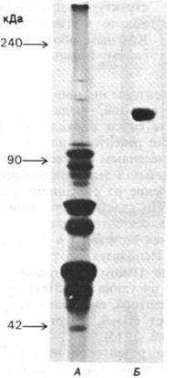
Insulin receptors account for a mere 4 • 10-3% of the total membrane protein in a liver homogenate. Consequently, to obtain 1 mg of pure receptor protein, one must process an amount of homogenate equivalent to 500 g of protein, which requires the livers of 200 rats. This calculation shows that isolating receptors in quantities sufficient for amino acid sequencing, X-Ray Diffraction Analysis,
and reconstitution experiments is a laborious yet feasible task.
It is likely that the interaction of insulin with receptors on the plasma membrane triggers many of the rapid effects of insulin, such as the activation of Amino Acid and glucose Transport Across the membrane. Another rapid effect of Insulin is the phosphorylation of a protein belonging to the ribosomal 40S subunit. None of these hormonal effects involve changes in cAMP levels. The intracellular messengers that mediate the Action of Insulin remain to be identified.
Diabetes — the name points to increased urination. Aretaeus, a Cappadocian physician of the second century AD, wrote: "The epithet 'diabetes' refers to a condition resembling the passage of Water through a siphon." With keen observation, he characterized diabetes as "a melting down of the flesh and limbs into urine."
Mellitus — (from Latin, meaning "sweetened with honey") indicates the presence of sugar in the urine of diabetic patients.
35.12. Insulin Deficiency Causes Diabetes
Diabetes Mellitus is a complex disorder affecting hundreds of millions of people. It is characterized by elevated levels of glucose in the blood and urine. Glucose excretion in the urine occurs when the blood glucose concentration exceeds the reabsorptive capacity of the renal tubules. Large amounts of water are excreted along with the glucose, so that an untreated patient in the acute stage of the disease suffers from intense hunger and thirst. The loss of glucose leads to the depletion of carbohydrate reserves, followed by The breakdown of fats and Proteins. The mobilization of fats generates large amounts of acetyl-CoA. If acetyl-CoA cannot be fully utilized in The Tricarboxylic Acid Cycle due to an insufficient supply of oxaloacetate, Ketone Bodies (acetoacetate, acetone, and β-hydroxybutyrate) are formed. Recall that animal organisms cannot synthesize oxaloacetate from acetyl-CoA; in animals, oxaloacetate is formed from glucose and Certain Amino Acids (Section 23.4). The excretion of ketone bodies disrupts acid-base balance and exacerbates dehydration. Ultimately, in the acute stage of diabetes, an untreated patient may develop a coma and die.
Diabetes mellitus is caused by insulin deficiency. However, the underlying causes of insulin deficiency are unknown in most cases. Diabetes can be reproduced
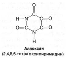
in experimental animals by surgical removal of a large portion of the Pancreas or by chemical destruction of β-cells. These insulin-producing cells are selectively destroyed by the administration of alloxan. Diabetes can also be induced by administering anti-insulin Antibodies. Further proof of the crucial role that insulin deficiency plays in The Development of diabetes is that insulin administration rapidly reverses the acute symptoms of the disease.
Why is The amount of insulin in a diabetic patient lower than the physiological needs of the Tissues? It is now established that the clinical condition known as diabetes mellitus can stem from a variety of distinct molecular defects. Let us consider some of the causes of diabetes.
1. Impaired conversion of proinsulin to insulin. Mutations affecting amino acid residues in the junction region between the A-chain (or B-chain) and the C-peptide in proinsulin can impair its conversion into insulin. Such patients exhibit high levels of proinsulin in their Blood Plasma (which lacks hormonal activity).
2. Altered molecular Structure of insulin. Another type of mutation leads to the substitution of an amino acid residue in a critically important region of the insulin molecule. For instance, if phenylalanine is replaced by leucine near the carboxyl terminus of the B-chain, the hormonal activity of such insulin is reduced 10-fold. Interestingly, this region of the insulin molecule has been evolutionarily conserved from primitive hagfish to humans.
3. Insulin receptor defects. Some patients secrete normal insulin, but its binding to target cells is impaired. Consequently,
in this case, There is a defect in the insulin receptors within the Plasma Membranes.
4. Impaired coupling of insulin receptors. In some cases, patients secrete normal insulin, their target cells contain a normal number of insulin receptors, and the hormone-binding parameters are also normal. Apparently, the defect in these patients is localized inside The Cell. Specifically, it is possible that there is a failure in coupling between the insulin-receptor complex and the next component in the hormonal signaling cascade.
35.13. Endorphins: Brain Peptides Acting Like Opiates
For centuries, opiates, particularly morphine, have been used as analgesics. In 1680, Thomas Sydenham wrote: "Among all the remedies which it has pleased Almighty God to give to man to relieve his sufferings, there is none so universal and so efficacious as opium." But why does the vertebrate Brain contain receptors for Alkaloids derived from poppy seeds? Neuropharmacologists hypothesized that opiate receptors are not intended for interaction with plant alkaloids, but rather for perceiving endogenous regulators of Pain Sensation. According to this view, morphine exerts its pharmacological effect solely because it mimics substances naturally occurring in the animal body. This question was finally resolved in 1975 when John Hughes isolated two Peptides with opiate-like activity from pig brain. These structurally similar pentapeptides, designated as Methionine-enkephalin and leucine-enkephalin, are abundant in certain nerve terminals. Apparently, they are involved in the integration of sensory information related to pain.
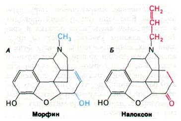
A year later, Roger Guillemin isolated longer peptides—endorphins—from the intermediate lobe of the Pituitary Gland. Endorphins possess virtually the same pain-relieving capacity as morphine (at equivalent concentrations). Administration of endorphins into the BRAIN VENTRICLES OF laboratory animals produces a remarkable effect. For instance, β-endorphin induces profound whole-body analgesia lasting several hours, accompanied by a drop in body Temperature. Moreover, the animals enter a stupor and lie motionless. After a few hours, The Effect of endorphins wears off, and the animals resume normal behavior. It was also discovered as a striking fact that the action of endorphins is abolished within seconds of administering naloxone (Fig. 35.17), a well-known morphine antagonist. Judging by the behavioral responses induced by endorphins, under normal conditions these peptides are involved in The regulation of emotional responses. Many Methods required to test this hypothesis have already been developed. For example, to detect extremely small quantities of peptides, such as endorphins, radioimmunoassay is used, combining the sensitivity of radioisotope methods with the Specificity of the immune reaction. Here we are witnessing
The Emergence of a new and promising field in neurobiology and neuropsychiatry.
Fig. 35.16. Amino acid sequences of methionine-enkephalin (A), leucine-enkephalin (B), and β-endorphin (C). The shared tetrapeptide sequence is highlighted in blue

Fig. 35.17. Structure of morphine, an opiate (A), and naloxone, a morphine antagonist (B)
Last update: 06/08/2026
Editorial and Educational Adaptation: This material has been compiled based on the primary/original source text. The project team performed an editorial review, corrected technical inaccuracies, structured sections, and adapted the content for an educational format.
What was processed:
- elimination of formatting defects (OCR errors, structural breaks, corrupted characters);
- editorial organization of content;
- standardization of terminology in accordance with academic sources;
- verification of factual statements against the original source text.
All mentions of the author, publication year, and origin of the primary text have been preserved in accordance with the source.