Review of Medical Physiology - William F. Ganong 2002
Endocrine System, Metabolism, and Reproduction
Endocrine Functions of the Pancreas and Regulation of Carbohydrate Metabolism
Consequences of Insulin Deficiency
The far-reaching Physiological effects of Insulin are best understood in light of the profound and widespread consequences of insulin deficiency.
In humans, insulin deficiency is a common and serious pathological condition. In animals, it can be induced by pancreatectomy; by administration of alloxan, streptozotocin, and Other toxins that selectively destroy the B Cells of the pancreatic islets in certain doses; by drugs that inhibit insulin secretion; or by administration of anti-insulin Antibodies. Several strains of mice, rats, hamsters, guinea pigs, and monkeys with a high incidence of spontaneous diabetes mellitus are also known and described.
Class="center">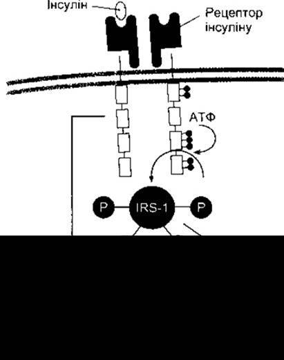
Fig. 19-7. Intracellular responses to insulin binding to its receptor. Dark circles and circles labeled with P indicate phosphate groups; IRS-1, Insulin Receptor substrate-1.
The constellation of abnormalities caused by insulin deficiency is called diabetes mellitus. Greek and Roman physicians used the term diabetes to describe conditions whose primary hallmark was a large volume of urine. They distinguished two types: diabetes mellitus, in which the urine had a sweet taste, and diabetes insipidus, in which the urine was tasteless. Today, the term diabetes insipidus is reserved for conditions resulting from a deficiency in the synthesis or action of vasopressin (see Chapter 14), while the word diabetes alone serves as a synonym for diabetes mellitus.
Diabetes is accompanied by polyuria, polydipsia, weight loss despite polyphagia (increased appetite), hyperglycemia, glucosuria, ketonemia, acidosis, and coma. Although a variety of biochemical abnormalities occur, the primary defects to which all others can be traced are, first, decreased glucose entry into various peripheral Tissues and, second, increased hepatic glucose output into the bloodstream. Consequently, there is an extracellular excess of glucose coupled with an intracellular glucose deficit in many cells—a condition aptly described as "starvation amidst plenty." In addition, amino acid uptake into Muscles decreases, and lipolysis increases.
It is now well established that diabetes involves an absolute or relative hypersecretion of Glucagon. This occurs even after total pancreatectomy, because glucagon is secreted not only by the Pancreas but also by other Regions of the digestive tract. Somatostatin reduces the secretion of both glucagon and insulin (see Chapter 14), and when administered to pancreatectomized animals, plasma glucose levels drop back to normal. It appears that the extracellular glucose excess is partly a consequence of hyperglucagonemia. However, some degree of hyperglycemia persists even when glucagon secretion is reduced to zero.
Glucose Tolerance
In diabetes, glucose accumulates in the Blood, especially after meals. When a diabetic individual is subjected to a glucose load, plasma glucose rises and returns to baseline much more slowly than in healthy subjects. The response to a standard oral test dose of glucose—the glucose tolerance test—is widely used in the clinical Diagnosis of diabetes (Fig. 19-8).
The impaired glucose tolerance seen in diabetes is partly the result of reduced cellular glucose entry (decreased peripheral utilization). In the absence of insulin, glucose uptake by skeletal, cardiac, and smooth Muscle, as well as other tissues, is markedly impaired (Fig. 19-9). Hepatic glucose uptake is also diminished, though this effect is mediated indirectly. Intestinal Glucose Absorption remains unchanged, as does renal tubular reabsorption of glucose from the ultrafiltrate in the proximal convoluted tubules. Glucose uptake by most of the Brain and by erythrocytes is also normal.
A second and major cause of hyperglycemia in diabetes is the disruption of the glucostatic function of the Liver (see Chapter 17). The liver normally extracts glucose from the blood and stores it as Glycogen; however, because it contains glucose-6-phosphatase, it also releases glucose back into the Circulation. Indeed, Claude Bernard referred to the liver as an endocrine gland that secretes glucose. Insulin promotes glycogen synthesis and inhibits hepatic glucose output. When plasma glucose is elevated, insulin secretion normally rises, and hepatic Gluconeogenesis declines. This regulatory response is lost in diabetes. Glucagon also contributes to the hyperglycemia; furthermore, during severe stress or illness, hepatic glucose release is further stimulated by catecholamines, cortisol, and Growth Hormone.
Effects of Hyperglycemia
Hyperglycemia alone can cause symptoms due to the hyperosmolarity of the blood. Furthermore, if the renal capacity for glucose reabsorption is exceeded, glucosuria ensues. The excretion of osmotically active glucose molecules is accompanied by the loss of large volumes of Water (osmotic diuresis; see Chapter 38). The resulting dehydration activates neural mechanisms that regulate water intake, leading to polydipsia. There is also a notable urinary loss of Na+ and K+. For every gram of glucose excreted, the body loses 4.1 kcal of energy. Increased food intake to compensate for these losses merely drives plasma glucose levels higher and intensifies glucosuria, without preventing the mobilization of endogenous protein and fat stores or halting weight loss.
When plasma glucose levels are chronically elevated, small amounts of Hemoglobin A undergo nonenzymatic glycosylation to form HbA1c (see Chapter 27). Rigorous management of diabetes with insulin lowers this level; consequently, HbA1c concentration is used clinically as an integrated index of glycemic control over the preceding four to six weeks.
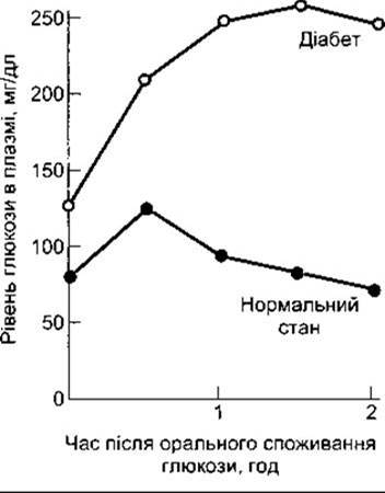
Fig. 19-8. Oral glucose tolerance test. Adults are given 75 g of glucose dissolved in 300 ml of water. In normal individuals, the fasting venous plasma glucose level is below 115 mg/dL, the two-hour post-load value is below 140 mg/dL, and no intermediate value exceeds 200 mg/dL. Diabetes mellitus is diagnosed if both the two-hour value and any other value exceed 200 mg/dL. Impaired glucose tolerance is diagnosed when values are higher than the upper limit of normal but below the diagnostic threshold for diabetes.
The Role of chronic hyperglycemia in The Development of long-term diabetic complications is discussed below.
Effects of Intracellular Glucose Deficiency
The extracellular excess of glucose in diabetes contrasts sharply with the intracellular shortage. Glucose Catabolism is normally the primary source of energy for cellular processes; in diabetes, however, energy demands must be met almost entirely through The breakdown of protein and fat reserves. This triggers pathways that drastically accelerate protein and fat catabolism, with ketonemia being a major consequence of enhanced lipid breakdown.
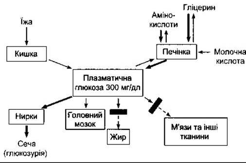
Fig. 19-9. Disturbances in plasma glucose Homeostasis during insulin deficiency. Compare with Fig. 17-14. Bold arrows indicate pathways described in the text. Bars across arrows denote blocked reactions.
Defective glucose utilization within the cells of the ventromedial nuclei of the Hypothalamus is likely the cause of hyperphagia in diabetes. If The activity of the satiety center in this Nucleus is suppressed owing to impaired cellular glucose utilization, the lateral feeding center operates unopposed, leading to increased food intake (see Chapter 14). However, other mechanisms have also been proposed to explain this phenomenon.
Glycogen depletion is a typical consequence of glucose deficiency. As expected, glycogen content in both The Liver and skeletal muscles of diabetic animals is significantly reduced.
Alterations in Protein METABOLISM
In diabetes, the rate at which Amino Acids are catabolized to CO2 and H2O is accelerated. In addition, greater amounts of amino acids are converted into glucose in the liver. These alterations are illustrated in Fig. 19-10, which also depicts other major anomalies of Intermediary Metabolism in the liver.
Some indication of The rate of gluconeogenesis in the fasting diabetic animal can be obtained by measuring The ratio of urinary glucose (dextrose) to nitrogen (the D:N ratio). In fasting animals, liver glycogen is depleted, and glycerol is converted to glucose in only very limited amounts, making protein virtually the sole source of plasma glucose (see Chapter 17). It can be calculated that the quantity of protein carbon equivalent to 1 g of urea nitrogen is sufficient to yield 8.3 g of glucose. Therefore, a D:N ratio of approximately 3, as observed in diabetes, indicates that about 33% of the carbon from metabolized protein is converted into glucose.
The increase in gluconeogenesis is driven by multiple factors. Glucagon stimulates gluconeogenesis, and hyperglucagonemia is commonly present in diabetes. Adrenal glucocorticoids also promote gluconeogenesis when their levels rise in severely ill diabetic patients. Furthermore, there is an increased supply of amino acids available for gluconeogenesis because, in the absence of insulin, less protein is synthesized in muscle, thereby elevating blood amino acid levels. Alanine is particularly readily converted into glucose. In addition, the activity of Enzymes that catalyze The conversion of Pyruvate and other two-carbon metabolic intermediates into glucose is enhanced. This applies notably to phosphoenolpyruvate carboxykinase, which facilitates the conversion of oxaloacetate to phosphoenolpyruvate (see Chapter 17), as well as fructose-1,6-bisphosphatase, which catalyzes the conversion of fructose bisphosphate to fructose-6-phosphate, and glucose-6-phosphatase, which controls the release of glucose from the liver into the blood. Elevated levels of acetyl-CoA stimulate pyruvate carboxylase activity, and insulin deficiency leads to increased acetyl-CoA production as Lipogenesis slows down. Pyruvate carboxylase catalyzes the conversion of pyruvate to oxaloacetate (see Fig. 17-9).
The combined effects of accelerated protein conversion to CO2, H2O, and glucose, alongside reduced Protein Synthesis in diabetes, result in a negative nitrogen balance, Protein deficiency, and wasting. Protein depletion of any Etiology is associated with lowered resistance to infections.
Lipid Metabolism IN Diabetes
The principal anomalies of lipid metabolism in diabetes are the acceleration of lipid catabolism with increased ketone body production, coupled with a decrease in the synthesis of Fatty acids and triglycerides. The manifestations of these lipid Metabolic Disorders are so prominent that diabetes is arguably better characterized as a disease of lipid metabolism rather than of Carbohydrate Metabolism.
Under normal conditions, 50% of ingested glucose is oxidized to CO2 and H2O; 5% is converted into glycogen; and 30% to 40% is converted into fat in adipose depots. In diabetes, less than 5% is converted into fat, even though the amount oxidized to CO2 and H2O is also diminished and the amount converted to glycogen is not increased. Consequently, glucose accumulates in the blood and spills over into the urine.
The roles of lipoprotein lipase and hormone-sensitive lipase in the Regulation of Lipid Metabolism are discussed in Chapter 17. In diabetes, the conversion of glucose to fatty acids for deposition is impaired due to intracellular glucose deficiency. Insulin inhibits hormone-sensitive lipase in adipose tissue; in the absence of this hormone, the plasma free fatty acid (FFA) level more than doubles. Elevated glucagon levels further contribute to FFA mobilization. Consequently, plasma FFA levels fluctuate in parallel with plasma glucose levels and serve as a somewhat better indicator of the severity of the diabetic state. In the liver and other tissues, Fatty acids are catabolized to acetyl-CoA. A portion of this acetyl-CoA is oxidized along with amino acid residues to form CO2 and H2O via The Citric Acid Cycle; however, the influx exceeds the capacity of the tissues to catabolize acetyl-CoA.
The hepatic processes occurring in diabetes are illustrated in Fig. 19-10. In addition to the aforementioned increase in gluconeogenesis and massive output of glucose into the blood, there is also a marked impairment in the conversion of acetyl-CoA to malonyl-CoA and subsequently to fatty acids. This defect arises from a deficiency of acetyl-CoA carboxylase, The enzyme catalyzing this conversion. The excess acetyl-CoA is instead diverted into The production of Ketone Bodies (see below).
In untreated diabetes, the plasma concentrations of triglycerides, chylomicrons, and FFAs are elevated—a condition referred to as lipemia (hyperlipemia). The accumulation of these constituents results from impaired clearance of triglycerides into adipose tissue depots. Reduced lipoprotein lipase activity contributes significantly to this decreased triglyceride removal.
Ketonemia
When there is an excess of acetyl-CoA in the body, a portion of it is converted into acetoacetyl-CoA and subsequently, in the liver, into acetoacetate. Acetoacetate and its derivatives—acetone and β-hydroxybutyrate—enter the bloodstream in substantial quantities (see Chapter 17). These circulating ketone bodies serve as an important energy source during fasting. It is estimated that in normal dogs subjected to fasting, half of the total Energy Metabolism is derived from ketone bodies. In diabetics, the rate of ketone body utilization is also significant. Studies indicate that in diabetic patients, the maximum rate at which fat can be catabolized without overt ketonemia is 2.5 g/kg of body weight per day. In untreated diabetes, production far exceeds this threshold, and ketone bodies accumulate in the bloodstream. There is some evidence that in severe diabetes, the rate of ketone body utilization may actually decline, exacerbating ketonemia, whereas insulin promotes the utilization of ketone bodies by muscle.
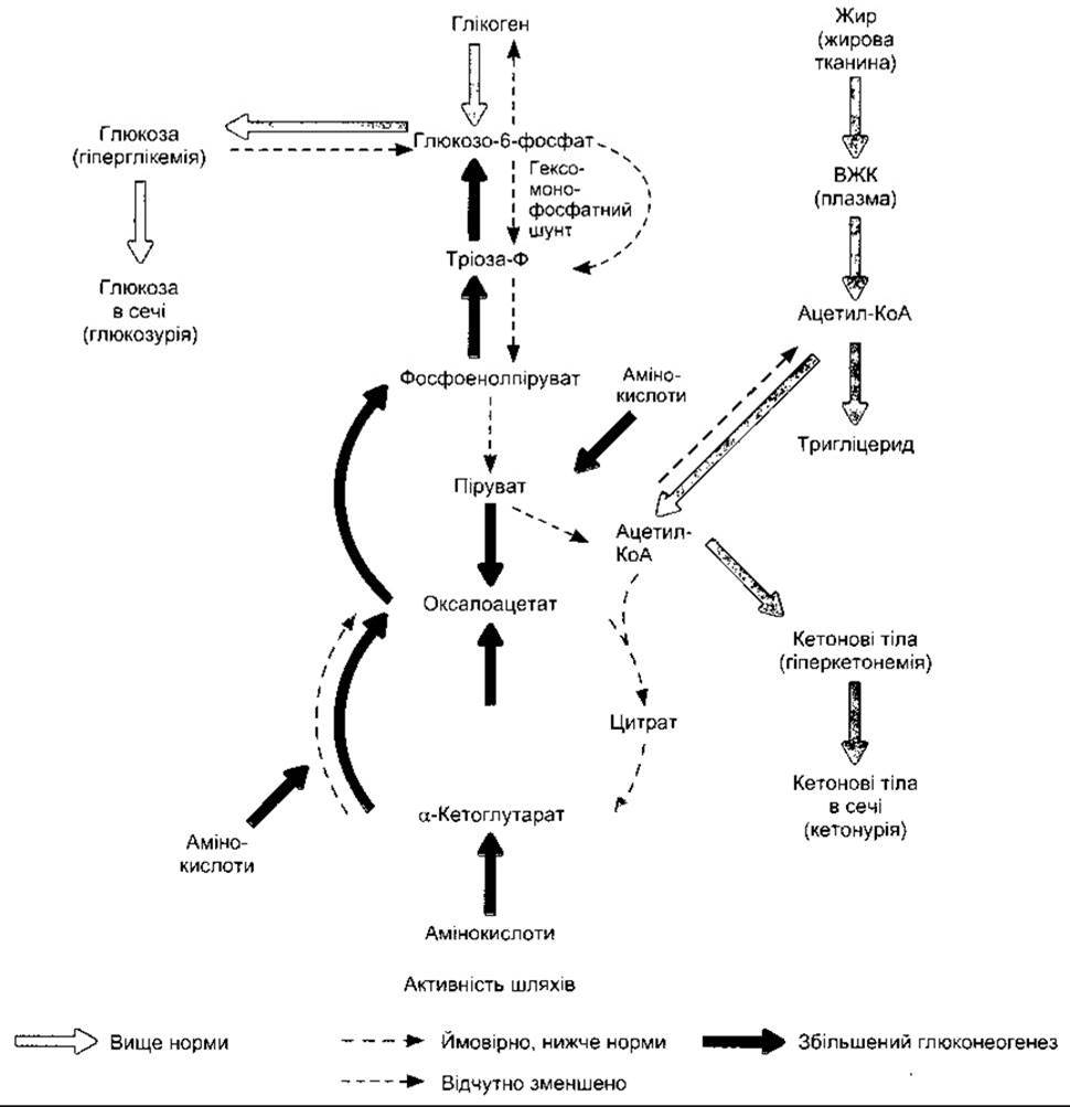
Fig. 19-10. Metabolic abnormalities in the liver in untreated diabetes (reproduced with permission from Murray RK et al: Harper's Biochemistry, 25th ed. McGraw-Hill, 2000).
Acidosis
Although most of the hydrogen ions released from acetoacetate and β-hydroxybutyrate are buffered, severe metabolic acidosis nonetheless develops. The resulting drop in blood pH stimulates the respiratory center, leading to the rapid, deep breathing described by Kussmaul as "air hunger" and named Kussmaul breathing in his honor. In this condition, the urine becomes acidic. Once the renal capacity to replace plasma cations accompanying organic anions with H+ and NH4+ is exceeded, Na+ and K+ are lost in the urine. The resultant loss of water and electrolytes leads to dehydration, hypovolemia, and hypotension. Ultimately, acidosis and dehydration depress consciousness to the point of coma. Although complicating infections can now be effectively controlled with Antibiotics, acidosis remains the most common cause of early death in clinical diabetes mellitus.
In pronounced acidosis, the total body sodium content is markedly diminished, and if sodium losses exceed water losses, the plasma Na+ concentration also falls. Total body potassium is likewise depleted, yet its plasma level is typically normal—partly due to the contraction of extracellular fluid (ECF) volume, and partly because K+ shifts out of cells into the ECF when extracellular H+ concentrations are high. Another factor maintaining plasma K+ levels is the absence of insulin-induced cellular uptake of K+.
The degree to which ketoacidosis complicates experimental diabetes varies among species. The size of the body's fat reserves is also a critical determinant of the metabolic response to diabetes. Prior to the isolation of insulin by Banting and Best in 1921, the primary Treatment for human diabetes was prolonged fasting (the Allen dietary regimen). This approach lowered not only plasma glucose levels but also reduced fat stores to a point where little fat remained available for mobilization.
Coma
Diabetic coma may ensue as a direct consequence of acidosis and dehydration. Alternatively, plasma glucose levels may rise to such extremes—independent of plasma pH—that severe plasma hyperosmolality induces a loss of consciousness (hyperosmolar coma). The accumulation of blood lactate (lactic acidosis) can also complicate diabetic ketoacidosis if tissues become hypoxic (see Chapter 33), potentially culminating in coma. Cerebral edema is observed in a significant number of patients with diabetic ketoacidosis and can likewise precipitate coma. Although the precise etiology of cerebral edema remains incompletely understood, it constitutes a serious complication with a poor prognosis.
Cholesterol Metabolism
Diabetes is characterized by elevated plasma cholesterol levels, which accelerate the development of atherosclerotic vascular disease—the major long-term complication of diabetes in humans. Cholesterol levels rise primarily due to increased concentrations of VLDL and LDL in the plasma (see Chapter 17), which may stem from enhanced hepatic VLDL production or impaired clearance of VLDL and LDL from the circulation.
Summary
The complexity of metabolic disturbances in diabetes warrants a Synthesis of the foregoing points. A hallmark of insulin deficiency (Fig. 19-11) is impaired glucose uptake across many tissues (reduced peripheral utilization). There is also a net increase in hepatic glucose output (enhanced production), partly driven by glucagon excess. Consequently, hyperglycemia leads to glucosuria and an osmotic diuresis resulting in dehydration, which in turn triggers polydipsia. Driven by intracellular glucose deprivation, appetite is stimulated; glucose is synthesized from Proteins (gluconeogenesis), while energy demands are met through accelerated protein and lipid metabolism. The clinical outcomes are weight loss, weakness, protein depletion, and wasting. Lipid catabolism is amplified, leaving the body with an excess of triglycerides and FFAs. Fat synthesis is suppressed, and overloaded Catabolic pathways cannot cope with the surplus of generated acetyl-CoA. In the liver, this acetyl-CoA is converted into ketone bodies. Because their rate of production outstrips the body's oxidative capacity, ketone bodies—which are predominantly organic acids—accumulate in the blood (ketonemia). The accumulation of ketone bodies precipitates metabolic acidosis. The depletion of Na+ and K+ is compounded by dehydration, as plasma cations are excreted alongside organic anions that fail to bind the H+ and NH4+ secreted by the Kidneys. Ultimately, the acidosis, hypovolemia, and hypotension of the depleted animal or patient culminate in coma—driven by the Toxic effects of acidosis, dehydration, and hyperosmolality on The Nervous system—and, if timely medical intervention is withheld, death.

Fig. 19-11. Effects of insulin deficiency (reprinted with permission from RJ Havel).
All of these abnormalities are corrected by insulin administration. Although immediate treatment of acidosis also involves parenteral alkalinization to combat the acidosis and rehydration to replenish the body's Na+ and K+ stores, only insulin cures the primary defects, enabling a return to the normal state.
Last update: 10/08/2026
Editorial and Educational Adaptation: This material has been compiled based on the primary/original source text. The project team performed an editorial review, corrected technical inaccuracies, structured sections, and adapted the content for an educational format.
What was processed:
- elimination of formatting defects (OCR errors, structural breaks, corrupted characters);
- editorial organization of content;
- standardization of terminology in accordance with academic sources;
- verification of factual statements against the original source text.
All mentions of the author, publication year, and origin of the primary text have been preserved in accordance with the source.