Protein Structure and Function. Application of Bioinformatics Methods - John Rigden 2014
Prediction of membrane protein structure
Structural classes
Alpha-helical bundles
There are two primary classes of Membrane Proteins: alpha-helices and Structure/56.html">Beta-barrels. Alpha-helical membrane proteins constitute the most abundant Class of TM proteins and are found in all types of Introduction/36.html">Biological Membranes, including the outer membranes of Bacteria. These proteins consist of one or more alpha-helices, each containing a stretch of hydrophobic Amino Acids, embedded within the membrane and connected to other helices via extramembrane loop regions. It is believed that alpha-helical membrane proteins can contain up to 20 TM helices, which accounts for their diverse topologies. Loop regions are known to have complex structures containing re-entrant loops and amphipathic helices. Re-entrant loops are short a-helices that enter and exit the membrane on the same side. Amphipathic helices lie parallel to the membrane plane and globular domains.
Alpha-helical TM proteins are subdivided into several subclasses. Subclass I proteins contain a single TM a-helix, with the N-terminus located on the extracellular side of the membrane and the C-terminus on the intracellular, or cytoplasmic, side. These proteins are further divided into two subclasses. Subclass Ia, which includes the majority of eukaryotic membrane proteins, contains cleavable signal sequences, whereas subclass Ib lacks such sequences. Subclass II membrane proteins, like those of subclass I, span the membrane only once. However, they exhibit the opposite orientation: the N-terminus is located on the cytoplasmic side of the membrane, and the C-terminus on the outer side.
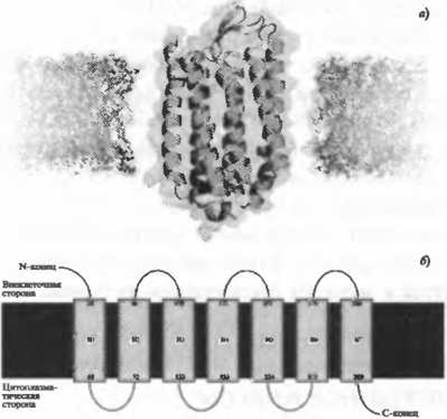
Fig. 4.1. (For the color version of the figure, see the insert.) a) Bacteriorhodopsin from Halobacterium salinarium. Seven transmembrane helices of a G-protein-coupled receptor are surrounded by a lipid bilayer. The receptor Functions as a proton pump, utilizing light energy to transport protons out of The Cell. PDB ID 1py6. Other G-protein-coupled receptors include halorhodopsin, a light-driven chloride pump. PDB ID 1e12. b) A simplified representation of bacteriorhodopsin topology
Subclass III membrane proteins consist of a single polypeptide chain folded into multiple transmembrane segments across certain regions. Proteins of this subclass are also divided into subclasses a and b: subclass IIIa proteins contain cleavable signal sequences, whereas subclass IIIb proteins have their N-termini directed toward the outer surface of the membrane and lack cleavable signal sequences. Subclass III membrane proteins include the family of G-protein-coupled receptors (GPCRs), whose members feature seven transmembrane helices (Fig. 4.1). G-protein-coupled receptors constitute a large family of receptor proteins that recognize extracellular molecules, activate signal Transduction pathways, and ultimately mediate cellular responses.
Subclass IV membrane proteins contain multiple domains forming an assembly whose elements repeatedly span the membrane. These domains may reside within a single polypeptide chain, though transmembrane proteins are frequently composed of multiple chains. This subclass includes Photosystem I, which consists of nine distinct chains (PDB ID 1jbO).
Last update: 06/08/2026
Editorial and Educational Adaptation: This material has been compiled based on the primary/original source text. The project team performed an editorial review, corrected technical inaccuracies, structured sections, and adapted the content for an educational format.
What was processed:
- elimination of formatting defects (OCR errors, structural breaks, corrupted characters);
- editorial organization of content;
- standardization of terminology in accordance with academic sources;
- verification of factual statements against the original source text.
All mentions of the author, publication year, and origin of the primary text have been preserved in accordance with the source.