Human Anatomy - Kotsan I. Y. 2009
Urogenital Apparatus
Genital Organs - Male Genital Organs
Seminal Vesicle
The seminal vesicle (vesicula seminalis) is a paired organ located in the pelvic cavity, lateral to the ampulla of the Ductus deferens, superior to the Prostate Gland, and lateral to the Urinary Bladder (Fig. 175). The Peritoneum covers only the upper PARTS OF THE seminal vesicle. The vesicle features an anterior surface (facies anterior), which faces the urinary bladder, and a posterior surface (facies posterior), which is adjacent to the rectum. The vesicle is approximately 5 cm long, 2 cm wide, and 1 cm thick. Its cavity consists of a convoluted canal with lateral pockets, creating a labyrinth-like appearance in cross-section. When the outer sheath of a seminal vesicle is partially removed and it is straightened out, it takes the form of a tube 10–12 cm long and 0.6–0.7 cm thick.
The wall of the seminal vesicle consists of three layers: an outer adventitial coat (tunica adventitia), a middle muscular coat (tunica muscularis), and an inner mucous coat (tunica mucosa). The mucous membrane of the vesicles produces a secretion—a clear, colorless, or slightly yellowish fluid that mixes with semen to dilute it, as well as to nourish and activate spermatozoa.
Each seminal vesicle comprises an upper expanded end (the base), a middle part (the body), and a lower narrowed end that transitions into the excretory duct (ductus excretorius). The excretory duct of the seminal vesicle joins the terminal part of the ductus deferens to form the ejaculatory duct (ductus ejaculatorius). The ejaculatory
duct pierces the prostate gland and opens via a slit-like orifice On the surface of the mucous membrane of the prostatic Urethra, lateral to the seminal colliculus (verumontanum). The length of the ejaculatory duct is about 2 cm, and its lumen width ranges from 1 mm at its origin to 0.3 mm near its opening into the urethra.
Blood supply to the seminal vesicle and ductus deferens is provided by Branches of the artery of the ductus deferens (a branch of the umbilical artery), the inferior vesical artery, and the middle rectal artery (branches of the internal iliac artery). Venous drainage occurs into the Veins OF THE ductus deferens (tributaries of the internal iliac vein), as well as the venous plexuses of the urinary bladder, rectum, and prostate gland. Lymphatic drainage leads to the internal iliac Lymph Nodes.
Class="center">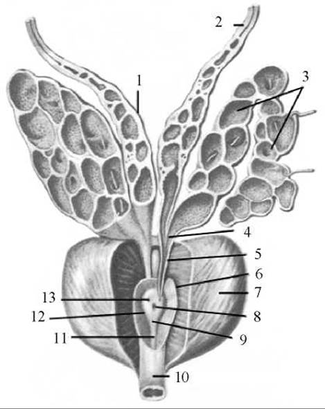
Fig. 175. Seminal Vesicles and prostate gland
1 — ampulla of ductus deferens; 2 — ductus deferens; 3 — seminal vesicle; 4 — excretory duct of seminal vesicle; 5 — ejaculatory duct; 6 — prostatic part of urethra; 7 — prostate gland; 8 — prostatic utricle; 9 — seminal colliculus; 10 — membranous part of urethra; 11 — urethral crest; 12 — prostatic sinus; 13 — opening of ejaculatory duct
Innervation (sympathetic and parasympathetic) of the seminal vesicle and ductus deferens is supplied by the plexus of the ductus deferens, formed by branches of the inferior hypogastric plexus.
Last update: 08/08/2026
Editorial and Educational Adaptation: This material has been compiled based on the primary/original source text. The project team performed an editorial review, corrected technical inaccuracies, structured sections, and adapted the content for an educational format.
What was processed:
- elimination of formatting defects (OCR errors, structural breaks, corrupted characters);
- editorial organization of content;
- standardization of terminology in accordance with academic sources;
- verification of factual statements against the original source text.
All mentions of the author, publication year, and origin of the primary text have been preserved in accordance with the source.