BIOCHEMISTRY - L. Stryer - 1984
VOLUME 1
PART I. CONFORMATION AND DYNAMICS
CHAPTER 5. MOLECULAR DISEASES: SICKLE-CELL ANEMIA
5.2. Deoxygenated Sickle-Cell Hemoglobin Has Lower Solubility
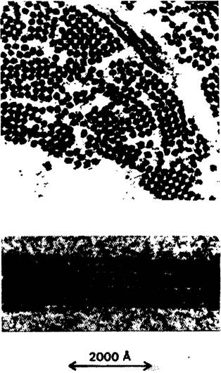
Herrick made a remarkably accurate suggestion regarding the localization of the primary defect responsible for Sickle-Cell Anemia. When oxygen concentration is reduced, THE RED Blood Cells of a patient assume a sickle shape on a Glass slide in vitro. It turned out that the Hemoglobin itself is defective in these cells. The solubility of sickle-cell deoxyhemoglobin is approximately 25 times lower than that of normal hemoglobin. Upon deoxygenation of a concentrated solution of sickle-cell hemoglobin, a fibrous precipitate forms (Fig. 5.3). It is precisely this precipitate that deforms the erythrocyte, giving it a sickle shape. Sickle-cell hemoglobin is commonly designated as hemoglobin S (Hb S), in contrast to hemoglobin A (Hb A), which is normal adult hemoglobin.
Class="center">Fig. 5.3. Electron micrograph of deoxygenated sickle-cell hemoglobin fibers. The top image is a cross-section, and the bottom image is a longitudinal section. Fiber thickness is -170 Å
5.3. Hemoglobin S Differs from Hemoglobin A in Electrophoretic Mobility
In 1949, Pauling and his coworkers investigated the PHYSICOCHEMICAL PROPERTIES OF hemoglobin in normal individuals, sickle-cell trait carriers, and patients with sickle-cell anemia. The experimental approach they used was as follows: they compared the mobility of the studied Hemoglobins in an electric field. The METHOD FOR DETERMINING mobility in an electric field is called Electrophoresis. The migration velocity (V) of a protein (or any other compound) in an electric field depends on the electric field strength (E), the net charge of the protein (Z), and the frictional coefficient (f). The frictional coefficient is determined by the size and shape of the protein. These quantities are related by the equation
 (1)
(1)
The isoelectric point of a protein is the pH value at which the protein carries no net electrical charge. At this pH value, the electrophoretic mobility is zero, since Z = 0 [equation (1)]. At a pH below the isoelectric point, the protein molecule is positively charged; at pH values above the isoelectric point, the protein is negatively charged. Sickle-cell hemoglobin differs from normal hemoglobin in its isoelectric point:
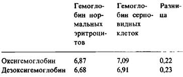
As can be seen from the table above, the difference between sickle-cell and normal hemoglobin is the same for both the oxidized and reduced forms.
The difference in electrophoretic mobility between hemoglobin A and S can be attributed to a change either in the net charge Z or in the frictional coefficient f. The friction effect itself (caused by A change in shape) would manifest as one substance moving slower than the other across the entire pH range. In the case under consideration, this did not occur, as judged by the fact that the curves of electrophoretic mobility versus pH had the same slope (Fig. 5.4). Furthermore, other physicochemical studies—specifically the determination of sedimentation velocity and free diffusion—showed that the frictional coefficients of the oxygenated forms of hemoglobin A and hemoglobin S were identical.
Fig. 5.4. pH dependence of the electrophoretic mobility of normal hemoglobin and sickle-cell hemoglobin
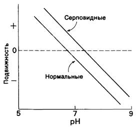
Based on these observations, it was concluded that the hemoglobins being compared differ in the number or type of ionized groups. How many such groups are there in each hemoglobin? The answer is provided by the acid-base titration curve of the hemoglobins. In the pH 7 region, this curve is nearly linear. A one-unit change in the pH of the hemoglobin solution produces a difference of nearly 13 charges. Therefore, a difference of 0.23 in the isoelectric points corresponds to approximately three charges in the hemoglobin molecule. It follows that sickle-cell hemoglobin differs from normal hemoglobin by the presence of 2 to 4 additional positive charges.
What is the cause of this difference: an altered polypeptide chain composition or a modified heme? Porphyrin was isolated from sickle-cell hemoglobin; according to melting point determinations and X-Ray Diffraction Analysis, it proved to be identical to the porphyrin of normal hemoglobin. This indicates that the difference between the two hemoglobins lies in the specific composition of their polypeptide chains.
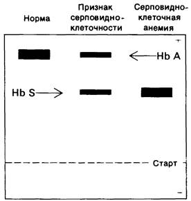
Patients with sickle-cell anemia (who are invariably homozygous for the defective Gene) possess hemoglobin S and lack hemoglobin A. At the same time, sickle-cell trait carriers (who are heterozygous for the sickle-cell gene) contain both hemoglobins in approximately equal amounts (Fig. 5.5). Thus, Pauling's work revealed an undeniable case of an altered protein molecule resulting from a change in the alleles of the gene encoding it. This was the first example of a molecular disease.
Fig. 5.5. Starch-gel electrophoresis at pH 8.6 of hemoglobin from a healthy individual, a sickle-cell trait carrier, and a patient with sickle-cell anemia
Last update: 06/08/2026
Editorial and Educational Adaptation: This material has been compiled based on the primary/original source text. The project team performed an editorial review, corrected technical inaccuracies, structured sections, and adapted the content for an educational format.
What was processed:
- elimination of formatting defects (OCR errors, structural breaks, corrupted characters);
- editorial organization of content;
- standardization of terminology in accordance with academic sources;
- verification of factual statements against the original source text.
All mentions of the author, publication year, and origin of the primary text have been preserved in accordance with the source.