BIOCHEMISTRY - L. Stryer - 1984
VOLUME 1
PART I. CONFORMATION AND DYNAMICS
CHAPTER 5. MOLECULAR DISEASES: SICKLE-CELL ANEMIA
In 1904, James Herrick, a Chicago physician, examined a 21-year-old Black student who was admitted to the hospital with a cough and fever. The patient complained of weakness, dizziness, and headaches. For the preceding year, he had suffered from bouts of palpitations and shortness of breath. Over the past few years, he had increasingly avoided sports and other physical activities. Three years prior to admission, he had experienced a six-month episode of pus discharge from his ear. Since childhood, he had frequently suffered from slow-healing ulcers on his legs.
Upon examination, the patient showed no significant deviations from normal physical and mental development. His sclerae had a yellowish tint, his mucous membranes were pale, and his Lymph Nodes were enlarged. About 20 scars were found on his shins and thighs. Significant cardiac abnormalities were observed, including an enlarged Heart border and a murmur. As Herrick noted, "...one got the impression that The Heart was acting under METABOLISM/18.html">The Influence of some powerful stimulant, although the patient had received no cardiac stimulants."
Laboratory workup included a thorough examination of the patient's stool to check for potential parasites, the likelihood of which is quite high for someone who grew up in the tropics. However, no parasites were found.
No tubercle bacilli were detected in his sputum. His urine contained cellular debris, indicating Kidney impairment. Blood tests revealed a drastic departure from normal:
Class="center">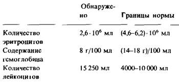
The patient was undoubtedly suffering from anemia, as his Hemoglobin level was half of normal. His erythrocytes varied greatly in size, with many much smaller than normal. In addition, A large number of nucleated red Blood Cells were present. Such nucleated cells serve as precursors to normal, mature, enucleated erythrocytes. Herrick described the patient's unusual red blood cells as follows: "THE RED BLOOD corpuscles showed a great variation in shape, but what especially attracted attention was the large number of thin, elongated, sickle-shaped, crescent-like cells. They were found in fresh blood samples regardless of how the blood was spread on the Glass slide. In blood samples taken simultaneously from other individuals and processed under identical conditions, such cells were not found. Undoubtedly, they were neither artifacts nor any form of parasitic Organism."
The patient was prescribed supportive Treatment: rest and a nutritious diet. After four weeks, he was discharged. He felt better, and his hemoglobin levels had increased. However, his blood still contained "peculiar crescent-shaped red blood cells, although in smaller numbers than before."
Herrick was struck by this clinical picture and laboratory data. Six years passed before he decided to publish the case, frankly admitting that the patient "could not even be given a definite Diagnosis." The author noted the chronic Nature of the disease and the variety of pathological findings observed during examination and laboratory testing: cardiac enlargement, generalized lymphadenopathy, jaundice, anemia, and signs of renal damage. He concluded that the Water/144.html">Origin of the disease could not be linked to an organic lesion of any single organ. Herrick singled out the hematological abnormality as the leading symptom and titled his case report *Peculiar Elongated and Sickle-shaped Red Blood Corpuscles in a Case of Severe Anemia*. Herrick suggested that "the primary cause of the disease might be some unknown alteration in the COMPOSITION OF THE corpuscles themselves."
5.1. Sickle-Cell Anemia: A Chronic Hereditary Hemolytic Disease
Shortly after Herrick's publication, other cases of this condition, termed "Sickle-Cell Anemia," were discovered. In fact, sickle-cell anemia is by no means a rare disease. In countries with sizable Black populations, it represents a significant public health challenge. Sickle-cell anemia occurs in approximately 4 out of every 1,000 Black individuals. In the past, sickle-cell anemia typically proved fatal, often before the age of 30, due to infection, renal failure, Heart Failure, or thrombosis.
Fig. 5.1. Sickle cells in the blood of a patient with sickle-cell anemia. Light Microscopy view
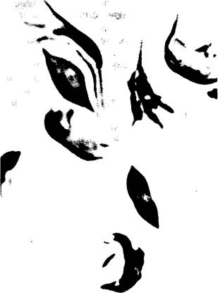
Fig. 5.2. Erythrocyte from a patient with sickle-cell anemia; scanning electron micrograph
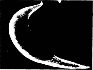
Sickle erythrocytes become trapped in the smallest Blood Vessels. As a result, blood supply is disrupted, leading to damage in various Organs, particularly the Kidneys and bones. Sickle cells are more fragile than normal cells. Because they lyse easily, their lifespan is shortened, resulting in severe anemia. The chronic course of the disease is punctuated by crises, during which the proportion of sickle cells in the blood increases sharply. During these crises, the patient may go into Shock.
Sickle-cell anemia is hereditary. Patients with sickle-cell anemia are homozygous for a defective Gene located on an autosome. Individuals who inherit one defective gene from one parent and one normal allele from the other are carriers of the sickle-cell trait. Such heterozygotes usually show no symptoms of the disease, as sickle erythrocytes account for only about 1% of the total red blood cell count in their venous blood; in homozygous individuals, the proportion of sickle erythrocytes reaches 50%. However, carrying the sickle-cell trait, which occurs in about one out of every ten Black individuals, is not entirely benign. For instance, carriers of the sickle-cell trait may be at risk during strenuous physical exertion at high altitudes, flights in unpressurized aircraft, or under general anesthesia. Why? We will explore this in the subsequent Structure/133.html">Discussion.
Last update: 06/08/2026
Editorial and Educational Adaptation: This material has been compiled based on the primary/original source text. The project team performed an editorial review, corrected technical inaccuracies, structured sections, and adapted the content for an educational format.
What was processed:
- elimination of formatting defects (OCR errors, structural breaks, corrupted characters);
- editorial organization of content;
- standardization of terminology in accordance with academic sources;
- verification of factual statements against the original source text.
All mentions of the author, publication year, and origin of the primary text have been preserved in accordance with the source.