Principles of Protein Structure - G. Schulz 1982
Mechanisms of polypeptide chain folding and association
Secondary structure
Linear groups
Secondary structures are the ordered arrangements of the polypeptide main chain, irrespective of the types and Conformations of side chains. All secondary structures are stabilized by Hydrogen Bonds between peptide amide and carbonyl groups.
The polypeptide backbone forms a regular one-dimensional array (line group) when its dihedral angles repeat. Any such array constitutes a helix. If the regularity is such that all sequentially arranged peptide units have identical relative orientations—that is, if all angles (∅, ψ) are equal (Fig. 2.2)—the polypeptide main chain forms a line group. Any line group is a helix that can be described by an axial Translation per repeat unit d, the number of units per turn n, and the distance r from a given point of a specified unit (here, the Ca atom) to the helical axis (Fig. 5.2). Since d is always considered positive, the handedness (chirality) of the helix is determined by the sign of n. Every helix is directional because the peptide unit, formed by the N- and C-termini of the Amino Acids, possesses a specific orientation.
Class="center">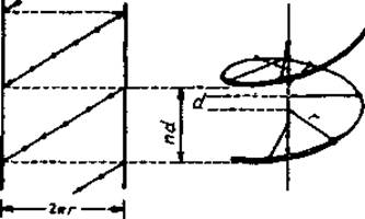
Fig. 5.2. A helix, its parameters, and surface projection (net). n is the number of residues per turn, d is the axial rise per residue, nd is the pitch of the helix, and r is the radius of the helix. To obtain the surface projection, the helix is projected onto a coaxially positioned cylindrical sheet of paper, which is then cut parallel to the helix axis and unrolled. This surface projection is also referred to as a cylindrical diagram. It clearly illustrates the geometric relationships between residues. Reversing the front side of the cut sheet leads to the mirror image of the same cylindrical diagram.
Fig. 5.3 shows the curves relating the dihedral angles (∅, ψ) (Fig. 2.2) to the helical parameters d and n. A helix with |n|<2 is geometrically impossible. There are only a few line groups that are free of steric hindrance and stabilized by hydrogen bonds either within the chain (e.g., a-helices) or between adjacent chains (e.g., the ß-pleated sheet, Collagen). The parameters of the most important line groups are listed in Table 5.1.
Line groups are also found in CARBOHYDRATES, Nucleic Acids, and protein aggregates. They have been identified in Polysaccharides whose repeating units are mono- or Oligosaccharides with or without side chains [178], as well as in RNA (and DNA), where ribose-phosphate diester units typically form a double helix through the intertwining of two antiparallel single strands [179]. Tobacco mosaic virus consists of a protein coat and RNA. The coat represents a line group whose repeating unit is a single protein bound to a trinucleotide [180]. Cylindrical Protein Assemblies found, for example, in microtubules [181], T4 phage tails [182], or F-Actin filaments [183], are often described as helices or line groups. Nevertheless, it should be kept in mind that such aggregates do not contain continuous single-stranded backbones, unlike RNA which links the protein subunits in tobacco mosaic virus into a specific helical Structure. Various approaches are used to describe such multi- and poly-helical architectures.
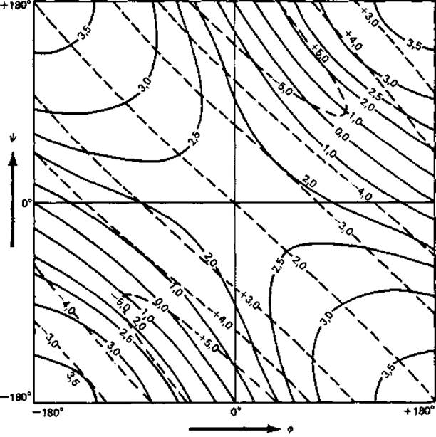
Fig. 5.3. Helical parameters d (solid lines) and n (dashed lines) as Functions of the dihedral angles — and ψ [29].
For a single helix, The values of — and ψ for all residues are identical.
Table 5.1 Line groups formed by polypeptide chains
|
Line group |
Occurrence |
Residues per turn n and chiralitya |
Axial rise per residue d, Å |
Helix radius r, Å |
|
Planar parallel ß-Structure |
Rare |
±2.0 |
3.2 |
1.1 |
|
Planar antiparallel ß-structure |
do. |
±2.0 |
3.4 |
0.9 |
|
Twisted parallel or antiparallel ß-structure |
Common |
—2.3 |
3.3 |
1.0 |
|
310-Helix |
Short segments |
+3.0 |
2.0 |
1.9 |
|
a-Helix (right-handed) |
Common |
+3.6 |
1.5 |
2.3 |
|
aL-Helix (left-handed) |
Hypothetical |
—3.6 |
1.5 |
2.3 |
|
п-Helix |
do. |
+4.3 |
1.1 |
2.8 |
|
In fibers |
-3.3 |
2.9 |
1.6 |
a Plus and minus signs denote right- and left-handed helices, respectively.
310-, α- and π-Helices
Regular helices are fully defined by a pair of dihedral angles. The structures of these helices are illustrated in Fig. 5.4, and their parameters are summarized in Table 5.1. The 310-, α-, and π-helices are stabilized by hydrogen bonds formed between the peptide amide and carbonyl groups of residues (i, i - 3), (i, i + 4), and (i, i - 5), respectively. Consequently, it is possible to form Helical structures featuring networks of consecutive hydrogen bonds between closely spaced chain elements. Being linear conformations, these helices are fully represented by a single point on the φ,ψ-map (Fig. 2.3). These points correspond to local energy minima, though minor deviations from these minima are possible.
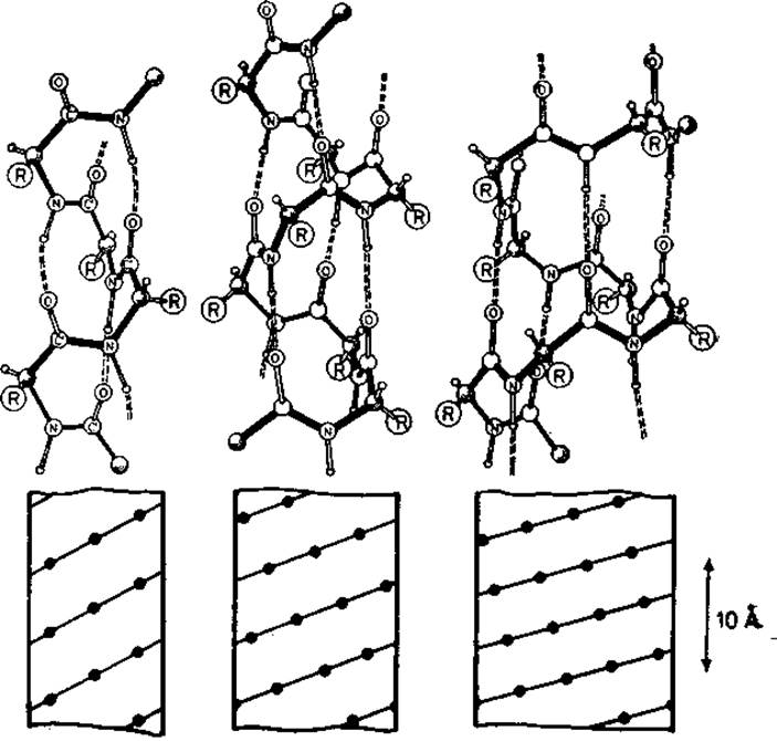
Fig. 5.4. Polypeptide chain helices stabilized by internal hydrogen bonds.
From left to right: 310-, α-, and π-helix. Bottom: cylindrical projections of these helices, showing the course of The polypeptide chains and the positions of Cα atoms.
The α-Helix is exceptionally stable and therefore the most prevalent. Because the α-helix is the most common Secondary structure in Proteins, its conformation must be remarkably stable. This is consistent with the Location OF THE α-helix in the center of the allowed region on the φ,ψ-map (Fig. 2.3), as well as with the fact that its Hydrogen bond dipoles are aligned linearly, which also corresponds to an energy minimum (Sec. 3.4). Furthermore, the radius of the helix (Table 5.1) is optimal for dispersion interactions between residues situated on opposite sides of the helical axis.
The α-helix was first postulated by Pauling et al. [25]. At the time, the idea of a helix with a non-integer value of n was met with skepticism, as it conflicted with the assumption of a high degree of molecular order. Direct confirmation of the postulate came from X-Ray Diffraction data of crystalline Hemoglobin [183]. Later, α-helices were discovered in numerous fibers [184], notably α-keratin and paramyosin, and in virtually all Globular proteins. In fact, the designations α and β for the α-helix and β-structure originated from the fact that all fiber X-ray patterns were classified into α- and β-diagrams, which were obtained primarily from samples of α-keratin and Silk Fibroin.
Fig. 5.5 illustrates the distribution of observed α-helix lengths in globular proteins. The average length is approximately 17 Å, which corresponds to 11 residues per 3 turns of the helix. The significant scatter in the distribution indicates that there are no preferred lengths, which is consistent with the expected properties of a linear polymer. Relative maxima for lengths of 7, 11, and 15 residues—corresponding to 2, 3, and 4 complete turns, respectively—are smaller than the expected error of a Poisson distribution (≈ 251/2= 5), making their significance rather questionable. α-Helices provide protein structures with quite robust "rods." Bends in these rods of about 20° are often achieved through the incorporation of Pro residues, as seen in Myoglobin [185] and adenylate kinase [186].
310-Helices are rare and form only very short segments. The 310-helix gets its name from the number of residues per turn (n = 3) and the number of atoms (10) in the ring formed by the hydrogen bond [187]. The hydrogen-bonding dipoles are not collinear, which does not correspond to an energy minimum (Section 3.4). The packing of side chains in this case is also rather unfavorable. As can be seen from Fig. 5.4, the side chains have identical azimuthal orientations, whereas in the α-helix they are staggered. On the (∅, ψ) map (Fig. 5.7), the 310-helix falls on the edge of the allowed region, indicating the presence of minor steric hindrance. This energetically unfavorable spatial conformation explains the scarcity of 310-helices in proteins; typically, these segments span about a single turn (sea lamprey hemoglobin contains two 310-helices, each two turns long [188, 189]). Such segments are usually found at the N- and C-termini of α-helices.
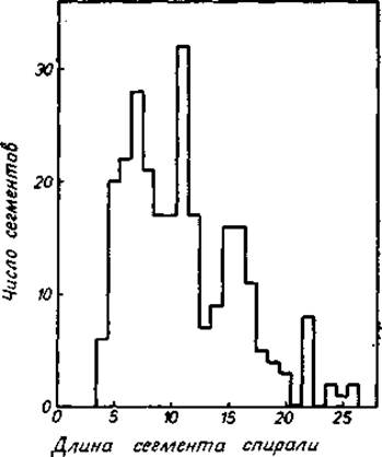
Fig. 5.5. Length distribution of α-helical segments for 23 proteins (data adapted from [323]).
Lengths are expressed in number of residues.
The π-helix is energetically unfavorable. The π-helix has never been observed experimentally and thus remains of purely academic interest. This helix lies at the edge of the allowed region (Fig. 2.3), pointing to the presence of steric clashes [29, 190]. The hydrogen-bonding dipoles are favorably aligned. Due to the large radius r (Table 5.1), backbone atoms located on opposite sides of the helical axis no longer form stabilizing contacts with one another. An axial cavity* is formed, which noticeably reduces the Van der Waals attraction energy. As evident from the cylindrical function in Fig. 5.4, the side chains of the π-helix are packed less favorably than in the α-helix, but significantly better than in the 310-helix. The absence of π-helices in real systems is presumably due to steric hindrance and the weakening of van der Waals interactions between opposing backbone elements.
Левые 310-, а- и п-спирали не были обнаружены. Если рассматривать только основную цепь полипептида, каждая спираль должна иметь энергетически эквивалентное зеркальное отображение. Однако взаимодействия боковых цепей приводят к энергетической невыгодностилевой аL-спирали, что и объясняет ее отсутствие в глобулярных белках. Это же относится и к левым 310- и п- спиралям.
Last update: 06/08/2026
Editorial and Educational Adaptation: This material has been compiled based on the primary/original source text. The project team performed an editorial review, corrected technical inaccuracies, structured sections, and adapted the content for an educational format.
What was processed:
- elimination of formatting defects (OCR errors, structural breaks, corrupted characters);
- editorial organization of content;
- standardization of terminology in accordance with academic sources;
- verification of factual statements against the original source text.
All mentions of the author, publication year, and origin of the primary text have been preserved in accordance with the source.