Biological Membranes - A. N. Ogurtsov 2012
Electrogenesis of Biomembranes
Mechanisms of Intercellular Signaling
Calcium Regulation of Neurotransmitter Exocytosis
Neurotransmitters are released into the synaptic cleft via regulated exocytosis, a process in which synaptic vesicles fuse with the axon terminal membrane (Figure 125). Neurotransmitter exocytosis from synaptic vesicles relies on the mechanisms of targeted Vesicular Transport and fusion characteristic of intracellular vesicular transport (Section 8.4).
Neurosecretion differs from other secretory mechanisms in that
1) neurosecretion occurs only when an Action Potential reaches the axon terminals,
2) after fusing with Cell/30.html">The Plasma Membrane and releasing their contents into the synaptic cleft, synaptic vesicles do not become part of the axon terminal plasma membrane, but are instead recycled to be refilled with neurotransmitters.
Figure 153 illustrates the complete vesicle functional cycle, in which a synaptic vesicle is filled with neurotransmitter molecules, releases its contents into the synaptic cleft, and is subsequently recycled. The complete cycle of such a synaptic vesicle takes about 1 minute.
Class="center">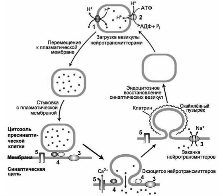
Figure 153 - Recycling of neurotransmitters and synaptic vesicles in axon terminals: 1 - H+-coupled neurotransmitter antiporter; 2 - V-type proton pump; 3 - Na+-coupled neurotransmitter symporters; 4 - Ca2+-sensitive docking Proteins; 5 - voltage-gated Ca2+ channels
Notably, acetylcholine, unlike other neurotransmitters, is not recycled According to the pathway shown in Figure 153.
Depolarization of the plasma membrane alone cannot force synaptic vesicles to fuse with it. To trigger this fusion process, the action potential must be converted into a chemical signal—specifically, a local increase in the cytosolic concentration of Ca2+ ions.
The transducers that convert the electrical action potential into a chemical activator signal for exocytosis are voltage-gated Ca2+ channels located in the presynaptic region of the synapse. Membrane depolarization caused by the arrival of an action potential opens these channels, allowing Calcium Ions to enter the axon terminal. The concentration of calcium cations rises from < 0.1 µM (at rest) to 1-100 µM. It is the binding of Ca2+ ions to proteins that anchor synaptic vesicles to the axonal plasma membrane (docking proteins (4) in Figure 153) that induces membrane fusion and subsequent neurotransmitter exocytosis.
The subsequent pumping of excess calcium ions out of the neuron Cytosol by plasma membrane Ca2+-ATPases rapidly lowers the cytosolic calcium level back to baseline, allowing the axon terminals to respond to incoming action potential impulses.
Two Types of synaptic vesicles are distinguished in axon terminals: (1) those "docked" to the plasma membrane and (2) those reserved in the "active zone" near the synaptic cleft. Each surge in Ca2+ ion concentration stimulates exocytosis in ~10% of the docked synaptic vesicles.
Those Membrane Proteins required for filling synaptic vesicles with neurotransmitters and docking them to the plasma membrane are returned to the synaptic vesicles via an endocytic mechanism analogous to retrograde transport (Figure 83), which is mediated by clathrin coats (Figure 84). Following the depolymerization of the clathrin coat, the endocytic vesicle is rapidly refilled with neurotransmitters. The fact that many Neurons are capable of firing more than 50 times per second indicates that the recycling of vesicle membrane proteins occurs very rapidly.
Immediately after neurotransmitters are released into the synaptic cleft and this chemical signal is transmitted to the postsynaptic cell, the neurotransmitter molecules must be cleared from the cleft; otherwise, the postsynaptic cell would be continuously stimulated by these neurotransmitters.
Signal termination can occur through the diffusion of neurotransmitter molecules out of the synaptic cleft. However, this is a very slow process and does not provide the physiologically necessary rate of cleft clearance.
Instead of simple diffusion, one or more faster mechanisms are employed to remove neurotransmitters from the synaptic cleft.
Signal transmission via acetylcholine is terminated by the Hydrolysis of acetylcholine molecules into Choline and acetic acid within the synaptic cleft by the enzyme acetylcholinesterase (see [9], Section 13.2). The resulting choline is transported back into the presynaptic axon by Na+/choline symporters and reused for the synthesis of acetylcholine. The operation of this symporter is similar to that of glucose symporters (Figure 103).
Unlike acetylcholine, all other neurotransmitters are not chemically modified; instead, they are removed from the synaptic cleft by being pumped back into the presynaptic axon, as illustrated in Figure 153.
The first to be studied in detail were four Na+/neurotransmitter symporters for GABA (gamma-aminobutyric acid), norepinephrine, dopamine, and serotonin. All these symporters share 60–70% sequence identity in their Amino acid sequences and each contain 12 transmembrane α-helices.
Just as with other Na+ symporters, the energy source for pumping neurotransmitters back into the presynaptic axon cytosol is the movement of Na+ ions down their electrochemical gradient across the plasma membrane. To maintain electroneutrality, The transport of Na+ ions and neurotransmitter molecules is typically accompanied by the transport of Cl- anions through Ion Channels.
Last update: 13/08/2026
Editorial and Educational Adaptation: This material has been compiled based on the primary/original source text. The project team performed an editorial review, corrected technical inaccuracies, structured sections, and adapted the content for an educational format.
What was processed:
- elimination of formatting defects (OCR errors, structural breaks, corrupted characters);
- editorial organization of content;
- standardization of terminology in accordance with academic sources;
- verification of factual statements against the original source text.
All mentions of the author, publication year, and origin of the primary text have been preserved in accordance with the source.