Human Biochemistry Volume 2 - Murray R. 1993
Special Topics
Contractile and Structural Proteins
Cell Motility and Cytoskeleton
It is quite evident that mechanical work—including amoeboid movement, morphogenesis, division, endocytosis, exocytosis, Intracellular Transport, and shape changes—is also performed by non-Muscle Cells. These cellular Functions are carried out by an extensive intracellular network of fibrous structures that make up the Cytoskeleton. The Cell Cytoplasm is not simply a bag of fluid, as was once believed. Virtually all Eukaryotic cells contain Three types of fibrous structures: Actin filaments (7–9.5 nm in diameter), microtubules (25 nm), and Intermediate filaments (10–12 nm). Each of these types can be distinguished using specific biochemical and electron microscopic techniques.
Non-Muscle Actin
The G-actin protein isolated from non-muscle cells has a Molecular Weight of approximately 43,000 and, like muscle actin (a-actin), contains N-methylhistidine residues. In the presence of magnesium and potassium chloride, this actin spontaneously polymerizes to form double-helical F-actin filaments, similar to muscle filaments. Non-muscle cells contain at least Two Types of actin: ß-actin and y-actin. Both may be present in the same cell and likely even co-polymerize into the same filament. In The Cell cytoplasm, actin forms microfilaments 7–9.5 nm in diameter, which often appear as bundles of tangled networks. Bundles of microfilaments lie beneath The Plasma Membrane of resting cells and are referred to as "stress" fibers. These fibers have a double-helical Structure and are decorated with Myosin S-1 fragments (Fig. 56.10). Stress fibers disappear upon an increase in cell motility, as well as during malignant cell transformation induced by chemical agents or Oncogenic Viruses.
Microfilaments are densely packed in a meshwork located beneath the leading edge or "ruffle" of a motile cell (Fig. 56.11). Actin microfilaments are found in all cellular microprojections, such as filopodia and microvilli. For example, the microvilli of intestinal mucosal cells contain 20–30 actin microfilaments arranged longitudinally, as shown in Fig. 56.12. These microfilaments, decorated with myosin S-1 fragments, exhibit uniform polarity (Fig. 56.12). At the Base of the microvilli are myosin filaments capable of retracting into the microvilli along with the actin filaments. The contraction process is not accompanied by any Changes in the length of actin or myosin and, thus, must occur—as in Muscles—via sliding filaments, i.e., by the mechanism formulated by Huxley. As in smooth muscles, the activation of the actin-myosin interaction and, consequently, the contraction process is mediated by phosphorylation of the myosin light chain.
Class="center">
Fig. 56.10. Electron micrograph of a cytoskeletal fragment to which myosin subfragment 1 (S-1) was added prior to rapid freezing. Almost all filaments of the longitudinal bundles and many lying between them have been converted into thick, rope-like structures (in the circle). However, some filaments that spontaneously arranged themselves between the bundles remained undecorated (arrow); these are predominantly intermediate filaments. x 70,000; in circle x 200,000. (Reproduced, with permission, from Heuser J. E., Kirschner M. W. Filament Organization revealed in platinum replicas of freeze-dried cytoskeletons. J. Cell Biol. 1980, 86. 212.)
Actin and myosin are also found between the spindle poles and Chromosomes, and along the Cleavage furrow in the telophase of mitosis.
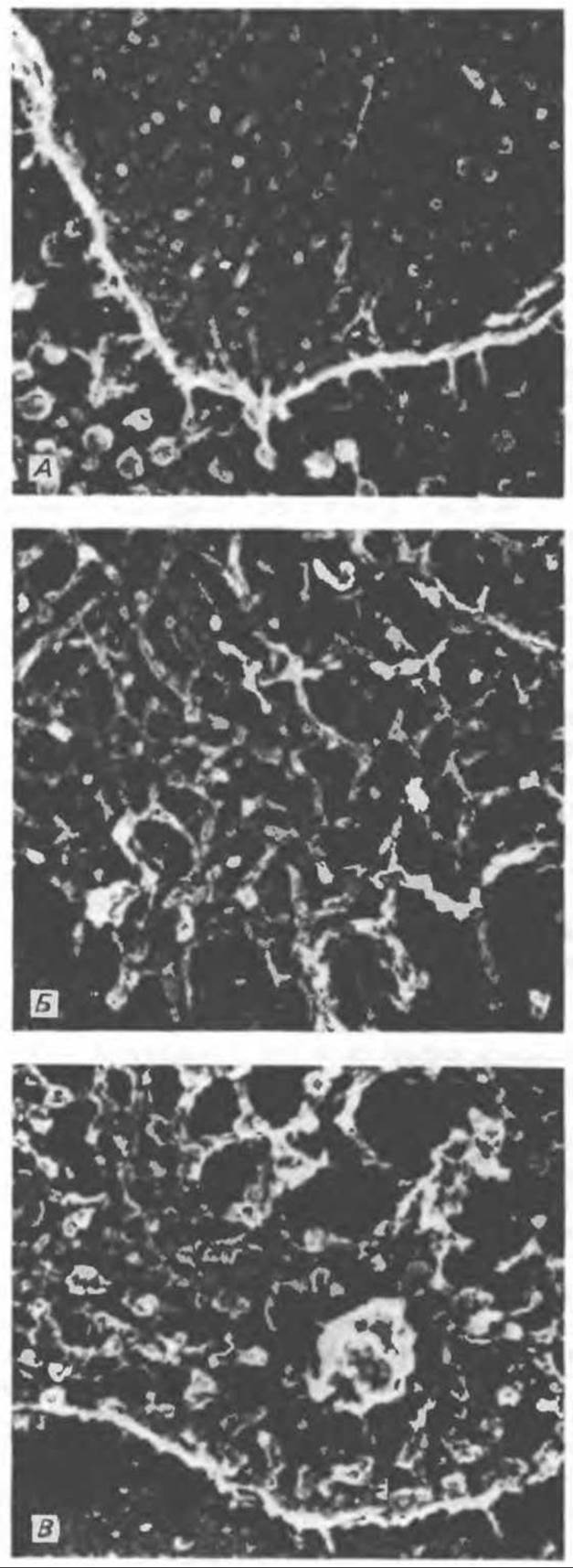
Fig. 56.11. Preparations of the ruffle or lamellipodia from fibroblasts (magnification). A. Complete fixation. B. Material extracted with Triton prior to fixation. C. Triton extraction performed after fixation. In the first case (photo A), the plasma membrane is intact, and the internal structure is not visible. In the second case (photo B), the plasma membrane has been removed, revealing the underlying interlocking "curly" filaments. Photo C—the plasma membrane was also removed, but only after aldehyde fixation. The fine network of underlying filaments appears coarser following chemical fixation. A, x 140,000; B and C, x 115,000. (Reproduced, with permission, from Heuser J. E., Kirschner M. W. Filament organization revealed in platinum replicas of freeze-dried cytoskeletons. J. Cell Biol., 1980, 86. 212.)
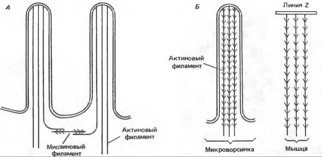
Fig. 56.12. Microvilli are small cytoplasmic projections of the epithelial cells lining the Small Intestine that increase the absorptive surface area. Microvilli contain both actin and myosin filaments and are capable of contracting like muscle cells. As shown in diagram A, bundles of actin filaments extend into each microvillus; myosin filaments are located at the base of the microvilli. In diagram B, the orientation of the actin filaments was determined by treating microvilli with isolated muscle myosin "heads" (heavy meromyosin); these fragments retain The ability to bind to actin filaments. This results in The formation of complexes resembling "arrowheads" that indicate the direction of filament movement. (Reproduced, with permission, from Lazarides E, Revel J. P.: The Molecular Bases of cell movement. Sci. Am. [May] 1979; 240, 100.)
Actin microfilaments in non-muscle cells are associated with other Proteins similar to those in muscle. a-Actinin is present at the plasma membrane attachment sites of microfilaments and at the tips of microvilli. The geodesic domes—the cytoskeletal "scaffolding" surrounding the nuclei of eukaryotic cells—consist of actin, a-actinin, and Tropomyosin. a-Actinin is also found within the actin microfilaments themselves.
As noted above, myosin is present at the base of microvilli in a state bound to actin microfilaments. It is also detected along actin fibers, though in the form of thinner and shorter filaments than those found in muscles. These filaments apparently play a role in maintaining the fibrous structure of actin.
Tropomyosin, as mentioned earlier, is involved in forming the domes surrounding the nuclei. Tropomyosin localized along actin microfilaments likely performs a structural rather than a motor function.
The function of non-muscle actin is apparently regulated by several specialized proteins. Profilin prevents the polymerization of G-actin even in the presence of sufficient concentrations of magnesium and potassium chloride. Filamin promotes the formation of an actin microfilament network. Tropomyosin enhances the bundling of actin stress fibers. α-Actinin facilitates the attachment of actin microfilaments to membranes, the substrate, and other cell Organelles.
The natural peptide cytochalasin disrupts microfilaments and prevents their polymerization. It is frequently used to verify the presence of microfilaments or test their function.
Cell motility is evidently determined by the ruffled membrane, or lamellipodium, which contains finger-like projections called filopodia. The ruffle attaches its apex to the substrate using filopodia, and the cell essentially flows forward. The ruffle is released and folds back up at the top of the cell until new filopodia anchor to the substrate (Fig. 56.13).
Microtubules
Microtubules are a vital component of the cellular cytoskeleton. They are essential for the formation and function of the mitotic spindle and, consequently, are present in all eukaryotic cells. These organelles also perform a variety of other cellular functions. They dictate the intracellular movement of endocytic and exocytic vesicles, serve as the primary structural component of Cilia and flagella, and act as the major protein constituent of axons and dendrites. Microtubules maintain their structure and participate in axoplasmic transport along these nerve processes.
Microtubules are cylinders composed of 13 longitudinally arranged protofilaments, each consisting of α- and β-tubulin dimers (Fig. 56.14). α-Tubulin (mol. wt. 53,000) and β-tubulin (mol. wt. 55,000) are closely related proteins. Tubulin dimers assemble into protofilaments, then into sheets, and finally into cylinders, as shown in Fig. 56.15. The assembly of tubulin into microtubules requires two GTP molecules per tubulin dimer. Two proteins, designated as high-molecular-weight protein (HMWP) and tau protein, promote microtubule formation, though they are not strictly required for assembly. Calmodulin and phosphorylation reactions may also be involved in microtubule assembly.

Fig. 56.13. An isolated cell from a tissue culture. The structure at the bottom right is a ruffle, or lamellipodium, indicating the leading edge of the cell. The cell is shown at an oblique angle as it migrates across the substrate, establishing new attachment points via the ruffle. (Reproduced, with permission, from Lazarides E., Revel J.P.: The Molecular Basis of cell movement. Sci. Am. [May] 1979; 240, 100.)
Many well-known Alkaloids are capable of preventing microtubule assembly. These include colchicine and its derivative demecolcine (used in the Treatment of acute gouty Arthritis), vinblastine (a Vinca alkaloid used in Cancer treatment), and griseofulvin (an antifungal agent).
Microtubules "grow" unidirectionally from specific organizing centers (centrioles) within the cell. Each chromatid of a chromosome (see Chapter 37) features a kinetochore from which microtubule growth originates. Many chromosome segregation defects result from structural or functional abnormalities of the kinetochores. Chromosome movement during anaphase of mitosis is microtubule-dependent, although the precise molecular mechanism remains to be elucidated.

Fig. 56.14. Electron micrograph of a microtubule with its structure deformed due to rapid freezing. The left half of the figure shows the outer surface of the microtubule with bundles of protofilaments spaced 55 nm apart. On the right, the microtubule has fractured, revealing its inner walls and angled bands separated by 40 nm intervals. The reticulum surrounding the microtubule is thought to consist of unpolymerized tubulin and microtubule-associated proteins. (Reproduced, with permission, from Heuser J. E., Kirschner M. W. Filament organization revealed in platinum replicas of freeze-dried cytoskeletons. J. Cell Biol. 1980, 86, 212.)
At the base of eukaryotic cilia and flagella lies a structure called the basal body; it is identical to the centriole and functions as the organizing center for the nine-doublet microtubule structure in cilia and flagella. These organelles are specialized for motility. Each member of a doublet shares a common wall of three protofilaments with its partner; the pairs are interconnected by a flexible protein, nexin. Movement is accomplished by the sliding of doublets relative to one another, which induces undulating bends in the cilia. Associated with one of the ciliary doublets is a large protein, dynein, which exhibits ATPase activity essential for the sliding of microtubule pairs.
Intermediate Filaments
Recent studies have demonstrated the existence of an intracellular system of fibrous filaments. Their diameter ranges from 8 to 10 nm, meaning that in thickness they occupy an intermediate position between microfilaments (6 nm in diameter) and microtubules (23 nm in diameter). These filaments are divided into 6 major groups that share common antigenic determinants.
Intermediate filaments are robust, relatively stable Components of the cytoskeleton that are not subject to rapid assembly and disassembly and do not disappear during mitosis. Data on some of the properties and localization of intermediate filaments are summarized in Table 56.3.
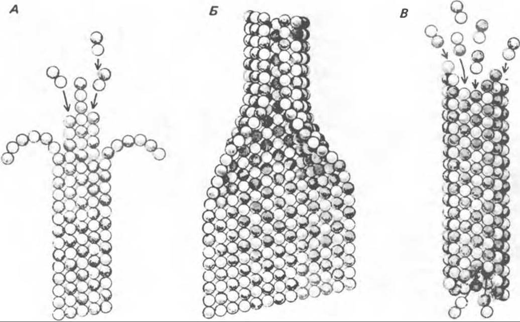
Fig. 56.15. In vitro assembly of microtubules begins with two globular protein molecules of a- and ß-tubulin. (They are likely more elongated in shape than depicted in the diagram.) Tubulins form dimers, or double molecules. When their concentration is high, they associate to form various intermediate structures (double rings, spirals, and combinations of rings). Depending on conditions, the equilibrium shifts either toward isolated dimers or intermediate structures. It can be hypothesized that the rings or spirals subsequently unroll to form strands (protofilaments) of linearly joined dimers, which zip up side-by-side to form a complete sheet (A); occasionally, the ends of the protofilaments curve. If the sheet is sufficiently wide, it presumably rolls up into a tube (B). This tube then elongates through The addition of dimers predominantly at one end (B). (Reproduced, with permission, from Dustin P. Microtubules. Sci. Am. [Aug] 1980, 243, 67.)
Intermediate filaments include type I and type II Keratins. They consist of 10–20 different Polypeptides that differ both from one another and from the polypeptides comprising the other four groups of intermediate filament proteins: desmin, vimentin, neurofilaments, and glial filaments. A high degree of Homology is observed among the latter four classes of proteins. A keratin filament contains at least two different keratin polypeptides, whereas the other four types of intermediate filaments are homopolymers. Each of them consists of four a-helical domains separated by regions of ß-pleated sheets and flanked on both sides by nonhelical terminal domains. The nonhelical terminal domains are involved in end-to-end elongation of protofilaments and in side-by-side interprotofibrillar interactions. The ends of keratin microfibrils can be cross-linked by Disulfide Bonds to form insoluble filaments resembling wool filaments.
Certain intermediate filaments, particularly those of muscle and mesenchymal origin, are present in A wide variety of Tissues.
Table 56.3. Types of Intermediate Filaments and Their Distribution
|
Proteins |
Mol. mass (thousands) |
Diameter (nm) |
Localization |
|
Type I and Type II keratin (tonofilaments) |
40-65 (10-20 major proteins) |
8 |
Epithelial cells (never found in cells of mesenchymal origin) |
|
Desmin |
50-55 |
10 |
Muscle (Z-line) |
|
Vimentin |
52 |
10 |
Mesenchymal and non-mesenchymal cells, such as muscle, glial, and epithelial cells |
|
Neurofilament |
200 150 70 |
10 |
|
|
Glial filament |
51 |
10 |
Glial cells |
Last update: 06/08/2026
Editorial and Educational Adaptation: This material has been compiled based on the primary/original source text. The project team performed an editorial review, corrected technical inaccuracies, structured sections, and adapted the content for an educational format.
What was processed:
- elimination of formatting defects (OCR errors, structural breaks, corrupted characters);
- editorial organization of content;
- standardization of terminology in accordance with academic sources;
- verification of factual statements against the original source text.
All mentions of the author, publication year, and origin of the primary text have been preserved in accordance with the source.