Human Biochemistry, Volume 2 - Murray R. 1993
Special Topics
Contractile and Structural Proteins
Collagen
Collagen, the major macromolecule of Connective Tissue, is the most abundant protein in the animal kingdom. It provides the extracellular framework in all Multicellular Organisms and is present in virtually every animal tissue. Mammalian Tissues contain at least five distinct types of collagen, which form a family of molecules sharing many common properties. The most characteristic feature of collagen molecules is their triple-helical Structure. Each subunit, or a-chain, is a left-handed helix with three amino acid residues per turn. Three such left-handed helices are further wound into a right-handed superhelix. As a result, a rigid, rod-shaped molecule is formed with a diameter of 1.4 nm and a length of about 300 nm. These collagen molecules then associate into fibrils (Fig. 56.16).
Collagen fibrils form staggered arrays that are slightly less than 1/4 the length of the triple helix. There is a gap between the end of one triple helix and the beginning of the next, which can serve as a nucleation site for the deposition of hydroxyapatite crystals during Bone Formation. The diameter of collagen fibrils ranges from 10 to 100 nm, and under the Microscope they appear as bundles within the Extracellular matrix of connective tissue.
Another characteristic feature of the collagen molecule is that every third residue of the triple-helical a-chain is Glycine. Glycine is the only amino acid whose size allows it to fit into the restricted central core of the triple-helical molecule. Thus, the central core of this molecule consists of glycine residues from all three a-subunits. This repeating structure can be represented as (Gly-X-Y)n, where X and Y are any Amino Acids other than glycine.
In mammalian collagen, about 100 X-positions are occupied by Proline and about 100 Y-positions by 4-hydroxyproline. These "rigid" amino acids restrict the Rotation of the polypeptide backbone and thereby increase the Stability of the triple helix. Hydroxyproline residues impart additional stability to the structure through The formation of extra intramolecular Hydrogen Bonds (mediated by surrounding Water molecules). Some X-positions of collagen also contain 3-hydroxyproline, and Y-positions contain 5-hydroxylysine.
Class="center">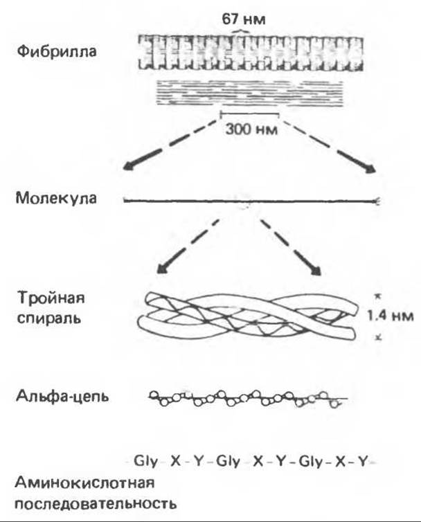
Fig. 56.16. Structural Features of collagen from primary sequence to fibrils. (Slightly modified and reproduced, with permission, from Eyre D. R: Collagen. Molecular diversity in the body’s protein scaffold. Science 1980, 207, 1315.)
The collagen triple helix is stabilized by numerous interchain cross-links between lysyl and hydroxylysyl residues. The Chemical Nature of these cross-links is discussed below. Mature collagen is a glycoprotein containing saccharides linked to hydroxylysine residues via O-glycosidic bonds.
Data on vertebrate collagens, their tissue distribution, and distinguishing features are summarized in Table 56.4.
Collagen Biosynthesis
Collagen is an extracellular protein, but it is synthesized as an intracellular precursor molecule that undergoes post-translational modification before the formation of mature collagen fibrils. Like all secreted Proteins, the collagen precursor undergoes Processing during its transit through the Endoplasmic reticulum and Golgi complex before appearing in the extracellular space. The earliest precursor of collagen is preprocollagen, which contains an N-terminal leader, or signal, sequence of approximately 100 amino acids. Preprocollagen is synthesized on Ribosomes attached to The endoplasmic reticulum. As the signal sequence enters the cisternal space of the endoplasmic reticulum, the leader sequence is cleaved, and the N-terminus of procollagen continues to advance within this space. Here, proline and Lysine residues in the Y-position of the (Gly-X-Y)n peptide are acted upon by prolyl 4-hydroxylase and lysyl hydroxylase, respectively. Prolyl 3-hydroxylase acts on proline residues in the X-positions immediately preceding the 4-hydroxyproline in the Y-positions.
Table 56.4. Genetically distinct vertebrate collagens. Higher animals possess at least five different molecules containing seven genetically distinct a-chains
|
Type |
Molecular formula |
Native polymer |
Tissue distribution |
Characteristic features |
|
I |
[а1(I)]2а2 |
Fibril |
Skin, tendons, bone, dentin, fasciae; abundant |
Low hydroxylysine content; few glycosylation sites; broad fibrils |
|
II |
[а1(II)]3 |
Fibril |
High hydroxylysine content; heavily glycosylated; fibrils generally thinner than those of type I |
|
|
III |
[а1(III)b |
Fibril |
High hydroxyproline content; low hydroxylysine content; few hydroxylysine glycosylation sites; interchain Disulfide Bonds between cysteines at the C-terminus of the helix; long carboxyl telopeptide |
|
|
IV |
[al(IV)], (approximate, debated) |
Basal lamina |
Renal glomeruli, lens capsule, Descemet's membrane; basal lamina of all epithelial and endothelial Cells (?) |
Very high hydroxylysine content; almost fully glycosylated; relatively rich in 3-hydroxyproline; low Alanine content; contains procollagen fragments |
|
V |
aA(aB)2 or (аА)3 and (аВ)3 |
Unknown |
Widely distributed in small amounts; basal lamina of blood vessels and smooth Muscle cells; exoskeleton of fibroblasts and other mesenchymal cells (?) |
High hydroxylysine content; heavily glycosylated; low alanine content; unable to form native fibrils in vitro |
The procollagen molecule contains Peptides at its N- and C-termini with molecular weights of approximately 20,000 and 30,000–35,000, respectively, which are absent in mature collagen. Both of these propeptides contain Cysteine residues. The N-terminal propeptide forms only intrachain disulfide bonds, whereas the C-terminal peptides form both intra- and interchain disulfide bonds. Following the formation of these bonds, the procollagen molecules assemble into a triple helix.
Once the triple helix is formed, further hydroxylation of proline and lysine residues becomes impossible. Glycosyltransferase, which transfers glucose or galactose to hydroxylysine residues, can also act only on procollagen a-chains that have not yet assumed a helical configuration.
Upon completion of this intracellular processing, the glycosylated procollagen molecules are transported to the outer Cell surface via the Golgi complex. Extracellular procollagen aminoprotease and carboxypeptidase remove the amino-terminal and carboxy-terminal propeptides, respectively. The newly formed collagen molecules contain approximately 1,000 amino acids per chain and spontaneously assemble into collagen fibrils indistinguishable from mature fibrils found in tissues.
However, these fibrils lack the tensile strength of mature collagen fibrils until they are cross-linked by a series of covalent bonds. An extracellular copper-containing enzyme, lysyl oxidase, catalyzes the Oxidative Deamination of the ε-amino groups of certain lysyl and hydroxylysyl residues in collagen, generating reactive aldehydes. These aldehydes can form Schiff bases with the ε-amino groups of other lysines, hydroxylysines, or even glycosylated hydroxylysines. These Schiff bases undergo chemical rearrangement to yield stable covalent cross-links. The aldehyde derived from hydroxylysine forms a more stable cross-link than the aldehyde from a lysine residue. Aldol Condensation products also contribute to the cross-linking of the molecules.
Data on the intracellular and extracellular Processing of the collagen precursor molecule are summarized in Table 56.5.
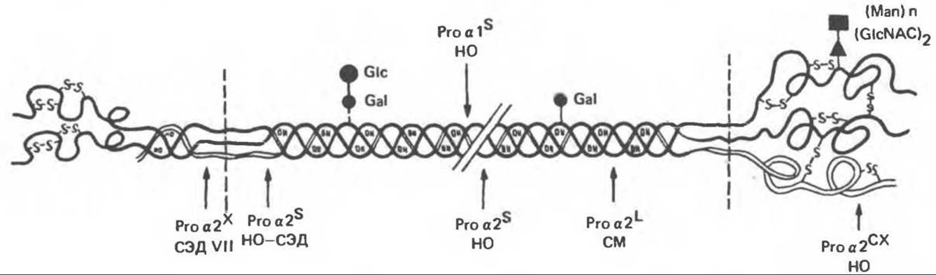
Fig. 56.17. Mutation sites in the type I procollagen molecule. EDS = Ehlers-Danlos syndrome; MFS = Marfan Syndrome; OI = Osteogenesis Imperfecta. (Reproduced, with permission, from Prockop D. J., Kivirikko K. I. Heritable diseases of collagen. N. Engl. J. Med. 1984, 311, 376.)
Table 56.5. Order and localization of processing steps for the collagen precursor (containing the [Gly-X-Y]n repeating structure)
|
Intracellular (endoplasmic reticulum) |
|
1) Cleavage of the signal peptide |
|
2) 4-Hydroxylation of Y-proline residues |
|
3) 3-Hydroxylation of X-proline, where Y is a 4-hydroxyproline residue |
|
4) 5-Hydroxylation of Y-lysine residues |
|
5) Glycosylation of hydroxylysine residues |
|
6) Formation of intra- and interchain S—S bonds |
|
7) Assembly of the procollagen triple helix |
|
Extracellular |
|
1) Cleavage of the NH2-terminal propeptide (MW 20,000) |
|
2) Cleavage of the COOH-terminal propeptide (MW 30,000–35,000) |
|
3) Fibril formation of immature collagen |
|
4) Oxidation of lysyl, hydroxylysyl, and glycosylated hydroxylysyl residues to aldehydes |
|
5) Cross-linking of chains and helical fibrillar molecules via Schiff bases and aldol condensation |
Cells that secrete collagen also secrete Fibronectin, a large glycoprotein present on The Cell surface, in the extracellular matrix, and in blood. Fibronectin binds to aggregating procollagen fibrils and alters The kinetics of fibril formation in the pericellular matrix. The Proteoglycans heparan sulfate and chondroitin sulfate are associated with fibronectin and procollagen in this matrix (see Chapter 54).
Cartilage is an extracellular matrix in which collagen provides tensile strength and proteoglycans provide pronounced elasticity.
Table 56.6. Molecular defects underlying four hereditary collagen disorders. (Reproduced, with permission, from Prockop D. J., Kivirikko K. I. Heritable diseases of collagen. N. Engl. J. Med., 1984, 311, 376.)
|
Disease |
Defect 1) |
Consequences |
|
Osteogenesis imperfecta |
Proa1 (I)0 |
50% reduction in type I collagen |
|
Type I |
Proa2 (I)s Others (unidentified) |
Likely abnormal fibrils |
|
Type II |
Proa 1 (I)s |
Unstable triple helix; increased synthesis of proa1 (III) |
|
Proa2(I)s and proa2(I)0 Others (unidentified) |
Unclear |
|
|
Type III |
Proa2 (I)cx |
Synthesis of proa1(I) trimers |
|
Proa I (I)сх or proa2(I)cx |
Increased mannose content in the C-propeptide and decreased solubility of type I procollagen |
|
|
Others (unidentified) |
||
|
Ehlers-Danlos syndrome variant |
Proa2 (I)s |
Insensitivity to procollagen N-proteinase and stability of pN-collagen2) |
|
Marfan syndrome |
Proa2 (I)L Others (unidentified) |
Likely impaired cross-linking |
|
Ehlers-Danlos syndrome |
Proa1 (III)+ |
Drastic reduction in type III collagen |
|
Type IV |
Proa1 (III)SM |
Unstable triple helix |
|
Proa1 (III)x |
Decreased rate of type III collagen secretion |
|
|
Others (unidentified) |
||
|
Type VI |
Lysyl hydroxylase deficiency Others (unidentified) |
Hydroxylysine-deficient collagen and impaired cross-linking |
|
Type VII |
Procollagen N-proteinase deficiency |
Stability of pN-collagen 2) |
|
Ргоа2 (I)x |
Insensitivity to procollagen N-proteinase and stability of pN-collagen 2) |
|
|
Type IX |
Copper METABOLISM disorder |
Lysyl oxidase deficiency and impaired cross-linking |
|
Menkes syndrome |
Copper metabolism disorder |
Lysyl oxidase deficiency and impaired cross-linking |
1) proa 1(I)0, proa2(I)0, and proa1 (III)+: non-functional or inefficiently functioning alleles of proa chains; proa 1 (I)s and proa2(I)s: shortened proa chains; proa2(I)L: elongated proa chains; proa1(I)cx and proa2(I)cx: Mutations altering The structure of the C-Polypeptides of proa chains; proa1 (III)SM: altered proa1 (III) chain exhibiting slow electrophoretic mobility in gel; proa1(III)x and proa2(I)x: uncharacterized mutations altering proa chain structure.
2) An intermediate in procollagen conversion reactions containing N-propeptides but lacking C-propeptides.
Hereditary defects of collagen and its assembly
Hereditary disorders caused by collagen abnormalities are collectively known as collagenoses. The most prominent among them are osteogenesis imperfecta, Marfan syndrome, Ehlers–Danlos syndrome, and Menkes syndrome (kinky Hair disease). Initially, these conditions were diagnosed solely on The basis of the proband's phenotype; however, as knowledge of collagen Structure and function accumulated, the molecular mechanisms underlying these syndromes became clear.
Many clinical symptoms can be explained by impaired processing of collagen precursors resulting from anomalies in the precursor itself or defects within the processing enzyme system. Understanding the molecular nature of these disorders makes it possible to predict the mode of Inheritance of the pathology (whether recessive or dominant).
Table 56.6 summarizes the molecular defects and their consequences in certain human hereditary collagenoses, while Fig. 56.17 indicates the defective sites within the type I procollagen molecule.
Adelstein R. S., Eisenberg R. Regulation and kinetics of Actin — Myosin ATP interaction, Annu. Rev. Biochem., 1980, 49, 921.
Adelstein R. S. et al. Phosphorylation of muscle contractile proteins, Fed. Proc., 1980, 39, 1544.
Barany Т. М. Barany К. Phosphorylation of the myofibrillar proteins, Annu. Rev. Physiol., 1980 42, 275.
Bornstein P., Sage H. Structurally distinct collagen types, Annu. Rev. Biochem., 1980, 49, 957.
Caplan A. Cartilage, Sсi. Am. (Oct.), 1984. 250, 84.
Clark M., Spudich J. A. Nonmuscle contractile proteins: The Role of Actin and myosin in Cell Motility and shape determination, Annu. Rev. Biochem., 1977, 46, 797.
DeCrombrugghe B., Pastan I. Structure and regulation of a collagen Gene. Trends Biochem. Sсi., 1982, 7, 11.
Dustin P. Microtubules, Sсi. Am. (Aug.), 1980, 243, 67.
Eyre D. R. et al. Cross-linking in Collagen and Elastin, Annu. Rev. Biochem., 1984, 53, 717.
Fuchs E., Hanukoglu I. Unraveling the structure of Intermediate filaments. Cell, 1983, 34, 332.
Heuser J. E., Kirschner M. W. Filament Organization revealed in platinum replicas of freeze-dried cytoskelctons. J. Cell Biol., 1980, 86, 212.
Kleinman H. K., Klebe R.J., Martin G.R. Role of collagenous matrices in the adhesion and growth of cells, J. Cell Biol., 1981, 88, 473.
Lazarides E. Intermediate filaments: A chemically heterogeneous, developmentally regulated class of proteins. Annu. Rev. Biochem., 1982. 51, 219.
Lazarides E., Revel J. P. The Molecular Basis of cell movement, Sсi. Am. (May), 1979, 240. 100.
Murray J. M., Weber A. The cooperative action of Muscle Proteins, Sс. Am. (Feb.), 1974, 230, 59.
Prockop D. J., Kivirikko К. I. Heritable diseases of collagen, N. Engl. J. Med., 1984. 311. 376.
Sandberg L. B., Soskel N. T., Leslie J. G. Elastin structure, biosynthesis, and relation to disease states, N. Engl, J. Med., 1981, 304, 566.
Last update: 06/08/2026
Editorial and Educational Adaptation: This material has been compiled based on the primary/original source text. The project team performed an editorial review, corrected technical inaccuracies, structured sections, and adapted the content for an educational format.
What was processed:
- elimination of formatting defects (OCR errors, structural breaks, corrupted characters);
- editorial organization of content;
- standardization of terminology in accordance with academic sources;
- verification of factual statements against the original source text.
All mentions of the author, publication year, and origin of the primary text have been preserved in accordance with the source.