Human Anatomy and Physiology (with Age-Related Features of the Child's Body) - Sapin M.R., Sivoglazov V.I. 2002
The Structure of the Human Body
Cells
The Human Body, functioning as a unified, integrated, and complex system, is composed of Organs and Tissues. Organs built from tissues are grouped into systems and apparatuses. Tissues, in turn, consist of various types of Cells and intercellular substance.
The Cell is the elementary, universal unit of living matter. It possesses an organized Structure, is capable of acquiring energy from the external environment, and utilizes it to perform Functions inherent to every cell. Cells actively respond to external stimuli (irritants), participate in METABOLISM, and exhibit capabilities for growth, regeneration, reproduction, transmission of Genetic information, and ADAPTATION TO ENVIRONMENTAL conditions.
Cells in the human body vary greatly in shape; they can be flat, spherical, ovoid, spindle-shaped, cuboidal, or stellate. Cellular shape is determined by their position within the Organism and their specific function. Cell sizes range from a few micrometers (such as a small lymphocyte) up to 200 µm (an ovum).
The intercellular substance is a product of cellular activity, consisting of a ground substance and various Connective Tissue fibers embedded within it.
Despite their vast diversity, All cells share common structural features, consisting of a nucleus and Cytoplasm enclosed within a cell membrane—the cytolemma (Fig. 3). The cell coat, or cell membrane (cytolemma, Plasmalemma), separates the cell from the external environment. The thickness of the cytolemma is 9–10 nm (1 nanometer equals 10-8 m or 0.002 µm). Composed of protein and lipid molecules, the cytolemma forms a trilaminar structure whose outer surface is covered by a delicate fibrillar glycocalyx. The glycocalyx contains various CARBOHYDRATES that form long, branching polysaccharide chains linked to the protein molecules embedded in the cytolemma. The outer and inner electron-dense lipid layers (leaflets) of the cytolemma are approximately 2.5 nm thick, while the middle electron-lucent layer (the hydrophobic region of lipid molecules) is about 3 nm thick. Protein molecules are situated within the bilipid layer of the cytolemma, with some spanning the entire thickness of The cell membrane.
The cytolemma does more than simply separate the cell from its external environment. It protects the cell, performs receptor functions (detecting external environmental signals), and mediates transport. The cytolemma facilitates The transfer of various substances (Water, low-molecular-weight compounds, ions) both into and out of the cell. Through the expenditure of energy (via ATP Hydrolysis), various organic substances (such as Amino Acids and sugars) are actively transported across the cytolemma.
Furthermore, the cytolemma forms Intercellular junctions (contacts) with neighboring cells, which can be simple or complex. Simple junctions appear as interlocking serrated seams, where projections (Teeth) of one cell's cytolemma interdigitate with those of the adjacent cell. An intercellular cleft with a width of 15–20 nm separates the cytolemmas of neighboring cells. Complex junctions are formed either by tightly apposed cell membranes of adjacent cells (tight junctions) or by the presence of a fine fibrillar substance between them (desmosomes). Conductive contacts include synapses and Gap Junctions (nexus junctions). In synapses, a cleft separates the cytolemmas of adjacent cells, allowing the transmission of excitation or inhibition in only one direction. In gap junctions, the intercellular space between adjacent cytolemmas is partitioned into distinct short segments by specialized protein structures.
Class="center">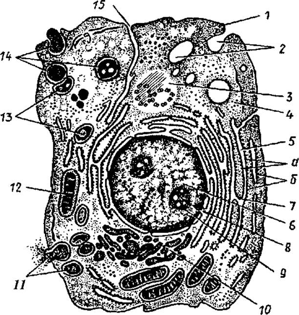
Fig. 3. Diagram of the ultramicroscopic structure of a cell: 1 — cytolemma (Plasma Membrane), 2 — pinocytotic vesicles, 3 — centrosome (cell center, cytocenter), 4 — hyaloplasm, 5 — Endoplasmic reticulum (a — membranes of The endoplasmic reticulum, b — Ribosomes), 6 — nucleus, 7 — connection between the perinuclear space and the cavities of the endoplasmic reticulum, 8 — nuclear pores, 9 — nucleolus, 10 — intracellular reticular apparatus (Golgi apparatus), 11 — secretory vacuoles, 12 — Mitochondria, 13 — Lysosomes, 14 — three consecutive stages of phagocytosis, 15 — connection between the cell membrane (cytolemma) and the membranes of the endoplasmic reticulum
The cytoplasm is heterogeneous in composition, comprising the hyaloplasm along with the Organelles and inclusions suspended within it.
The hyaloplasm (from Greek hyalinos — transparent) forms the cytoplasmic matrix, constituting its internal environment. Externally, it is bounded by the cell membrane, or cytolemma. Appearing as a homogeneous substance, the hyaloplasm is a complex colloidal system consisting of Proteins, Nucleic Acids, Polysaccharides, Enzymes, and other substances.
A critical function of the hyaloplasm is to integrate all intracellular structures and facilitate their chemical interactions. Proteins essential for cellular viability and function are synthesized within the hyaloplasm. Additionally, the hyaloplasm stores Glycogen, lipid inclusions, and serves as an energy reservoir containing adenosine triphosphate (ATP) molecules.
Resident within the hyaloplasm are general-purpose organelles found in all cells, as well as non-permanent structures known as cytoplasmic inclusions. Organelles include mitochondria, the internal reticular apparatus (Golgi apparatus), the cytocenter (cell center), granular and agranular endoplasmic reticula, ribosomes, and lysosomes. Inclusions encompass glycogen, proteins, Lipids, Vitamins, pigment granules, and other structures.
Organelles are permanent cytoplasmic structures found in cells that perform specific, vital functions. They are categorized into membranous and non-membranous organelles. Certain specialized tissues contain unique organelles, such as myofibrils within Muscle tissue structures.
Membranous organelles are closed, microscopic cavities—either isolated or interconnected—separated from the surrounding hyaloplasm by a membrane. Membranous organelles include mitochondria, the internal reticular apparatus (Golgi apparatus), the endoplasmic reticulum, lysosomes, and Peroxisomes. The endoplasmic reticulum is subdivided into granular (rough) and agranular (smooth) types. Both are formed by cisternae, vesicles, and tubules enclosed by a membrane approximately 6–7 nm thick. The endoplasmic reticulum bearing ribosomes attached to its membranes is termed the granular (rough) endoplasmic reticulum, whereas the absence of surface ribosomes designates the smooth endoplasmic reticulum.
Endoplasmic reticulum membranes participate in intracellular substance transport. Protein Synthesis occurs on the ribosomes of the rough endoplasmic reticulum, while glycogen and lipids are synthesized on the membranes of the smooth endoplasmic reticulum.
The internal reticular apparatus (Golgi apparatus) is formed by the membranes of closely stacked flat cisternae accompanied by numerous small vesicles located along their periphery. Clusters of these membranes are termed dictyosomes. A single dictyosome comprises 5–10 flat membranous cisternae separated by layers of hyaloplasm. The membranes of the internal reticular apparatus function to accumulate and chemically modify substances synthesized by the endoplasmic reticulum. Within the cisternae of the Golgi complex, polysaccharides are synthesized and complexed with proteins. The Golgi complex also participates in exporting synthesized substances beyond the cell boundary and serves as the source for The formation of cellular lysosomes.
Mitochondria possess a smooth outer membrane and an inner membrane folded inward to form cristae. This folding of The inner mitochondrial membrane significantly increases its internal surface area. A narrow intermembrane space separates the outer and inner mitochondrial membranes. The mitochondrial matrix, possessing a fine-grained structure, fills the cavity between the cristae and contains DNA (deoxyribonucleic acid) molecules along with mitochondrial ribosomes. Mitochondria average about 0.5 µm in diameter and reach lengths of 7–10 µm. Their primary function is The oxidation of Organic compounds and the utilization of the released energy for the synthesis of ATP molecules.
Lysosomes are membrane-bound, spherical structures ranging from 0.2 to 0.4 µm in diameter. The presence within lysosomes of hydrolytic enzymes (Hydrolases) that break down various Biopolymers indicates their involvement in intracellular Digestion processes.
Peroxisomes (Microbodies) are small membrane-bound vacuoles measuring 0.3–1.5 µm containing a granular matrix. This matrix contains catalase, which degrades hydrogen peroxide generated during the enzymatic Oxidative Deamination of amino acids.
Non-membranous organelles include ribosomes, microtubules, centrioles, microfilaments, and other structures. Ribosomes function as the elementary machinery for the synthesis of protein and polypeptide molecules. They consist of ribonucleoprotein granules (20–25 nm in diameter) formed by proteins and RNA molecules. Alongside solitary ribosomes, cells frequently contain clusters of ribosomes known as polyribosomes or Polysomes.
Microtubules are located within the Cell Cytoplasm as hollow cylinders measuring approximately 24 nm in diameter. They are composed of tubulin proteins. Microtubules form the Cytoskeleton and participate in cellular motility. They maintain cell shape and facilitate directed Intracellular Transport. Furthermore, microtubules contribute to The structure of centrioles, the mitotic spindle, basal bodies, flagella, and cilia.
Centrioles are hollow cylinders approximately 0.25 µm in diameter and up to 0.5 µm in length. Their walls are constructed from interconnected microtubule triplets (9×3). Two centrioles oriented at a right angle to each other form a diplosome. Surrounding the centrioles (diplosome) is the centrosphere, appearing as an amorphous, dense halo with radiating fine fibrils.
Together, the centrioles and centrosphere constitute the cell center (centrosome). In preparation for mitotic division, the number of centrioles within the cell doubles.
Centrioles participate in forming the mitotic spindle and cellular motility apparatuses—Cilia and flagella. Cilia and flagella are cylindrical cytoplasmic projections containing a central system of microtubules.
Microfilaments are thin (5—7 nm) protein filaments arranged in bundles or layers, primarily in the peripheral Regions of the cell. They are composed of various contractile proteins, including Actin, Myosin, and Tropomyosin. Microfilaments provide structural support and facilitate cell motility. Intermediate filaments, or microfibrils, are about 10 nm thick and vary in composition across different cell types. In epithelial cells, these filaments are composed of Keratins; in muscle cells, of desmin; and in Nerve Cells, of neurofilament proteins. Intermediate filaments also serve as structural scaffolding for cells.
Cytoplasmic inclusions are temporary structures formed As a result of cellular activity. They are classified into trophic, secretory, and pigment inclusions. Trophic inclusions can be proteinaceous, lipid, or carbohydrate-based; they serve as nutrient reserves accumulated by the cell. Secretory inclusions are products of glandular cell activity, containing BIOLOGICALLY ACTIVE SUBSTANCES required by the body. Pigment inclusions are colored substances essential to the body that accumulate within the cell. Pigments can be of exogenous origin (such as Dyes) or endogenous origin (including melanin, Hemoglobin, bilirubin, and lipofuscin).
The Cell Nucleus. The Nucleus is an essential component of the cell; it contains genetic information and regulates protein synthesis. Genetic information is encoded in deoxyribonucleic acid (DNA) molecules. During Cell Division, this information is transmitted in equal amounts to the daughter cells. The nucleus possesses its own protein synthesis machinery that controls synthetic processes in the cytoplasm. Various types of ribonucleic acid (RNA)—messenger, transfer, and ribosomal—are transcribed from DNA templates within the nucleus.
The nucleus of a non-dividing (interphase) cell is typically spherical or ovoid and consists of Chromatin, the nucleolus, and karyoplasm (nucleoplasm), which are separated from the cytoplasm by the nuclear envelope.
Chromatin in the interphase nucleus represents chromosomal material, which consists of loose, decondensed Chromosomes. Decondensed chromosomes are referred to as euchromatin. Thus, chromosomes in cell nuclei can exist in two Structural and functional states. In their decondensed form, chromosomes are in an active, functional state. During this time, they participate in the Transcription and Replication (from Latin replicatio — repetition) of nucleic acids (RNA, DNA). Chromosomes in the condensed (compact) state are inactive; they are involved in the distribution and transfer of genetic information to daughter cells during cell division. In the early phases of mitotic cell division, chromatin condenses, forming visible chromosomes. In humans, somatic cells contain 46 chromosomes: 22 pairs of homologous chromosomes and two sex chromosomes. In females, the sex chromosomes are homologous (XX chromosomes), whereas in males, they are non-homologous (XY chromosomes).
The nucleolus is a dense, intensely staining, rounded structure within the nucleus, measuring 1—5 µm in size. It consists of filamentous structures—Nucleoproteins and intertwined RNA strands—as well as ribosomal precursors. The nucleolus serves as the site of ribosome assembly, which are then used to synthesize polypeptide chains in the cytoplasm.
Nucleoplasm is the electron-transparent part of the nucleus, representing a colloidal protein solution that surrounds the chromatin and nucleolus.
The nuclear envelope (nucleolemma) consists of an outer nuclear membrane and an inner nuclear membrane separated by the perinuclear space. The nuclear envelope contains pores occupied by protein granules and fibrils (the pore complex). Selective transport of proteins occurs through these nuclear pores, enabling the passage of macromolecules into the cytoplasm and facilitating Metabolic exchange between the nucleus and the cytoplasm.
Last update: 10/08/2026
Editorial and Educational Adaptation: This material has been compiled based on the primary/original source text. The project team performed an editorial review, corrected technical inaccuracies, structured sections, and adapted the content for an educational format.
What was processed:
- elimination of formatting defects (OCR errors, structural breaks, corrupted characters);
- editorial organization of content;
- standardization of terminology in accordance with academic sources;
- verification of factual statements against the original source text.
All mentions of the author, publication year, and origin of the primary text have been preserved in accordance with the source.