HUMAN MEDICAL BIOLOGY, ANATOMY, PHYSIOLOGY AND PATHOLOGY - Ya.I.Fedoniuk 2010
BIOLOGY
CHAPTER 1. BIOLOGICAL FOUNDATIONS OF HUMAN VITAL ACTIVITY
1.3. MOLECULAR-GENETIC AND CELLULAR LEVELS OF LIFE ORGANIZATION
1.3.2. Structural, chemical, and functional organization of eukaryotic cells
Methods of Cell study
To study cell Structure and function, Cytology employs microscopic Methods based on The Use of microscopes. Depending on the state of the studied specimen, these methods are subdivided into vital (or supravital) methods, which examine living (or surviving) Cells, and post-vital methods, which examine dead specimens specially prepared by fixation. Cells in this case can be stained or unstained. The primary method for studying all cells is light Microscopy using a Light Microscope (Fig. 1.30).
Class="center">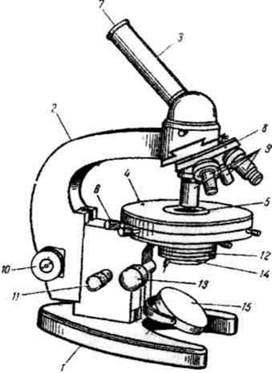
Fig. 1.30. Structure of a light microscope
To study unstained cells, Special Methods of light microscopy are used, such as phase-contrast, dark-field, and fluorescence (luminescent) microscopy. The Phase-contrast microscopy method is based on the fact that individual areas of an otherwise transparent cell differ from one another in density and refractive index. In a phase-contrast microscope, a phase plate is built into the objective lens, which causes an additional phase shift of light waves passing through it. This creates a light-and-dark contrast image of the specimen. THE PRINCIPLE OF the fluorescence method is that A number of substances have the property of emitting light when they absorb light energy. Autofluorescence is exhibited by certain pigments (chlorophyll), Vitamins (A, B2), and Hormones. When plant cells are viewed under a fluorescence microscope, brightly glowing red granules—METABOLISM/14.html">Chloroplasts—are visible against a dark blue Background.
Microsurgical methods are employed in the Study of Living cells. Using a micromanipulator device, cells can be dissected, parts of them extracted, and substances injected. This method makes it possible to transplant nuclei from one cell to another and study alterations in heredity.
In modern cytology, histochemical, autoradiographic, immunomorphological, and cytospectrophotometric Research Methods have gained widespread use.
The histochemical method makes it possible to determine the localization of specific chemical substances within various Structural components of Cells and Tissues. The autoradiographic method is based on the use of radioactive isotopes and compounds labeled with them. Such compounds are introduced into the body of an experimental animal, and the radioactive substances are subsequently detected in histological sections using a photographic emulsion applied over the specimen and then developed. In areas where the photographic emulsion contacts the radioactive substance, exposed spots—tracks—remain. Immunohistochemical methods rely on antigen-antibody reactions. Cytospectrophotometry is a method for quantitatively measuring the content of various substances in a cell based on The Study of their Light absorption spectra.
A major breakthrough was the invention and application of the Electron microscope. An electron microscope uses a beam of electrons to "illuminate" the specimen; this beam has a much shorter wavelength compared to the visible light used in a light microscope. As a result, the resolution limit, which equals 1/3 of the wavelength, is 0,2 µm for a light microscope (theoretically), whereas for an electron microscope it is 0,002 nm. In practice, the resolution limit of the best electron microscopes reaches 0,1-0,7 nm.
Last update: 08/08/2026
Editorial and Educational Adaptation: This material has been compiled based on the primary/original source text. The project team performed an editorial review, corrected technical inaccuracies, structured sections, and adapted the content for an educational format.
What was processed:
- elimination of formatting defects (OCR errors, structural breaks, corrupted characters);
- editorial organization of content;
- standardization of terminology in accordance with academic sources;
- verification of factual statements against the original source text.
All mentions of the author, publication year, and origin of the primary text have been preserved in accordance with the source.