Cytology, General Histology and Embryology - V. K. Napkhanyuk 2002
Microscopes. Microscopy Techniques
Microscopy Techniques
Phase-contrast microscopy
Many specimens, particularly unstained live biological objects, do not absorb light and appear colorless and transparent. The phase-contrast method makes it possible to obtain high-contrast images of these specimens, which are nearly invisible under conventional observation Methods. This eliminates The Need for specimen staining, which invariably has harmful effects on living objects.
Class="center">
Fig. 2. Schematic diagram of a fluorescence Microscope: 1 — light source; 2 — collector field Diaphragm; 3, 9 — light filters; 4 — illuminator mirror; 5 — condenser; 6 — specimen; 7 — objective lens; 8 — eyepiece
The phase-contrast method is based on the principle that specific structural areas of a transparent specimen differ from the surrounding medium in their refractive index. Consequently, light passing through them undergoes a phase delay equal to
s = h (n0 - nс)
where h is the thickness of the object;
n0, nс are the refractive indices of the object itself and the surrounding medium, respectively.
As a result, the light wave passing through the specimen undergoes various phase changes, acquiring what is known as a «phase relief». However, this relief cannot be directly perceived by the human eye or a photographic plate, both of which are sensitive only to changes in light intensity depending solely on the amplitude of light oscillation. The phase-contrast method makes it possible to convert phase changes into amplitude changes, thereby replacing the «phase relief» with an «amplitude relief». This yields a visible, so-called phase-contrast image of the specimen, in which the distribution of illumination corresponds to the distribution of phases.
Due to the aforementioned principles, the design of a phase-contrast microscope has some fundamental differences compared to a standard Light Microscope (Fig. 3): an annular diaphragm is installed in the front focal plane of the condenser instead of the standard iris aperture diaphragm. A phase Glass plate with an etched annular groove (phase ring) is positioned within the objective lens. The diameter of this ring equals the diameter of the condenser diaphragm image; thus, the phase ring blocks all light that passes directly through the specimen and the objective (solid lines). The action of the phase ring is twofold. First, it absorbs a significant portion of the directly transmitted rays, for which the bottom of the groove is coated with a semitransparent metal film. Second, the phase ring shifts the phase of the light oscillations by a quarter of the light wavelength. Accordingly, the depth of the groove in the plate must be approximately 0.5 µm. Light scattered by the specimen (dashed lines in the figure) bypasses the phase ring and undergoes no attenuation or phase shift.
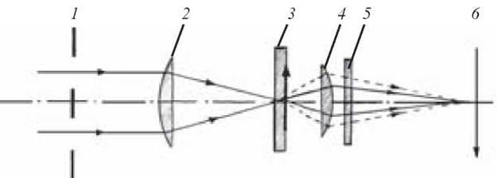
Fig. 3. Optical diagram of a phase-contrast microscope:
1 — annular diaphragm; 2 — condenser; 3 — specimen; 4 — objective lens; 5 — phase plate; 6 — real image of the specimen
The phase plate is designed such that rays unscattered by the object pass through a slightly thinner layer of glass in the plate compared to the diffracted rays. Consequently, the phase of the directly transmitted rays leads the phase of the diffracted rays. This forms a so-called positive phase-contrast image of the specimen in the image plane. Small particles whose refractive index is higher than that of the surrounding medium appear dark against a light Background. For larger objects, only the edges become dark, while the central portion remains light. All dark images are surrounded by broad bright halos. Particles with a refractive index lower than that of the surrounding medium produce images that appear brighter than the background.
Last update: 10/08/2026
Editorial and Educational Adaptation: This material has been compiled based on the primary/original source text. The project team performed an editorial review, corrected technical inaccuracies, structured sections, and adapted the content for an educational format.
What was processed:
- elimination of formatting defects (OCR errors, structural breaks, corrupted characters);
- editorial organization of content;
- standardization of terminology in accordance with academic sources;
- verification of factual statements against the original source text.
All mentions of the author, publication year, and origin of the primary text have been preserved in accordance with the source.