LEHNINGER PRINCIPLES OF BIOCHEMISTRY - VOL. 1. THE FOUNDATIONS OF BIOCHEMISTRY: STRUCTURE AND CATALYSIS - 2011
PART I. STRUCTURE AND CATALYSIS
4. THE THREE-DIMENSIONAL STRUCTURE OF PROTEINS
4.2. Protein Secondary Structure
The term Secondary Structure refers to any chosen segment of a polypeptide chain and describes the local spatial conformation of its backbone atoms, without regard to the conformation of side chains or interactions with other segments. A regular secondary structure occurs when the torsion angles (φ and Ψ) remain constant or nearly constant throughout a specific segment of the chain.
The most well-known types of secondary structure are α-helices and β-sheets; β-turns also occur quite frequently. When no regular structures are found, the region is referred to as having an unordered secondary structure or a random coil. However, this latter term is somewhat misleading, as the polypeptide backbone in virtually any protein is far from random; moreover, it is typically fixed and specific to the Structure and function of a given protein. Here, we will focus on the most common regular structures.
The α-Helix Is a Widespread Protein Secondary Structure
Protein Architecture—The α-Helix. Pauling and Corey recognized the crucial role of Hydrogen Bonds in orienting polar chemical groups, such as C=O and N–H, within the peptide group. They were also aware of the work by William Astbery, who in the 1930s first performed X-Ray Diffraction Analysis on Proteins. Astbery showed that the protein forming The basis of Hair and porcupine quills (the fibrous protein α-keratin) possesses a regular structure, specifically consisting of repeating units with a periodicity of 5.15–5.20 Å. (The unit of length, the angstrom [Å], is named in honor of the Swedish physicist Anders Jonas Ångström; 1 Å = 0.1 nm. Although not an SI unit, it is widely used in structural chemistry and biology to describe atomic dimensions and interatomic distances.) Armed with this information, their own data on the geometry of the peptide bond, and precisely constructed models, Pauling and Corey set out to determine the likely Conformations of protein molecules.
The simplest way to arrange a polypeptide chain—given its rigid peptide bonds and free-rotating single bonds—is a helical structure, which Pauling and Corey named the α-helix (Fig. 4–4). In this structure, the polypeptide backbone coils tightly around the long axis of the molecule, while The amino acid R-groups protrude outward from the helical backbone. The repeating unit corresponds to a single turn of the helix, with a pitch of approximately 5.4 Å, which is only slightly larger than the value Astbery deduced from X-ray diffraction analysis of hair keratin. The amino acid residues in an α-helix have torsion angles of φ = -57° and Ψ = -47°, with each turn containing 3.6 amino acid residues. Some deviation from these ideal torsion angle values is frequently observed in the α-helical regions of proteins, and variations can occur even within a single segment, allowing the helix to form slight bends and loops. In all proteins, the α-helix is right-handed (Box 4–1). The α-helix has proved to be the predominant structure in α-Keratins. Furthermore, about a quarter of all amino acid residues in polypeptide chains are incorporated into α-helices (varying somewhat among different proteins).
Class="center">Fig. 4–4. Models of the α-helix illustrating various structural features, (a) Ball-and-stick model of a right-handed α-helix showing intra-chain hydrogen bonds. The pitch of the helix corresponds to 3.6 amino acid residues, (b) End-on view of an α-helix looking down its long axis (PDB ID 4TNC). Note the arrangement of the R-groups, depicted here in purple. This ball-and-stick model creates the false impression that the interior of the helix is hollow; however, the sphere sizes do not correspond to Van der Waals radii, (c) As seen in the space-filling model, the atoms in the core of the α-helix are very tightly packed, (d) Helical net projection. Surfaces with distinct properties are highlighted in color. For example, hydrophobic residues are shown in yellow; these are typically located at the interface between this helix and another part of the same or a different polypeptide. Red and blue residues illustrate potential Electrostatic Interactions between positively and negatively charged side-chain groups separated by three residues in the sequence.

Why does the α-helix form more readily than other possible structures? To a large extent, this is because the α-helix maximizes internal hydrogen bonding. The structure is stabilized by hydrogen bonds formed between the hydrogen atom attached to the electronegative nitrogen of a peptide bond and the electronegative carbonyl oxygen of the fourth amino acid along the chain (Fig. 4–4a). Every peptide group in the α-helix (except those near the ends) participates in these hydrogen bonds. Consequently, each successive turn is linked to the preceding one by three or four hydrogen bonds, rendering this polypeptide conformation quite stable.
Subsequent model experiments demonstrated that the α-helix can form from either L- or D-Amino Acids, but all residues within a given helix must belong to the same stereochemical series. Incorporating a D-amino acid would disrupt a regular structure composed of L-amino acids, and vice versa. In principle, naturally occurring L-Amino acids can form both right-handed and left-handed helices, but extended left-handed helices have not been observed in proteins.
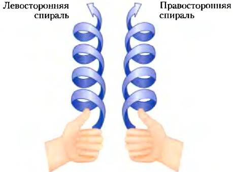
Box 4–1. Methods: How to Distinguish Between Right-Handed and Left-Handed Helices
There is a simple visual trick to distinguish between right-handed and left-handed helices. First, curl the four fingers of each hand toward your palms, leaving your thumbs extended straight up (see illustration). Now look at your RIGHT HAND AND imagine that the four fingers wrap around the thumb in a helical path pointing in the direction of the thumb (i.e., counterclockwise). This represents a right-handed helix. Do the same with your left hand: the four fingers form a left-handed helix winding around the thumb and pointing upward (i.e., clockwise).
Worked Example 4–1. Protein Secondary Structure and Dimensions
What is the length of a polypeptide consisting of 80 amino acid residues that forms a continuous single α-helix?
Solution. One turn of an ideal α-helix contains 3.6 amino acid residues and has a length of 5.4 Å. Therefore, the axial rise per amino acid residue is 1.5 Å. Consequently, the total length of the polypeptide chain is: 80 residues × 1.5 Å/residue = 120 Å.
Amino Acid Sequence Affects α-Helix Stability
Not all Polypeptides can form stable α-helices. Each amino acid residue in a polypeptide chain has a characteristic propensity to form an α-helix (Table 4–1), which depends on The chemical properties of its R-group and the ability of adjacent backbone atoms to adopt the required φ and Ψ angles. In most experimental model systems, Alanine exhibits the highest α-helix-forming propensity.
Table 4–1. Amino Acid Propensities for α-Helix Formation
Amino Acid |
∆∆G° (kJ/mol)* |
Amino Acid |
∆∆G° (kJ/mol)* |
Ala |
0 |
Leu |
0,79 |
Arg |
0,3 |
Lys |
0,63 |
Asn |
3 |
Met |
0,38 |
Asp |
2,5 |
Phe |
2,0 |
Cys |
3 |
Pro |
4 |
Gln |
1,3 |
Ser |
2,2 |
Glu |
1,4 |
Thr |
2,4 |
Glv |
4.6 |
Tyr |
2,0 |
His |
2,6 |
Trp |
2.0 |
lle |
1,4 |
Val |
2,1 |
*∆∆G° represents the free-energy change for adopting an α-helical conformation relative to alanine.
THE POSITION OF an amino acid residue relative to its local environment is also critically important. Interactions between amino acid side chains can either stabilize or destabilize the helical structure. For instance, if a polypeptide chain contains a long stretch of contiguous glutamate residues, it cannot form an α-helix at pH 7.0. This is because the negatively charged carboxylate groups of adjacent Glu residues repel each other strongly enough to prevent helix formation. For the same reason, stretches featuring closely spaced Lys and/or Arg residues, whose R-groups carry a positive charge at pH 7.0, undergo electrostatic repulsion that hinders helix formation. Neighboring Asn, Ser, Thr, and Cys residues can likewise destabilize the α-helix, in these cases primarily due to the bulk and shape of their R-groups.
The turns of the α Helix allow interactions between the side chain of one Amino Acid and that of another located three or four residues away (Fig. 4-4d). Frequently, positively charged Amino acids are positioned three residues apart from negatively charged ones, which facilitates ion-pair formation. Two aromatic amino acids are often arranged spatially in a similar manner, enabling hydrophobic interactions.
Another obstacle to α-helix formation is the presence of Pro or Gly residues, which have the lowest propensity to form an α helix. In Proline, the nitrogen atom is part of a rigid ring (Fig. 4-7b), ruling out any rotation around the N–Cα bond. As a result, Pro residues introduce destabilizing kinks into the α helix. Moreover, the nitrogen atom in a Pro residue lacks a hydrogen atom capable of participating in hydrogen bonding with another amino acid residue. Consequently, proline is exceedingly rare in sequences organized as an α helix. Glycine is uncommon in α helices for a different reason: it possesses greater conformational flexibility than other amino acid residues, causing polymers of glycine to adopt Helical structures distinct from α helices.
Finally, the stability of a polypeptide's α-helical structure is significantly influenced by the specific amino acid residues located at the ends of the helical segments. Each peptide bond possesses a small electric dipole (Fig. 4-2a). These dipoles are aligned through the hydrogen bonds spanning the helix, causing the entire structure to act as a macroscopic dipole whose magnitude increases with the length of the helix (Fig. 4-5). Four amino acids at each end of the helix are not fully engaged in hydrogen bonding. The partial positive and partial negative charges of the dipole are typically localized on the peptide amino group and carbonyl group near the N- and C-termini of the helix, respectively. For this reason, negatively charged amino acids are frequently found at the N-terminus of a helical segment, where they can neutralize the positive charge of the dipole. Conversely, the presence of positively charged amino acids at this end exerts a destabilizing effect. All of the above (with inverted charge signs) applies equally to the C-terminus of the helix.
Fig. 4-5. The α-helix dipole. The electric dipole inherent to each peptide bond (see Fig. 4-2a) extends across the entire length of the α-helical segment via hydrogen bonds. In the diagram, the amino and carbonyl groups of each peptide bond are designated with + and - signs, respectively. The N- and C-terminal amino and carbonyl residues of the peptide groups that are not fully engaged in hydrogen bonding are highlighted in red.

Thus, the Stability of the α helix is governed by five distinct types of factors: (1) the intrinsic α-helix-forming propensity of the amino acid residues; (2) interactions between R groups, particularly those spaced three (or four) amino acid residues apart; (3) the close proximity of bulky amino acid R groups; (4) the presence of Gly and Pro residues; and (5) interactions between the amino acid residues at the N- and C-termini of the helical segment and the electric dipole generated within the helix. Therefore, the ability of a specific segment of a polypeptide chain to form an α helix depends on which amino acids are present and how they are ordered within that segment.
Polypeptide segments with a β conformation form β sheets
Protein architecture—the β sheet. In 1951, Pauling and Corey predicted the existence of a second type of regular peptide Organization—the β structure—in which the chains adopt a more extended conformation, as confirmed by X-ray diffraction data. In this case, the polypeptide backbone forms a zigzag rather than a helical structure (Fig. 4-6). These zigzag polypeptide chains can be arranged side by side to form a series of pleats. In this architecture, known as β sheets, hydrogen bonds form between adjacent segments of polypeptide chains. Typically, β sheets are formed by segments located relatively close to each other in the polypeptide chain, but they can also involve relatively distant sequence regions or even segments from entirely different polypeptide chains. The amino acid R groups project outward from both sides of the zigzag structure, which is clearly visible in a side-projected view of the chain (Fig. 4-6).
Fig. 4-6. The β conformation of a polypeptide chain. Top and side views of The polypeptide chains reveal the projecting R groups of the amino acid side chains and emphasize the pleated structure of β sheets, often referred to as β-pleated sheets. Hydrogen bonds between adjacent strands are depicted as short blue dashes. The orientation of the N- and C-termini of neighboring strands (indicated by arrows) can be identical or opposite, forming either (a) an antiparallel β sheet or (b) a parallel β sheet.

Adjacent polypeptide chains in β sheets can run either parallel or antiparallel (i.e., possessing the same or opposite N-to-C orientations). The resulting structures are quite similar yet differ slightly in their repeat unit dimensions (6.5 Å for parallel strands and 7.0 Å for antiparallel strands), and their hydrogen-bonding patterns are organized differently. In idealized structures, the dihedral angles are: φ = -119°, Ψ = +113° (parallel arrangement) and φ = -139°, Ψ = +135° (antiparallel arrangement). In real proteins, these values vary somewhat, leading to structural deviations much like those described earlier for the α helix.
Certain Amino acid sequences restrict The formation of β sheets. Specifically, for two or more β sheets to pack closely together within a protein, the contacting R groups of the amino acid residues must be relatively small. Such β keratins, including silk and spider fibroins, contain a very high proportion of Gly and Ala residues, which possess the smallest R groups. In Silk Fibroin, for example, Gly and Ala residues alternate throughout extensive Regions of the sequence.
β-Turns frequently occur in protein structures
Protein architecture: the β-turn. In Globular proteins, which are characterized by a compact packing of polypeptide chains, about one-third of all amino acid residues are found in turns and loops that arise when the polypeptide chain reverses its folding direction (Fig. 4-7). These structural elements connect adjacent segments of α-helices and β-sheets. The most common are β-turns, which link the ends of two neighboring anti-parallel β-sheet segments. This structure forms a 180° loop involving four amino acid residues, where the carbonyl oxygen of the first residue forms a Hydrogen bond with the amino hydrogen of the fourth. The peptide groups of the two central residues do not participate in hydrogen bonding with any other residues. Gly and Pro residues frequently appear in these structures: the former due to its small size and flexibility, and the latter because the peptide bond involving the imine nitrogen of proline readily adopts a cis-conformation that facilitates loop formation (Fig. 4-7, b). Fig. 4-7, a illustrates the Two Types of β-turns most commonly found in proteins. These structures are typically located near the protein surface, where the two central amino acids of the four that form the turn can participate in hydrogen bonds with Water molecules. Considerably less frequent are γ-turns consisting of three amino acid residues, in which the hydrogen bond forms between the first and third residues.
Fig. 4-7. STRUCTURE OF THE β-turn. a) Type I and type II β-turns are the most prevalent in proteins, with type I occurring more than twice as often as type II. In type II β-turns, a Gly residue is always present at the third position. Note the hydrogen bonds between the first and fourth residues of the loop (individual amino acid residues are schematically represented as large blue circles and numbered). b) trans- and cis-isomers of the peptide bond involving proline. Peptide bonds between amino acid residues in proteins are 99.95% in the trans-configuration. An exception is found in peptide bonds involving Pro: 6% of the peptide bonds it forms adopt the cis-configuration, and many of these are located within β-turns.

Protein secondary structures are characterized by specific Bond Angles
The most prevalent secondary structures in most protein molecules are α-helices and β-sheets, although certain specialized proteins feature other secondary structures (Collagen, for example, is shown in Fig. 4-12). Each type of secondary structure can be fully described by the φ and ψ torsion angles formed by the bonds within the amino acid residues. As seen in the Ramachandran plot, α-helices and β-sheets fall within a fairly narrow range of allowed conformations (Fig. 4-8, a). Most of the φ and ψ values derived from known protein structures fall into this required range, with the vast majority corresponding specifically to α-helical and β-sheet conformations (Fig. 4-8, b). Glycine is the only amino acid whose characteristics fall outside this standard range. Owing to The small size of its side chain (a single hydrogen atom), the glycine residue can adopt A wide variety of conformations that are sterically forbidden for Other Amino Acids.
Fig. 4-8. Ramachandran plot for various protein structures. a) The φ and Ψ values for various secondary structures are plotted on the graph from Fig. 4-3. Although left-handed α-helices several amino acids long can theoretically exist, no such structures have been found in proteins. b) The φ and Ψ values for all amino acid residues (except glycine) of rabbit Pyruvate kinase are mapped onto the plot of allowed conformations (Fig. 4-3). Small Gly residues, capable of adopting various configurations, are excluded from consideration because they frequently fall outside the allowed range (shown in blue).

Secondary structures can be identified using circular dichroism spectroscopy
Molecules with an asymmetric structure absorb the right- and left-circularly polarized components of plane-polarized light to different extents. Measuring these differences is the basis of circular dichroism (CD) spectroscopy. Ordered structures, such as folded proteins, exhibit absorption spectra with characteristic peaks and troughs. Protein spectra are typically recorded in the far-ultraviolet region (190 to 250 nm). The absorbing (chromophoric) unit in this case is the peptide bond; the signal originates from peptide bonds embedded in the environment of a folded protein. The difference in molar extinction coefficients (see Box 3-1) for left- and right-circularly polarized light (Δε) is plotted as a function of wavelength.
α-Helices and β-sheets have characteristic CD spectra (Fig. 4-9). CD spectroscopy can be used to determine whether a protein is properly folded, to estimate the fraction of the protein adopting a particular conformation, and to monitor transitions between the folded and unfolded states.
Fig. 4-9. Circular dichroism spectra of polylysine. Polylysine is shown entirely in the α-helical, β-sheet, or denatured random coil conformation. The y-axis represents units most commonly used in CD experiments. Because the spectra for the α-helix, β-sheet, and denatured random coil differ significantly, the CD spectrum of a given protein can be used to estimate roughly the proportion of the protein adopting the two most common types of secondary structure. The CD spectrum of a native protein can serve as a control corresponding to the fully folded protein, which is particularly useful when studying Denaturation or Conformational changes induced by alterations in solution conditions.

Summary of Section 4-2 Protein Secondary Structure
■ Secondary structure refers to the ordered conformation of amino acid residues within a segment of a polypeptide chain, where each residue adopts a strictly defined spatial orientation relative to the others.
■ The most prevalent regular secondary structures in proteins are $\alpha$-helices, $\beta$-sheets, and $\beta$-turns.
■ The Introduction/11.html">Secondary structure of a polypeptide segment is fully defined by the set of $\phi$ and $\psi$ torsion angles for all amino acid residues within that region.
■ Circular dichroism spectra make it possible to identify domains with Major Types of secondary structure in proteins and to monitor the folding process.
Last update: 06/08/2026
Editorial and Educational Adaptation: This material has been compiled based on the primary/original source text. The project team performed an editorial review, corrected technical inaccuracies, structured sections, and adapted the content for an educational format.
What was processed:
- elimination of formatting defects (OCR errors, structural breaks, corrupted characters);
- editorial organization of content;
- standardization of terminology in accordance with academic sources;
- verification of factual statements against the original source text.
All mentions of the author, publication year, and origin of the primary text have been preserved in accordance with the source.