LEHNINGER PRINCIPLES OF BIOCHEMISTRY - VOL. 1. THE FOUNDATIONS OF BIOCHEMISTRY: STRUCTURE AND CATALYSIS - 2011
PART I. STRUCTURE AND CATALYSIS
Class="center">Perhaps the most remarkable property of (Myoglobin) is its complexity and lack of Symmetry. Its Organization appears utterly devoid of the regularity one instinctively expects to find, and proves to be far more complex than any Cell/13.html">Protein Structure theory predicts.
John Kendrew. From an article in Nature, 1958
4. THE THREE-DIMENSIONAL STRUCTURE OF PROTEINS
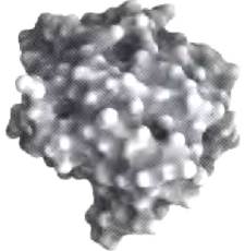
The backbone of a typical protein is formed by hundreds of covalent bonds. Because free rotation is possible around many of these bonds, a protein molecule can theoretically assume countless different Conformations. However, every protein carries out a specific chemical or structural function, which requires it to maintain a strictly defined three-dimensional structure (Fig. 4-1). In the late 1920s, crystals of certain Proteins were obtained, including Hemoglobin (Mr = 64,500) and the enzyme urease (Mr = 483,000). Given that an ordered arrangement of molecules in a crystal is generally possible only when those molecules are identical, the crystalline state of many proteins demonstrates that even the largest proteins consist of discrete chemical units with a defined structure. This Conclusion completely revolutionized the understanding of proteins and their Functions at the time.
Fig. 4-1. The globular structure of Chymotrypsin. To give a sense of scale, a Glycine molecule is shown next to the chymotrypsin molecule. Known three-dimensional protein structures are stored in specialized data banks (Protein Data Bank, PDB; see Box 4-4). Image taken from the PDB data bank (6GCH).
In this chapter, we will discuss how the Amino Acid Sequence of a polypeptide chain determines the three-dimensional structure of a protein. To this end, we will examine five fundamental questions. First, the three-dimensional structure of a protein is determined by its amino acid sequence. Second, protein function depends on structure. Third, an isolated protein typically exists in one or more stable structural forms. Fourth, the most important contribution to the stabilization of a specific protein structure comes from noncovalent interactions rather than covalent ones. Finally, fifth, among the vast number of unique protein structures, certain common types can be distinguished that help elucidate the principles of protein architecture.
In presenting these topics, we do not mean to imply that proteins have a static, unchanging three-dimensional structure. Protein function often involves the mutual interconversion of two or more of its spatial forms. The dynamic aspects of protein structure are discussed further in chapters 5 and 6. Understanding all LEVELS OF STRUCTURAL organization in proteins is essential for discussing their functions, which we will turn to in subsequent chapters.
4.1. Overview of Protein Structure
The spatial arrangement of atoms in a protein molecule is called its conformation. Protein conformations encompass all the structural states that a protein can adopt without breaking covalent bonds. Conformational changes can occur, for example, As a result of the relative rotation of groups about single bonds. Out of the theoretically possible variety of conformations for a protein with hundreds of single bonds, only one or a few options are realized in biological systems under normal conditions. The existence of only a few stable conformations of a protein molecule reflects the necessity of the changes that occur in most proteins upon binding to other molecules or during catalytic reactions. The realized conformations are generally the most thermodynamically favorable and are characterized by the lowest Gibbs Free energy (G). A protein in any of its functional conformations is referred to as a native protein.
What determines the stability of a particular protein conformation? We will arrive at an understanding of this gradually—starting from a Discussion of Primary Protein Structure (Chap. 3) through a subsequent familiarization with secondary, tertiary, and quaternary structures. We will Supplement this traditional approach to studying proteins with new material on the Classification of so-called Supersecondary structures formed during protein folding. We will begin by introducing some fundamental principles.
Protein Conformation is Largely Stabilized by Weak Interactions
When discussing protein structure, protein stability can be defined as its ability to maintain its native conformation. Native proteins are stable only conditionally under physiological conditions; The change in free energy, ΔG, from the folded to the unfolded protein does not exceed 20 to 65 kJ/mol. Any polypeptide chain can theoretically adopt a countless number of different conformations, as a result of which the unfolded protein is characterized by high conformational Entropy. This entropy, along with Hydrogen Bonds between numerous groups in the polypeptide chain and solvent molecules (Water), tends to keep the protein in an unfolded state. Among the chemical interactions that counteract these forces and stabilize the native conformation are Disulfide Bonds and the weak (noncovalent) interactions described in Chapter 2: hydrogen bonds, hydrophobic interactions, and ionic interactions.
Many proteins lack disulfide bridges. The conditions inside most Cells are strongly reducing, which prevents The formation of -S-S- bonds. In eukaryotes, disulfide bonds are found primarily in secreted extracellular proteins (for example, in the Insulin molecule). Disulfide bonds are also rare in bacterial proteins. However, thermophilic Bacteria and archaea generally contain many proteins with disulfide bonds that stabilize these structures; this is likely one of the adaptations for surviving at elevated temperatures.
In most organisms, weak intermolecular interactions are particularly important when a polypeptide chain forms secondary and tertiary structures within an intracellular protein. The formation of quaternary structure from multiple Polypeptides also depends on weak interactions.
Breaking a single covalent bond requires between 200 and 460 kJ/mol, whereas disrupting weak interactions requires only 4 to 30 kJ/mol. Individual covalent bonds involved in maintaining the native protein conformation—such as disulfide bonds holding together PARTS OF THE polypeptide chain—are obviously much stronger than individual weak interactions. However, due to their multiplicity, it is precisely weak interactions that make the primary contribution to stabilizing protein structure. Typically, the conformation of a protein with the lowest free energy (i.e., the most stable one) is distinguished by the maximum number of realized weak interactions.
Protein stability is not determined simply by the sum of the free energies of Formation of the many weak interactions within it. Each group participating in hydrogen bonding in the folded protein was already hydrogen-bonded (with approximately the same strength) to water before the protein folded, but those bonds were broken. The overall contribution of a specific weak interaction to protein stability, or the free energy difference between the folded and unfolded protein, is approximately zero. Ion interactions can also be stabilizing or destabilizing. Thus, to understand why the native conformation of a protein is favored, other factors must also be taken into account.
When examining the contributions of weak interactions to protein stability, we find that hydrophobic interactions generally predominate among them. Pure water is a network of hydrogen-bonded H2O molecules. No other molecules have such a high potential for hydrogen bonding, and therefore any other molecule in aqueous solution disrupts the hydrogen-bonding network of water. Any hydrophobic molecule in an aqueous solution is surrounded by a structured shell of water molecules (solvation shell) arising from the optimal organization of hydrogen bonds (see Fig. 2-7). The increasing order of water molecules in the solvation shell is characterized by a thermodynamically unfavorable decrease in entropy. However, if nonpolar solute groups come together and form clusters, the size of the solvation shell decreases because not all of the nonpolar group's surface is exposed to the solution in that case. The thermodynamic result of this is an increase in entropy. As shown in Chapter 2, entropy is the main driving force causing hydrophobic groups to associate in aqueous solution. Thus, hydrophobic amino acid side chains tend to cluster within the protein molecule to stay away from water.
Under physiological conditions, hydrogen bonding and ionic interactions are largely the result of this same entropic factor. Polar groups are generally capable of forming hydrogen bonds with water, which confers water solubility. However, the number of hydrogen bonds per unit mass in pure water is much higher than in any other liquid or solution, so There is a limit to the solubility of even the most polar molecules because their presence reduces the number of hydrogen bonds per unit mass of solution. Consequently, a solvation shell of structured water molecules forms even around polar molecules. Even if the energy of intramolecular hydrogen bonding or ionic interactions between two polar groups in a macromolecule is largely offset by the disruption of similar interactions between those same groups and water, the release of structured water molecules upon the formation of intramolecular bonds is a driving force that promotes folding. Thus, the greatest fraction of the free energy gain resulting from weak interactions within a protein in aqueous solution is associated with the increase in entropy due to the disappearance of hydrophobic surfaces. This gain is sufficient to compensate for the loss of conformational entropy associated with the protein adopting a single unique conformation.
Clearly, hydrophobic interactions are extremely important for stabilizing a specific conformation, which is usually characterized by the presence of a tightly packed Hydrophobic core consisting of amino acid side chains inside the folded protein molecule. It is also crucial that all polar or charged groups inside the protein molecule have suitable partners for hydrogen bonding and ionic interactions. A single Hydrogen bond does not make a large contribution to the Stability of the native structure; however, the presence of an unpaired polar or charged group in the hydrophobic core can have such a strong destabilizing effect that such a conformation may prove thermodynamically unviable. The free energy gain associated with the pairing of such a group with another group or molecule in solution can exceed the difference between the free energy values of the folded and unfolded protein. Furthermore, the formation of hydrogen bonds between protein groups in repeating secondary structures is a cooperative process (i.e., the formation of one bond increases the probability of forming the next bond), as shown below. Thus, hydrogen bonds often make an important contribution to the protein folding process.
Interactions between oppositely charged groups forming ion pairs (salt bridges) can also stabilize the native conformation of certain proteins. As in the case of hydrogen bonds, in an unfolded protein molecule, charged amino acid side chains interact with water and salts, so the cessation of these interactions must be accounted for when evaluating THE CONTRIBUTION OF salt bridges to the stability of the packed protein. However, the strength of salt bridges increases in environments with a lower dielectric constant (see p. 77): from polar aqueous environments (ε around 80) to the nonpolar environment inside the protein (ε around 4). Thus, salt bridges, especially those that are partially or fully buried, can play a significant role in stabilizing protein structure. This fact explains the widespread occurrence of buried salt bridges in proteins of thermophilic organisms. Ionic interactions also restrict the mobility of the Protein Structure and impart a specific uniqueness to it that nonspecific hydrophobic interactions cannot provide.
Most types of protein structures discussed in this chapter demonstrate two simple rules: 1) hydrophobic amino acid residues are largely tucked into the interior of the protein molecule to minimize contact with water; 2) the maximum possible number of hydrogen bonds is realized within the protein molecule. Insoluble and membrane-associated proteins (which are the subject of discussion in Chapter 11) follow somewhat different rules due to their functions and environment, but weak interactions also exert a decisive influence on their structure.
Peptide Bonds Exhibit Rigidity and a Planar Configuration
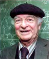
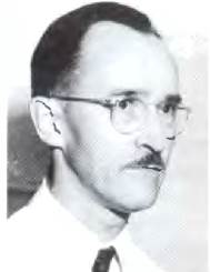
Protein Architecture—Primary Structure. Covalent bonds also restrict the number of possible conformations a polypeptide can adopt. In the late 1930s, Linus Pauling and Robert Corey initiated a series of investigations that laid the foundation for our current understanding of protein structure. They began with a meticulous Study of the peptide bond.
Linus Pauling, 1901-1994
Robert Corey, 1897-1971
It is known that the α-carbons of two adjacent Amino Acids are separated by three covalent bonds arranged in the sequence Cα—C—N—Cα. X-Ray Diffraction analyses of amino acid crystals and simple dipeptides revealed that the C—N peptide bond is somewhat shorter than the C—N bond in simple amines, and that the atoms participating in the peptide bond are coplanar (i.e., lying in a single plane). This indicates Resonance or partial delocalization of two electron pairs between the carbonyl oxygen and the amide nitrogen (Fig. 4-2a). The oxygen carries a partial negative charge and the nitrogen a partial positive charge, resulting in a weak electric dipole. The six atoms constituting the peptide group lie in a single plane, with the carbonyl oxygen and the amide hydrogen in a trans configuration relative to each other. Based on these findings, Pauling and Corey concluded that the C—N peptide bond does not permit free rotation because of its partial double-bond character. Rotation is possible only around the Cα-C and N-Cα bonds. Thus, the polypeptide backbone can be envisioned as a series of rigid planes capable of rotation about the Cα atoms (Fig. 4-2b). The rigidity of the peptide bond severely limits the number of conformations the peptide chain can adopt.
Fig. 4-2. Planar STRUCTURE OF THE peptide bond. (a) Due to resonance, each peptide bond has a degree of double-bond character and therefore cannot rotate. (b) Adjacent α-carbons in a polypeptide chain are separated by three bonds. The N—C peptide bond allows no free rotation; the N-Cα and Cα-C bonds are rotatable, and their dihedral angles are designated by the Greek letters ɸ and  , respectively. Rotation about other single bonds may be restricted by the bulk or charge of the R-group. (c) Atoms and planes defining the angle
  . (d) By convention, the ɸ and
  angles are 180° (or -180°) when the first and fourth atoms are as far apart as possible and the peptide chain is fully extended. Looking down (in either direction) the bond undergoing rotation, the ɸ and
  angles increase when the fourth atom is rotated clockwise relative to the first atom. In a protein, certain conformations shown here (e.g., an angle of 0°) are forbidden due to steric clashes. In panels (b) through (d), the atom spheres are drawn smaller than their Van der Waals radii for clarity.

Conformational Changes in proteins are described using three torsion, or dihedral, angles—referred to as φ (phi), ψ (psi), and ω (omega)—which specify the rotation around each of the three repeating bonds in the peptide backbone. A torsion angle is the angle of intersection between two planes. In Peptides, these planes are defined by the bond directions within the backbone. Two consecutive bonds define a plane; three consecutive bonds describe two planes (with the central bond shared by both planes; see Fig. 4-2c), and the angle between these planes defines the protein conformation.
Key Conventions.
The important torsion angles in peptides are described by three vectors connecting four consecutive backbone atoms (Fig. 4-2b): the angle φ is defined by the C-N-Cα-C bonds (rotation is possible around the N-Cα bond), and the angle ψ is defined by the N-C-Cα-N bonds. Both angles are ±180° when the peptide chain is fully extended and all peptide groups are coplanar (Fig. 4-2d). Looking at the central bond along the vector direction (as shown for angle φ in Fig. 4-2c), the value of the torsion angle increases as the fourth (distant) atom is rotated clockwise (Fig. 4-2d). The torsion angle increases from -180° to 0°, at which point the first and fourth atoms eclipse each other. Rotation can continue from 0° to +180° (the same physical position as -180°), returning the structure to its starting point. The third torsion angle, ω, is less frequently discussed; it is defined by the Cα-C-N-Cα bonds. Here, the central bond is the peptide bond, which has restricted rotation. The peptide bond is normally (99.6% of the time) in the trans configuration, Setting the ω angle to ±180°. In the rare instances of a cis peptide bond, ω = 0°. ■
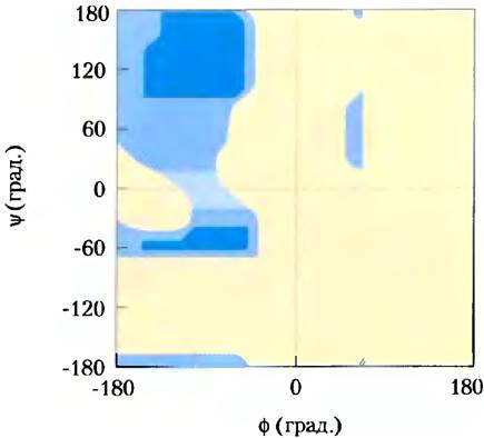
In principle, the φ and ψ angles can assume any value between -180° and +180°, but many states are forbidden due to steric hindrance caused by The amino acid side-chain atoms. For instance, conformations where both φ and ψ equal 0° are not realized (Fig. 4-2c). This particular conformation is used purely as a reference point (zero degrees) for measuring rotational angles. The allowable values for φ and ψ can be mapped on a ψ versus φ plot known as a Ramachandran plot (Fig. 4-3), named after G. N. Ramachandran.
Fig. 4-3. Ramachandran plot for an L-Ala residue. Introduction/10.html">Peptide Conformation is defined by The values of φ and ψ. Conformations that involve no significant steric hindrance—calculated using known van der Waals atomic radii and Bond Angles—are realized. Dark blue areas represent fully allowed conformations with no steric overlap. Lighter blue areas correspond to conformations that are permissible if minor unfavorable interatomic contacts occur. The lightest cyan areas represent conformations made possible by slight stretching of bond angles. Yellow areas indicate forbidden conformations. The Asymmetry of the plot reflects the L-Stereochemistry of the amino acid residues. Plots for other L-amino acids with unbranched side chains look very similar. The range of allowable angles for amino acids with branched side chains (Val, Ile, Thr) is slightly narrower than for Ala. Conversely, the range of allowed conformations for glycine, which has the minimal steric constraints, is much wider. The conformational freedom of Pro is severely restricted because its φ values are confined between -35° and -85° due to the cyclic structure of its side chain.
Summary of Section 4.1 Overview of Protein Structure
■ Every protein possesses a unique three-dimensional structure intricately linked to its biological function.
■ The Spatial Structure of a protein is stabilized by a multitude of weak interactions. Hydrophobic interactions provide the primary driving force for the stabilization of the globular structure in most soluble proteins. Thermodynamically most stable structures feature optimal organization of Hydrogen bonds and ionic interactions.
■ The covalent bonding geometry within the polypeptide chain imposes constraints on protein structure. The peptide bond has partial double-bond character, locking the six atoms of the peptide group rigidly into a single plane. The N—Cα and C—Cα bonds permit limited rotation, with their respective rotation angles designated by the Greek letters φ and ψ.
Last update: 06/08/2026
Editorial and Educational Adaptation: This material has been compiled based on the primary/original source text. The project team performed an editorial review, corrected technical inaccuracies, structured sections, and adapted the content for an educational format.
What was processed:
- elimination of formatting defects (OCR errors, structural breaks, corrupted characters);
- editorial organization of content;
- standardization of terminology in accordance with academic sources;
- verification of factual statements against the original source text.
All mentions of the author, publication year, and origin of the primary text have been preserved in accordance with the source.