LEHNINGER PRINCIPLES OF BIOCHEMISTRY - VOLUME 1. THE FOUNDATIONS OF BIOCHEMISTRY: STRUCTURE AND CATALYSIS - 2011
PART I. STRUCTURE AND CATALYSIS
3. AMINO ACIDS, PEPTIDES, AND PROTEINS
Questions and Problems
1. Absolute configuration of citrulline.
What is the configuration (D or L) of citrulline isolated from watermelon, whose formula is shown in the figure? Explain your answer.
Class="center">
2. Relationship between The titration curve and the acid-base properties of Glycine.
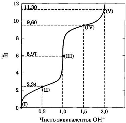
A glycine solution (100 mL, concentration 0.1 M, pH 1.72) was titrated with a 2 M NaOH solution. During the titration, changes in pH were monitored, and a graph was plotted based on the measurement results (see below). Key points on the graph are labeled with numbers from I to V. Which of these points should be specified in the answer to the questions below? Explain your answer.
a) Glycine predominantly exists in the H3N-CH2-COOH form.
б) Средний суммарный заряд молекул глицина
c) Half of the Amino groups are ionized.
d) The pH value is equal to the pKa of the carboxyl group.
e) The pH value is equal toвало значению рКа протонированной аминогруппы.
f) Glycine exhibits maximum buffer capacity.
g) The average net charge of glycine molecules is zero.
h) The carboxyl group is completely titrated (first equivalence point).
i) Glycine is completely titrated (second equivalence point).
j) Glycine predominantly exists in the +H3N-CH2-COO- form.
k) The average net charge of glycine molecules is -1.
l) Half of the glycine molecules are predominantly in the +H3N-CH2-COOH form, and half are in the +H3N-CH2 COO- form.
m) What is the isoelectric point of glycine?
n) At what point is the titration complete?
o) Indicate the regions with the minimum buffer capacity of glycine.
3. What fraction of all molecules consists of completely uncharged Alanine forms?
At a pH value equal to the isoelectric point of alanine, the net charge of the molecule is zero. The figure illustrates two structures, neither of which carries a net charge; however, the predominant form of alanine at pH = pI is the zwitterion.

a) Why does alanine predominantly exist as a zwitterion rather than an uncharged species at pH = pI?
b) What fraction of alanine molecules bears no net charge at the isoelectric point? Explain your answer.
4. Ionization state of Amino Acids.
Each ionizable group of an amino acid can exist in one of two states—charged or neutral. The electrical charge on a functional group is determined by the relationship between the pH of the solution and the pKa of that group. This relationship is described by the Henderson-Hasselbalch equation.
a) Histidine has three ionizable functional groups. Write the equations for the three corresponding ionization processes and indicate approximate pKa values for each reaction. Draw The Structure of histidine in all ionization states. What is the net charge carried by a histidine molecule in each of these states?
b) Draw the ionic structures of histidine that predominate at pH 1, 4, 8, and 12. Note that the ionization state can be determined by treating each ionizable group independently of the others.
c) What is the net charge of a histidine molecule at pH 1, 4, 8, and 12? Toward which electrode will a histidine molecule move in an electric field at each of these pH values—the cathode (-) or the anode (+)?
5. Separation of amino acids by Ion-exchange chromatography.
Analysis of an amino acid mixture begins with the separation of its components using ion-exchange chromatography. After application to a cation-exchange resin Column containing -SO3- groups (Fig. 3-17, a), the amino acids move down the column at different rates because their migration is influenced by two factors: 1) electrostatic attraction between the -SO3- groups of the support and positively charged Functional groups of the amino acids; 2) hydrophobic interactions between amino acid side chains and the highly hydrophobic polystyrene matrix of the support. For each of The amino acid pairs given below, determine which amino acid will elute from the column first when the column is eluted with a buffer at pH 7.0.
a) Asp and Lys
b) Arg and Met
c) Glu and Val
d) Gly and Leu
e) Ser and Ala
6. Designation of stereoisomers of isoleucine.
The structural formula of the amino acid isoleucine is shown below.

a) How many chiral centers does an isoleucine molecule have?
b) How many optical isomers can isoleucine have?
c) Draw the stereochemical formulas for all optical isomers of isoleucine.
7. Comparison of pKa values of alanine and polyalanine.
The titration curve of the amino acid alanine reflects the presence of two ionizable functional groups with pKa values of 2.34 and 9.69, which correspond to The ionization of the carboxyl group and the protonated amino group, respectively. The titration curves of alanine di-, tri-, and oligopeptides also exhibit only two ionizable groups, although their experimentally determined pKa values differ from those of the functional groups of free alanine (see table).
Amino acid or peptide |
pK1 |
pK2 |
Ala |
2.34 |
9.69 |
Ala-Ala |
3.12 |
8.30 |
Ala-Ala-Ala |
3.39 |
8.03 |
Ala (Ala)n-Ala, n ≥ 4 |
3.42 |
7.94 |
a) Draw the structural formula of Ala-Ala-Ala. Indicate the functional groups in this molecule corresponding to pK1 and pK2.
b) Why does the value of pK1 increase with The addition of each successive alanine residue?
c) Why does the value of pK2 decrease with the addition of each successive alanine residue?
8. Protein Size.
What is the approximate Molecular Weight of a protein consisting of 682 amino acid residues joined in a single polypeptide chain?
9. Number of Tryptophan Residues in Bovine Serum Albumin.
Quantitative Amino acid analysis showed that bovine serum albumin (BSA) contains 0.58% (by mass) tryptophan (Mr = 204).
a) Calculate the minimum molecular weight of BSA (i.e., assuming the protein molecule contains only a single tryptophan residue).
b) Based on Gel filtration results, the molecular weight of BSA is 70,000. How many tryptophan residues are contained in its molecule?
10. Subunit Composition of a Protein.
According to gel filtration data, the molecular weight of a protein is 400 kDa. Sodium dodecyl sulfate (SDS) polygel Electrophoresis revealed three protein bands corresponding to masses of 180, 160, and 60 kDa. In the presence of both SDS and dithiothreitol, three bands were also observed, this time corresponding to molecular weights of 160, 90, and 60 kDa. Determine the subunit COMPOSITION OF THE protein.
11. Net Electrical Charge of a Peptide Molecule.
The peptide has the sequence Glu-His-Trp-Ser-Gly-Leu-Arg-Pro-Gly.
a) What is the net charge of this molecule at pH 3.8 and 11? (Use the pKa values for side-chain functional groups as well as the terminal carboxyl and amino groups given in Table 3-1.)
b) Estimate the pI value of this peptide.
12. Isoelectric Point of Pepsin.
Pepsins are a group of digestive Enzymes secreted as larger precursor Proteins by the gastric glands. These glands also secrete Hydrochloric acid, which dissolves food particles present in the diet, thereby allowing pepsin to enzymatically cleave individual protein molecules. The resulting mixture of food, HCl, and digestive enzymes, known as chyme, has a pH of about 1.5. What must the pI value of pepsin be? What functional groups must pepsin contain to ensure such a pI value? Which amino acids in proteins contain such groups?
13. Isoelectric Point of Histones.
Histones are proteins found in The Nucleus of Eukaryotic Cells that are tightly bound to DNA molecules, which are rich in phosphate groups. The pI value of histones is very high, around 10.8. Which amino acid residues must be present in large amounts in histone molecules? How do these residues ensure the tight binding of histones to DNA?
14. Solubility of Polypeptides.
One method for separating polypeptides is based on differences in their solubility. The solubility of large polypeptides in Water depends on the relative polarity of their R groups, particularly on the number of ionizable groups: the more ionizable groups a polypeptide has, the more readily it dissolves in water. Which polypeptide in each pair below has better solubility at the specified pH?
a) (Gly)20 or (Glu)20 at pH 7.0
b) (Lys-Ala)3 or (Phe-Met)3 at pH 7.0
c) (Ala-Ser-Gly)3 or (Asn-Ser-His)5 at pH 6.0
d) (Alu-Asp-Gly)5 or (Asn-Ser-His), at pH 3.0
15. Enzyme Purification.
A biochemist investigates and isolates a novel enzyme. The purification results are summarized in the table below.
Purification step |
Total protein (mg) |
Activity (units) |
1. Crude extract |
20 000 |
4 000 000 |
2. Salt precipitation |
5 000 |
3 000 000 |
3. pH precipitation |
4 000 |
1 000 000 |
4. Ion-exchange chromatography |
200 |
800 000 |
50 |
750 000 |
|
6. Gel filtration |
45 |
675 000 |
a) Using the data provided in the table, calculate the specific activity of the enzyme after each purification step.
б) Which purification step was the most effective (i.e., resulted in the greatest relative increase in purity)?
c) Which step was the least effective?
d) Is there any indication in the given data that the protein is completely pure after step 6? What else needs to be done to assess the purity of the enzyme preparation?
16. Dialysis.
The purified protein was obtained in Hepes buffer (N-(2-hydroxyethyl)piperazine-N'-(2-ethanesulfonic acid)), pH 7, containing 500 mM NaCl. A 1 mL sample of the protein solution is placed in a dialysis tubing and dialyzed against 1 L of the same buffer, but without NaCl. Small molecules and ions (such as Na+, Cl-, and Hepes) can pass through the membrane, whereas the protein cannot.
a) What is the concentration of NaCl in the protein sample after dialysis equilibrium is reached? Assume that the volume of the protein solution does not change during dialysis.
b) What would the salt concentration in the protein solution be if the same 1 mL protein sample were dialyzed successively twice against 100 mL of the same Hepes buffer?
17. Peptide Purification.
Three Peptides with the amino acid compositions given below are eluted from a column packed with a cation-exchange resin at pH 7.0. In what order will the peptides elute from the column?
Peptide A: Ala 10%, Glu 5%, Ser 5%, Leu 10%, Arg 10%, His 5%, Ile 10%, Phe 5%, Tyr 5%, Lys 10%, Gly 10%, Pro 5%, and Trp 10%.
Peptide B: Ala 5%, Val 5%, Gly 10%, Asp 5%, Leu 5%, Arg 5%, Ile 5%, Phe 5%, Tyr 5%, Lys 5%, Trp 5%, Ser 5%, Thr 5%, Glu 5%, Asn 5%, Pro 10%, Met 5%, and Cys 5%.
Peptide C: Ala 10%, Glu 10%, Gly 5%, Leu 5%, Asp 10%, Arg 5%, Met 5%, Cys 5%, Tyr 5%, Phe 5%, His 5%, Val 5%, Pro 5%, Thr 5%, Ser 5%, Asn 5%, and Gln 5%.
18. Sequencing of Brain-Derived Leucine Enkephalin.
A group of peptides influencing Nerve Impulse transmission in certain Regions of the brain was isolated from normal brain tissue. These peptides are termed opioids because they bind to specific receptors that also bind opiates (opium Alkaloids) such as morphine and naloxone. Thus, opioids mimic certain properties of opiates. Some scientists view these substances as the brain's own built-in painkillers. Using the data below, determine the Amino Acid Sequence of the opioid leucine enkephalin. Explain how your proposed structure is consistent with each of the results (a–c).
a) Complete Hydrolysis with 6 M HCl at 110 °C followed by amino acid analysis indicated the presence of Gly, Leu, Phe, and Tyr in a molar ratio of 2:1:1:1.
b) Chromatographic analysis performed after treating the peptide with 1-fluoro-2,4-dinitrobenzene followed by complete hydrolysis revealed the presence of a 2,4-dinitrophenyl derivative of Tyrosine. No free tyrosine was detected.
c) Complete Digestion of the peptide with pepsin followed by chromatographic separation showed the presence of a dipeptide containing Phe and Leu residues, and a tripeptide containing Tyr and Gly in a 1:2 ratio.
19. Structure of a Polypeptide Antibiotic Isolated from Bacillus brevis.
Extracts obtained from a bacterial culture of Bacillus brevis contain a peptide with antibiotic properties. This peptide forms complexes with Metal Ions and apparently disrupts the ion transport system across The Cell membrane in other bacterial species, leading to their death. The STRUCTURE OF THE peptide was determined based on the results below.
a) Complete acid hydrolysis of the polypeptide followed by amino acid analysis demonstrated the presence of equimolar amounts of Leu, Orn, Phe, Pro, and Val. Orn is the abbreviation for Ornithine, an amino acid not found in proteins but present in certain peptides. Ornithine has the following structure:

b) The molecular mass of the peptide was found to be approximately 1200.
c) The peptide was not cleaved by Treatment with the enzyme carboxypeptidase. This enzyme catalyzes the removal of any C-terminal residues, except for Pro, as well as cases where the residue lacks a free carboxyl group for any reason.
d) Treatment of the original peptide with 1-fluoro-2,4-dinitrobenzene followed by complete hydrolysis and chromatographic separation revealed the presence of free amino acids only, along with a derivative of the following structure:

(Hint: note that the 2,4-dinitrophenyl group is attached not to the usual α-nitrogen atom, but to the amino group of the side chain.)
e) Partial hydrolysis of the peptide, chromatographic separation of the products, and their amino acid analysis revealed the presence of di- and tripeptides of the following structure (amino-terminal residues are always on the left): Leu-Phe, Phe-Pro, Orn-Leu, Val-Orn, Val-Orn-Leu, Phe-Pro-Val, Pro-Val-Orn. Using this information, determine the amino acid sequence of the peptide antibiotic. Explain your reasoning. Explain how your proposed structure is consistent with all experimental data.
20. Peptide sequencing efficiency.
A peptide with the Primary Structure Lys-Arg-Pro-Leu-Ile-Asp-Gly-Ala was sequenced by the Edman Degradation method. Each cycle stage was performed with an efficiency of 96%. Determine what fraction of all amino acids released in the fourth cycle is leucine? What will this value be if the efficiency of each cycle is 99%?
21. Sequence comparison.
Proteins called molecular chaperones (see Chapter 4) are involved in protein folding. Proteins of a single chaperone class, found in organisms ranging from Bacteria to mammals, are called heat Shock protein 90 (Hsp90). All Hsp90 chaperones contain 10 characteristic Amino acid sequences that allow these proteins to be easily identified in database searches. Two Examples of such sequences are presented below.

a) Which amino acid in this sequence is invariant (conserved across all species)?
b) At which position(s) do you find only amino acids with positively charged side chains? For each such position, determine which amino acid occurs most frequently.
c) At which position(s) do you find only amino acids with negatively charged side chains? For each such position, determine which amino acid occurs most frequently.
d) At which position can any amino acid be located (although one amino acid is still much more common than the others)? What is this position, and which amino acid occurs here most frequently?
22. Biochemical protocol: your first Protein Purification.
Starting your first job in a biochemistry laboratory, you spend the first few weeks learning to wash glassware and label test tubes. Then you are trusted with preparing buffer and stock solutions needed for various laboratory tasks. Finally, you take responsibility for purifying your first protein. This is citrate synthase, an enzyme of The Citric Acid Cycle localized in the Mitochondria. You carry out all the steps listed below, following a well-known purification protocol. Your supervising senior student asks questions at each stage of purification regarding the rationale for performing that step. Provide your Answers. (Hint: for osmolarity information, see Chapter 2; for cell organelle isolation, see p. 23).
a) From a local slaughterhouse, you obtained 20 kg of bovine hearts. You transported them on ice and performed every purification step on ice or in a cold room. Using a specialized homogenizer, you homogenized the hearts at pH 7.2 in a buffer containing 0.2 M sucrose. Why do you use bovine Heart as the starting material, and in such a large quantity? Why must the tissue be kept on ice and disrupted at pH 7.2 in the presence of sucrose? What happens to the tissue during homogenization?
b) You subject the tissue homogenate (dense and opaque) to differential centrifugation. Why do you do this?
c) For further experiments, you take the supernatant, which mainly contains intact mitochondria. Next, you subject the mitochondria to osmotic lysis. The lysate has a lower density than the homogenate, but is still opaque. It consists of mitochondrial membranes and internal mitochondrial contents. To this lysate, you add a highly soluble salt—ammonium sulfate—at a specific concentration, centrifuge your mixture, decant the supernatant, and discard the pellet. To the supernatant, which looks clearer than the lysate, you add the next portion of ammonium sulfate and centrifuge the sample again. But this time, you keep the pellet because it contains the protein of interest. Why must the salt be added in two steps?
d) You dissolve the pellet obtained from the second ammonium sulfate precipitation and dialyze it overnight against a large volume of buffer at pH 7.2. Why does the dialysis buffer not contain ammonium sulfate? Why do you use a buffer rather than water?
e) You apply the dialyzed solution to a gel filtration column. Following the protocol, you collect the first protein fraction eluted from the column and discard all other proteins. The presence of protein in the fractions is determined by measuring the ultraviolet absorbance of the solution (at 280 nm). What does the fact that your protein elutes first from the column signify? Why is absorbance at 280 nm a good indicator of protein presence in solution?
f) You apply the fraction collected from the previous step to a cation-exchange chromatography column. You discard all the initial solution that passed through the column and begin washing the column with a solution of higher pH. You collect the first eluted protein fraction. Explain your actions.
g) You apply a sample from your fraction—which is now greatly reduced in volume and looks nearly transparent (perhaps with a faint pinkish tint)—to an isoelectric focusing gel. After separation and gel staining, you see three broad bands on the gel. According to the protocol, your protein of interest has a pI of 5.6. But you want to confirm the purity of the protein using another method. You cut out the band with pI 5.6 from the gel and place it on a polyacrylamide gel for electrophoresis in the presence of SDS. Why are you not certain of the purity of the protein in the band you selected? What can the results of SDS-Polyacrylamide gel electrophoresis tell you? Why is it important to perform SDS-PAGE after isoelectric focusing?
Analysis of Experimental Data
23. Determination of the Amino Acid Sequence of Insulin.
Figure 3-24 illustrates the amino acid sequence of the insulin hormone. Its structure was determined by Frederick Sanger and his co-workers. Most of this work was published in the Biochemical Journal between 1945 and 1955.
When Sanger and his team began this series of studies, it was known that insulin is a small protein consisting of two or four polypeptide chains linked by disulfide bridges. The researchers managed to develop several straightforward Methods FOR STUDYING protein sequencing.
Reaction with FDNB. FDNB (1-fluoro-2,4-dinitrobenzene) reacts with free amino groups (but not with amido or guanidino groups) in a protein to form dinitrophenyl (DNP) Amino Acid Derivatives:

Acid hydrolysis. Boiling a protein with 10% HCl for several hours leads to the hydrolysis of all peptide and amide bonds. Brief treatment yields short peptides; the longer the treatment, the more complete The breakdown of the protein into its constituent amino acids.
Cysteine oxidation. Treating a protein with performic acid cleaves all Disulfide Bonds, converting all cysteine residues into cysteic acid residues (Fig. 3-26).
Paper chromatography. This most primitive precursor to Thin-Layer Chromatography (see Fig. 10-24) is designed to separate substances based on differences in their chemical properties, allowing for the identification of individual Amino Acids and, in some cases, dipeptides. Thin-layer chromatography enables the separation of larger peptides.
In his first paper (1945), Sanger treated insulin with FDNB and then subjected the resulting product to hydrolysis. He discovered many free amino acids, but only three DNP derivatives: α-DNP-glycine (with the DNP group attached to the α-amino group of the amino acid), α-DNP-phenylalanine, and ε-DNP-Lysine (with the DNP group attached to the ε-amino group). Sanger interpreted this result to mean that insulin consists of two protein chains: one has Gly at its N-terminus, and the other has Phe at its N-terminus. Additionally, one of the two chains contains a Lys residue, though not at the N-terminus. Sanger designated the chain starting with the Gly residue as chain A, and the chain starting with the Phe residue as chain B.
a) Explain how Sanger arrived at these Conclusions.
b) Do these results align with the structure of insulin known today (Fig. 3-24)?
In a later paper (1949), Sanger described how this method allowed him to identify several amino acid residues at the beginning of each insulin chain (from the N-terminus). For instance, when analyzing The sequence of chain B, he performed the following steps:
1. Oxidation of insulin to separate chains A and B.
2. Preparation of a pure sample of chain B using paper chromatography.
3. Reaction of chain B with FDNB.
4. Mild acid hydrolysis of the product to obtain several short peptides.
5. Separation of DNP-peptide derivatives from peptides lacking DNP groups.
6. Isolation of four DNP-peptides, designated B1, B2, B3, and B4.
7. Complete hydrolysis of each DNP-peptide to yield free amino acids.
8. Identification of the hydrolysis products of each peptide using paper chromatography.
The following results were obtained:
B1: α-DNP-phenylalanine only;
B2: α-DNP-phenylalanine and valine;
B3: aspartic acid, α-DNP-phenylalanine, and valine;
B4: aspartic acid, glutamic acid, α-DNP-phenylalanine, and valine
c) Based on these results, name the first four amino acids at the N-terminus of peptide B. Explain your reasoning.
d) Do these results agree with the known sequence of insulin (Fig. 3-24)? Explain any discrepancies.
Sanger and his colleagues used their method to determine the complete sequence of the A and B chains. They established the following sequence for the A chain (N-terminus on the left):

Because acid hydrolysis converts all Asn residues into Asp and all Gln residues into Glu, these residues had to be designated as Asx and Glx, respectively (as it was impossible to identify them precisely). Sanger solved this problem by fragmenting the sequences into shorter peptides using proteases that cleave peptide bonds without affecting the amide bonds in Asn and Gln residues. Next, he determined the number of amide groups in each peptide by measuring the concentration of NH4+ released upon acid hydrolysis of the peptide. Below are some of the results obtained for the A chain. These peptides may not have been completely free of impurities, so these numbers are not absolutely exact, but they are sufficiently accurate for the problem Sanger set out to solve.
Peptide name |
Amino acid sequence |
Number of amide groups in the peptide |
Ac1 |
Cys-Asx |
0,7 |
Ap15 |
Tyr-Glx-Leu |
0,98 |
Ap14 |
Tyr-Glx-Leu-Glx |
1,06 |
Ap3 |
Asx-Tyr-Cys Asx |
2,10 |
Ap1 |
Glx-Asx-Tyr-Cys-Asx |
1,94 |
Ap5pa1 |
Gly-Ile-Val-Glx |
0,15 |
Ap5 |
Gly-Ile-Val-Glx-Glx-Cys-Cys- Ala- Ser-Val - Cys- Ser- Leu |
1,16 |
e) Based on these data, determine the amino acid sequence of the A chain. Explain your answer and compare it with the data presented in Fig. 3-24.
Last update: 06/08/2026
Editorial and Educational Adaptation: This material has been compiled based on the primary/original source text. The project team performed an editorial review, corrected technical inaccuracies, structured sections, and adapted the content for an educational format.
What was processed:
- elimination of formatting defects (OCR errors, structural breaks, corrupted characters);
- editorial organization of content;
- standardization of terminology in accordance with academic sources;
- verification of factual statements against the original source text.
All mentions of the author, publication year, and origin of the primary text have been preserved in accordance with the source.