Practical Protein Chemistry - A. Darbre 1989
Determination of amino acid sequence using 4-dimethylaminoazobenzene-4'-isothiocyanate (manual procedure)
Identification of DABTC-amino acids
Thin-layer chromatography
All DABTC-Amino Acids, except for Ile and Leu, are identified by Thin-Layer Chromatography on polyamide plates (25x25 mm). Samples are applied to the corner of the plate at a distance of 6 mm from the adjacent edges using a 1 µl micropipette (H. E. Pederson) with a fine, carefully polished spherical tip. The spot diameter should not exceed 1 mm; a cool-air hairdryer is used during application. Two-dimensional chromatography is carried out in closed chambers (30x80x80 mm) using an ascending solvent flow. Phase saturation is optional. Solvent 1 (first dimension): Water – acetic acid (2:1); solvent 2 (second dimension): toluene – n-hexane – acetic acid (2:1:1).
Class="center">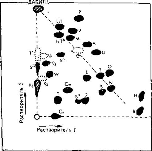
FIG. 14.3. Polyamide Separation of DABTC-Amino Acid Derivatives and reaction byproducts.
Solvent 1: water – acetic acid (2:1); solvent 2: toluene – n-hexane – acetic acid (2:1:1). DABTC derivatives are designated by the single-letter code of the corresponding amino acids. Dark spots represent red-colored derivative spots; dotted-line circles indicate blue spots; hatched spots indicate orange spots; e denotes the diethylamine DABTC derivative; U represents byproduct decomposition products of the excess reagent. For detailed information on the DABTC derivatives of Serine, Threonine, and Lysine, see [3].
Separation of Ile and Leu derivatives is achieved by one-dimensional chromatography on silica gel plates in a chloroform – ethanol (100:3) system [9]. After drying, the plate is exposed to HCl vapors, which turns the yellow spots into red or blue (Fig. 14.3). Successful identification of DABTC-amino acid derivatives largely depends on the experimenter's skill in performing chromatography and interpreting the obtained results [2, 10].
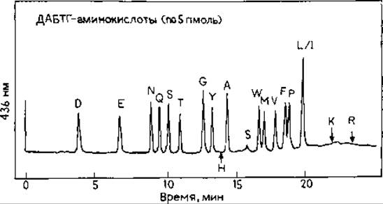
FIG. 14.4. HPLC separation of a mixture of DABTC-amino acid derivatives (5 pmol each) on a Zorbax ODS Column. Chromatographic conditions: solvent A, 35 mM acetate buffer (pH 5.0); solvent B, acetonitrile. Gradient from 45% to 70% of buffer B over a time interval of 0–10 min; 70% B for 10–12 min; 70–80% B for 12–14 min; 80% B for 14–22 min; 80–45% B for 22–25 min. Column Temperature 22 °C. Chart recorder speed 40 cm/h. Detection at 436 nm, scale 0.005 AU.
Extensive practice is required to correctly identify the derivatives of such Amino acids as serine, threonine, and lysine, each of which produces multiple spots on the chromatogram [3, 10].
Last update: 06/08/2026
Editorial and Educational Adaptation: This material has been compiled based on the primary/original source text. The project team performed an editorial review, corrected technical inaccuracies, structured sections, and adapted the content for an educational format.
What was processed:
- elimination of formatting defects (OCR errors, structural breaks, corrupted characters);
- editorial organization of content;
- standardization of terminology in accordance with academic sources;
- verification of factual statements against the original source text.
All mentions of the author, publication year, and origin of the primary text have been preserved in accordance with the source.