BIOCHEMISTRY - L. Stryer - 1984
VOLUME 1
PART I. CONFORMATION AND DYNAMICS
Class="center">Illustrated through the relationship between protein Spatial Structure and biological activity
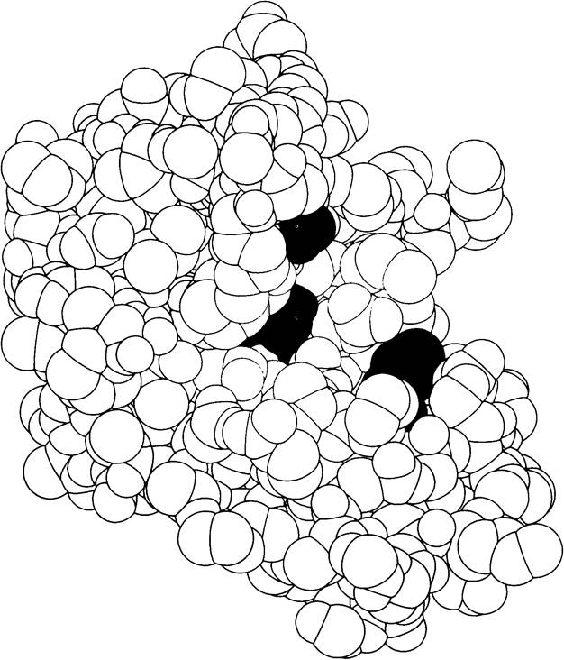
A model of Ribonuclease S, an enzyme that hydrolyzes Ribonucleic Acids. Amino Acids in the Active Site critical for catalysis are highlighted in color. The three-dimensional structure of this enzyme was elucidated by Frederic Richards and Harold Wyckoff (based on a drawing kindly provided by Dr. F. Richards, S. Anderson, and A. Perlo).
CHAPTER 2. BASIC CONCEPTS OF PROTEIN STRUCTURE AND FUNCTION
Proteins play a decisive role in virtually all biological processes. The significance and remarkable diversity of their Functions are evident from the following Examples.
1. Enzymatic Catalysis. In biological systems, almost all reactions are catalyzed by specific macromolecules called Enzymes. Some of these reactions, such as the Hydration of carbon dioxide, are very simple. Others, such as the Replication of an entire chromosome, are extremely complex. Nearly all enzymes are powerful catalysts, increasing reaction rates by at least a million-fold. Thus, in the absence of enzymes, chemical transformations in vivo are rarely observed. To date, several thousand enzymes have been characterized, many of which have been isolated in crystalline form. It is a striking fact that all known enzymes are proteins. Consequently, it is proteins that determine the course of chemical transformations in biological systems.
2. TRANSPORT AND STORAGE. The transport of many small molecules and ions is carried out by specific proteins. For example, Hemoglobin, contained in red Blood Cells, transports oxygen to Tissues, whereas the related protein Myoglobin stores oxygen in Muscles. In Blood Plasma, iron is transported as a complex with transferrin, and in the Liver it is stored as a complex with another protein, ferritin.
3. Coordinated movement. Proteins are the primary component of Muscle. Muscle contraction is driven by the sliding of Two Types of protein filaments past one another. Coordinated movements at the microscopic level, such as the Separation of Chromosomes during mitosis or the propulsion of a sperm Cell by its flagellum, are also mediated by contractile structures composed of proteins.
4. Mechanical support. The high elasticity of Skin and bones is due to the presence of the fibrous protein Collagen.
Fig. 2.1. Micrograph of a hexokinase crystal—a key enzyme in glucose utilization

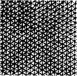
Fig. 2.2. Transmission electron micrograph of an insect flight muscle cross-section. Protein filaments of two types can be seen forming hexagonal arrays
5. Immune protection. Antibodies are highly specific proteins capable of recognizing and binding foreign objects such as Viruses, Bacteria, and cells from other organisms. Thus, proteins play a vital role in distinguishing self from non-self.
Fig. 2.3. Electron micrograph of a collagen fiber

6. Generation and transmission of nerve impulses. The response of Nerve Cells to specific stimuli is mediated by receptor proteins. For example, rhodopsin is the photoreceptor protein found in retinal rod cells. Receptor molecules, triggered by specific low-molecular-weight substances such as acetylcholine, ensure the transmission of nerve impulses at synapses, i.e., the junctions between nerve cells.
Fig. 2.4. Micrograph of a ganglion showing nerve proliferation following The addition of nerve growth factor, which is a protein complex
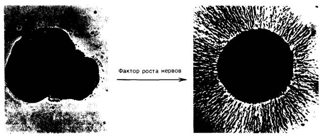
7. Regulation of growth and differentiation. Strict Regulation of the sequence of Gene Expression is crucial for orderly cell growth and differentiation. At any given time in an Organism's life, only a small fraction of The Cell's genome is expressed. In bacteria, the primary regulatory elements are repressor proteins that silence specific regions of cellular DNA. The regulation of Cell Differentiation by proteins occurs through an entirely different mechanism, as illustrated by nerve growth factor, a protein complex that drives The formation of the neuronal network in higher organisms.
2.1. Proteins Are Built from Amino Acids
The fundamental structural Building Blocks of proteins are amino acids. Each amino acid consists of an amino group, a carboxyl group, a hydrogen atom, and a distinctive R-group attached to a carbon atom called the α-carbon (Fig. 2.5). R-groups are referred to as side chains for reasons that will become clear in subsequent discussions. At neutral pH, amino acids in solution do not exist as uncharged molecules, but predominantly as dipolar ions (zwitterions). In this state, the amino group is protonated (—NH3+) and the carboxyl group is dissociated (—COO-). Amino acid ionization is pH-dependent (Fig. 2.6). In acidic solutions (e.g., at pH 1), the carboxyl group is in its un-ionized form (—COOH), while the amino group is ionized (—NH3+). In alkaline solutions (e.g., at pH 11), conversely, the carboxyl group is ionized (—COO-) and the amino group is un-ionized (—NH2). Concepts concerning The Role of pH and the acid-base properties of Amino acids are discussed further in the Appendix to this chapter.
The tetrahedral arrangement of four different chemical groups around the α-carbon atom determines the Optical Properties of amino acids. Structures that are mirror images of each other are called L- and D-isomers (Fig. 2.7). Proteins contain exclusively L-amino acids. Therefore, in the subsequent Discussion, we will omit the isomer designation, assuming that whenever Protein Structure is discussed, L-amino acids are implied (unless explicitly stated otherwise).
Fig. 2.5. Structure of an amino acid in unionized and zwitterionic forms
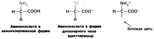
Fig. 2.6. Dependence of amino acid ionization on pH value
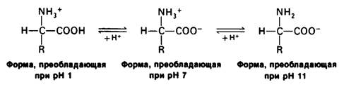
Fig. 2.7. Absolute configurations of L- and D-amino acid isomers

Proteins feature 20 types of amino acid side chains, which vary in size, shape, hydrogen-bonding capacity, and chemical reactivity. It is worth emphasizing that all proteins across every species of living organisms—from bacteria to humans—are built from the exact same set of 20 amino acids. This universal protein alphabet has existed for about 2 billion years. The ability of proteins to perform diverse functions stems from the variety and flexibility of The properties of their 20 constituent structural units. In the following chapters, we will see how complex three-dimensional structures arise from this alphabet, enabling proteins to participate in such a vast array of biological processes.
Let us examine this set of amino acids. The simplest among them is Glycine, which has a hydrogen atom in place of a side chain. In Alanine, the side chain is a methyl group. Valine, leucine, isoleucine, and Proline contain hydrocarbon side chains. Proline, however, differs from the other 19 standard amino acids by possessing a secondary rather than a primary amino group. Strictly speaking, proline is an imino (rather than an amino) acid. Its side chain is bonded to both the amino group and the α-carbon, resulting in a cyclic structure.
Two amino acids—Serine and Threonine—contain aliphatic hydroxyl groups.
There are three widespread aromatic amino acids: phenylalanine, Tyrosine, and Tryptophan.
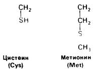
All the aforementioned Amino acids have uncharged side chains at physiological pH values. Let us now turn to amino acids with charged side chains. At neutral pH, Lysine and Arginine carry a positive charge, whereas Histidine is either positively charged or neutral depending on its microenvironment. The side chains of glutamic and aspartic acids carry a negative charge. Hereinafter, we will refer to these Amino acids as glutamate and aspartate, respectively, highlighting the fact that they bear a negative charge at physiological pH. Uncharged derivatives of glutamate and aspartate also exist—glutamine and asparagine—which contain an amide group instead of a terminal carboxyl group. Finally, two amino acids contain a sulfur atom in their side chain: Methionine and Cysteine (Fig. 2.15). As will be shown later, cysteine plays a special role in many proteins by facilitating the formation of cross-linking Disulfide Bonds.
Fig. 2.15. Cysteine and methionine contain sulfur-bearing side chains
Last update: 06/08/2026
Editorial and Educational Adaptation: This material has been compiled based on the primary/original source text. The project team performed an editorial review, corrected technical inaccuracies, structured sections, and adapted the content for an educational format.
What was processed:
- elimination of formatting defects (OCR errors, structural breaks, corrupted characters);
- editorial organization of content;
- standardization of terminology in accordance with academic sources;
- verification of factual statements against the original source text.
All mentions of the author, publication year, and origin of the primary text have been preserved in accordance with the source.