Biochemistry, Vol. 1 - A. Lehninger 1985
Biomolecules
Water
Almost all amino acids contain an asymmetric carbon atom
As shown in Fig. 5-2, all standard Amino Acids except one contain an asymmetric carbon atom at the a-position, bonded to four different substituent groups: a carboxyl group, an amino group, an R-group, and a hydrogen atom. Thus, the asymmetric a-carbon atom serves as a chiral center (Section 3.5). As we know, compounds with a chiral center exist in two different isomeric forms that share identical chemical and physical properties, with one exception—the direction in which they rotate the plane of polarization of plane-polarized light passing through them. The angle of rotation is measured using a polarimeter (Section 3.5). With the exception of Glycine, which lacks an asymmetric carbon atom (Fig. 5-3), the other 19 amino acids obtained from the mild Hydrolysis of Proteins are optically active, meaning they can rotate the plane of light polarization in one direction or the other. Because the valence bonds around the a-carbon atom in Amino acids have a tetrahedral arrangement, the four different substituent groups can be arranged in space in two distinct ways, allowing the molecule to exist in two configurations that are non-superimposable mirror images of each other (Fig. 5-4). These two molecular forms are called optical isomers, enantiomers, or stereoisomers. A solution of one stereoisomer of a given amino acid rotates the plane of light polarization to the left (counterclockwise); such a stereoisomer is called levorotatory [denoted by the minus sign (—) placed before its name]. The other stereoisomer rotates the plane of polarization by the exact same angle but to the right (clockwise) and is called dextrorotatory [denoted by the plus sign (+) placed in front]. An equimolar mixture of the (+)- and (—)-forms is optically inactive and does not rotate the plane of light polarization. Since all amino acids (except glycine) isolated from proteins under mild conditions exhibit optical activity, it is clear that they occur within protein molecules in only one specific stereoisomeric form.
Class="center">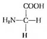
Fig. 5-3. Glycine, the only amino acid that lacks an asymmetric carbon atom. The R-group, which consists of a single hydrogen atom, is highlighted in red.

Fig. 5-4. A. Two optical isomers of Alanine. The carboxyl groups, used as reference points, are positioned at the end of the bond extending vertically from the chiral center. The L- and D-alanine structures are non-superimposable mirror images of each other. B and C. Two different Methods for depicting the spatial configuration of optical isomers. In perspective formulas, bonds projecting above the plane of the drawing are represented by solid wedges, while bonds extending behind the plane are indicated by dashed lines. In projection formulas, horizontal bonds are assumed to project above the plane of the drawing, whereas vertical bonds extend behind it. However, projection formulas are not always strictly interpreted and are frequently used independently of the actual stereochemical configuration of the molecule.
The optical activity of a stereoisomer is quantitatively expressed by its specific rotation, which can be determined by measuring the angle of Rotation of the plane of polarization as light passes through a solution of the pure stereoisomer at a known concentration within a polarimeter tube of a specified path length:
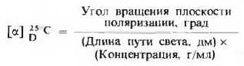
Table 5-2. Specific Rotation of Selected Amino Acids Isolated from Proteins
All of these amino acids have the L-configuration, yet some are dextrorotatory while others are levorotatory.
|
Amino acid |
Specific rotation, [а]D |
|
L-alanine |
+ 1,8 |
|
L-Arginine |
+ 12,5 |
|
- 38,5 |
|
|
L-glutamic acid |
+ 12,0 |
|
L-isoleucine |
+ 12,4 |
|
L-Lysine |
+ 13,5 |
|
L-Proline |
- 86,2 |
|
L-Serine |
- 7,5 |
|
- 28,5 |
|
|
L-phenylalanine |
- 34,5 |
The optical path length is expressed in decimeters, and the Temperature and wavelength of the light used must be specified (typically the D-line of the sodium spectrum, λ = 589 nm). Table 5-2 lists the specific rotation values for several amino acids; note that they include both levorotatory and dextrorotatory Examples.
Last update: 06/08/2026
Editorial and Educational Adaptation: This material has been compiled based on the primary/original source text. The project team performed an editorial review, corrected technical inaccuracies, structured sections, and adapted the content for an educational format.
What was processed:
- elimination of formatting defects (OCR errors, structural breaks, corrupted characters);
- editorial organization of content;
- standardization of terminology in accordance with academic sources;
- verification of factual statements against the original source text.
All mentions of the author, publication year, and origin of the primary text have been preserved in accordance with the source.