Practical Protein Chemistry - A. Darbre 1989
Separation of protein and peptide mixtures by high-performance liquid chromatography
Gel chromatography
Chemical factors
Polysaccharide- or polyacrylamide-based gels used in standard Gel filtration exhibit very weak interactions with Proteins AND Peptides. A different picture emerges when working with the stationary phases employed in HPLC.
The surface of silica-based sorbents contains acidic silanol groups that can either interact with positively charged protein groups, causing adsorption, or with negatively charged groups, resulting in electrostatic repulsion of the protein molecules. These interactions reduce the efficiency of molecular penetration into the gel pores. To prevent this, silanol groups are modified with neutral compounds containing hydrophilic chains. Typically, this modification is incomplete; consequently, unreacted silanol or other charged groups affect protein elution, with the extent of this effect depending on the Mobile phase composition and the pI of the given protein.
Currently, columns from two manufacturers are most commonly used: Waters (protein analysis columns I-60, I-125, I-250) and Toyo Soda TSKSW series (2000, 3000, 4000), which are marketed under license by various companies. In addition, there are Synchropak GPC (Synchrom), Lichrosorb and Lichrosphere DIOL (Merck), Glycophase GPC (Pierce), Aquapore-OH (Brownlee), Superose (Pharmacia), and Spheron (Lachema) columns.
Column evaluation was carried out taking into account METABOLISM/18.html">The Influence of chemical factors, particularly with respect to the Synchropak sorbent [19]. The results of a detailed study on Lichrosphere DIOL columns have been published [27]. The Conclusions drawn from these columns are applicable to varying degrees to columns from other manufacturers. It should be borne in mind that to prevent support degradation, silica-based columns should only be used at 2<рН<7.7.
The Effect of Ionic strength on the elution volumes of four proteins with different pI values is shown in Fig. 6.1. Chromatographic Separation was performed on Pepsin (pI 1), Lysozyme (pI 11.0), Ovalbumin (pI 4.7), and chymotrypsinogen (pI 9.5) using Lichrosphere DIOL at pH 5. As the ionic strength increased, the effect of ionic repulsion on the acidic protein pepsin and ionic adsorption on the basic proteins chymotrypsinogen and lysozyme decreased, whereas the elution volume of ovalbumin (pI 4.7) remained unchanged. For salts yielding higher ionic strength (at identical molar concentrations), the suppression of support-mediated ionic effects is more pronounced than for lower ionic strength solutions (for instance, phosphate is more effective than acetate).
Class="center">
FIG. 6.1. Effect of ionic strength on protein elution volumes with various pI values during gel filtration Chromatography on a Lichrosorb DIOL column. Ionic strength was adjusted using sodium acetate and sodium sulfate at pH 5 [27].
In the absence of interactions between the sorbents and the protein, a linear relationship should be observed between the logarithm of the molecular weight and the elution volume, which is confirmed for the Lichrosphere DIOL column (Fig. 6.2) [27]. In this case, the analysis was performed in a solution with an ionic strength of μ = 0.36 mol/L. As seen from the data presented in Fig. 6.2, under the specified ionic strength, lysozyme and chymotrypsinogen elute at volumes that deviate from the general trend [27]. This deviation can be partly explained by hydrophobic interactions, since The addition of Ethylene glycol to the mobile phase reduces Ve. The observed anomalous elution of cytochrome c and Hemoglobin is associated with protein aggregation. Thus, for cytochrome c (0.05 M sodium phosphate, 0.1 M sodium chloride, pH 7.5), Ve corresponds to an M of 12.5∙103, whereas at pH 5 (0.1 M sodium acetate) Ve correlates with an M of 20∙103. For comparison, calibration plots for the separation of several reference proteins (indicating separation conditions) on Synchropak GPC, Waters (series I), and Toyo Soda columns are also provided (Figs. 6.3–6.5).

FIG. 6.2. Correlation between elution volumes and apparent molecular weights of various proteins in gel permeation chromatography. For analytical conditions, see the caption to Fig. 6.1 (ionic strength 0.36 mol/L) [27].
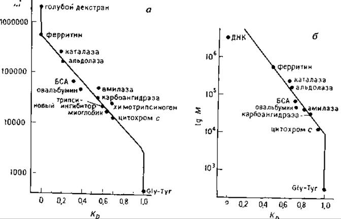
FIG. 6.3. Calibration plots obtained by gel filtration chromatography of proteins on Synchropak TPC 100 (a) and 500 (b) columns. Eluent: 0.1 M KH2PO4 (pH 7); flow rate 0.5 mL/min [19].
Knowing The Nature of the Physical and Chemical interaction forces between proteins and the support surface, and taking into account the Factors affecting the apparent molecular weights M of proteins in solution, one can determine the apparent M of proteins on columns (under non-denaturing conditions) with an accuracy of ~ 10%. Under these conditions, protein biological activity is preserved while maintaining sufficiently high recovery yields.
Last update: 06/08/2026
Editorial and Educational Adaptation: This material has been compiled based on the primary/original source text. The project team performed an editorial review, corrected technical inaccuracies, structured sections, and adapted the content for an educational format.
What was processed:
- elimination of formatting defects (OCR errors, structural breaks, corrupted characters);
- editorial organization of content;
- standardization of terminology in accordance with academic sources;
- verification of factual statements against the original source text.
All mentions of the author, publication year, and origin of the primary text have been preserved in accordance with the source.