Practical Protein Chemistry - A. Darbre 1989
Separation of protein and peptide mixtures by high-performance liquid chromatography
Gel chromatography
Determination of true molecular weights
Determination of the molecular weight (M) of a protein is impossible until its molecules adopt a random coil conformation. This requires breaking the Disulfide Bonds either by reduction, performic acid oxidation, or sulfitolysis. Chromatography can be performed in a 0.1% aqueous SDS solution, but optimal results are achieved using 6 M guanidine hydrochloride (Gu∙HCl). The Effect of these detergents on Separation is illustrated in Figs. 6.6 and 6.7 [7–9]. A strong correlation is observed between the M values and the protein distribution coefficients, KD. The performance characteristics of TSK-SW 3000 and 4000 columns (Toyo Soda) in 6 M Gu∙HCl have been studied in detail [28]. The data obtained were in good agreement with the results of separation in agarose gels [10]. Generally, The Use of Gu∙HCl in chromatographs is discouraged because chloride ions cause stainless steel corrosion. However, if all steel parts that have come into contact with the Gu∙HCl solution are thoroughly rinsed after use, the solvent can safely be used in chromatographic systems. As an alternative Mobile phase, the authors used a solution of 6 M urea containing 0.2 M formic acid instead of Gu∙HCl [31]. A good correlation between M and KD was observed, but the exclusion limit of the Column (in this case, I-125) decreased to M 65,000 due to polypeptide chain unwinding. Urea solutions are useful in structural studies to suppress protein-support interactions and to solubilize poorly soluble Proteins. Urea is removed by adsorbing the Peptides on a Sep-Pak cartridge and washing the cartridge with 0.08% trifluoroacetic acid (TFA) or 10 mM ammonium acetate. The peptides are then eluted with 50% aqueous acetonitrile containing 10 mM ammonium acetate or 0.08% TFA. If the peptides are sufficiently large and not retained by dialysis tubing, urea can be removed using Spectra/Por tubing. Most samples are effectively purified using tubing with a molecular weight cut-off (MWCO) of 1,000–3,500.
Class="center">
FIG. 6.4. Calibration plots for gel chromatography of proteins on Waters I-125 (a) and I-250 (b) columns.
a — two Waters I-125 columns connected in series; buffer: 0.1 M KH2PO4, pH 7, flow rate 2 mL/min [6]; b — buffer: 0.05 M NaH2PO4, pH 6.8. (Reprinted with permission from Waters Associates.)

FIG. 6.5. Protein separation by Gel filtration on TSK-SW sorbents with various pore sizes. Standard protein mixtures contained:
1 — thyroglobulin, 2 — BSA, 3 — ß-lactoglobulin, 4 — Myoglobin, 5 — cytochrome c, 6 — tetraglycine [7].
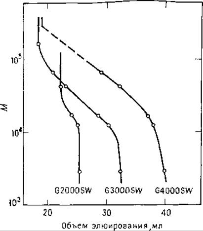
FIG. 6.6. Calibration plots for protein separation by gel filtration in an SDS solution on TSK-SW columns. Buffer: 0.1 M NaH2PO4, pH 7, containing 0.1% SDS [8].
Columns designed for gel filtration can also be operated in partition or adsorption chromatography modes. In this case, incomplete Modification of the support is exploited for protein sorption. Protein separation using these Methods is discussed below.
Last update: 06/08/2026
Editorial and Educational Adaptation: This material has been compiled based on the primary/original source text. The project team performed an editorial review, corrected technical inaccuracies, structured sections, and adapted the content for an educational format.
What was processed:
- elimination of formatting defects (OCR errors, structural breaks, corrupted characters);
- editorial organization of content;
- standardization of terminology in accordance with academic sources;
- verification of factual statements against the original source text.
All mentions of the author, publication year, and origin of the primary text have been preserved in accordance with the source.