Practical Protein Chemistry - A. Darbre 1989
Separation of protein and peptide mixtures by high-performance liquid chromatography
Adsorption and partition of polypeptides and proteins
Reverse-phase HPLC (RP-HPLC) of Peptides and Proteins has been widely used since 1970 [4, 18]. Attempts to adapt the Separation methodology established for small peptides to the Chromatography of larger Polypeptides have largely met with limited success. When separating protein or polypeptide mixtures on conventional RP sorbents, researchers frequently encounter difficulties stemming from the inherent properties of these complex molecules.
Protein molecules often feature multiple surface patches with contrasting physicochemical properties. These regions interact differently with the stationary phase, and many of these interactions lead to a loss of biological activity; the necessity of preserving this activity severely restricts the choice of chromatographic conditions. During Protein Isolation, interactions between the sample components and both the support matrix and organic Solvents should be minimized, while extreme pH values and drastic shifts in salt concentration must be avoided. However, these constraints are largely negligible when proteins are isolated specifically for amino acid sequencing.
Class="center">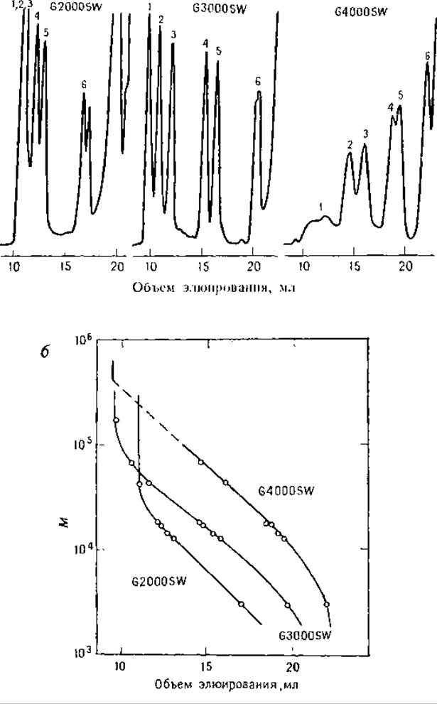
FIG. 6.7. Gel filtration of proteins on TSK SW columns in Gu*HCl solutions. Proteins were reduced and carboxyamidomethylated with iodoacetamide [9]. (a) Separation using three TSK SW columns; the eluent was 6 M Gu*HCl containing 0.1 M NaH2PО4, pH 6. The standard protein mixtures contained: 1 — thyroglobulin, 2 — BSA, 3 — Ovalbumin, 4 — Myoglobin, 5 — cytochrome c, 6 — Insulin; (b) calibration plots for proteins separated in 6 M GuHCl on TSK SW columns.
It is frequently observed that a protein or polypeptide exists in multiple distinct forms that (often for unclear reasons) resolve during RP-HPLC. Such unexpected occurrences naturally complicate fractionation. Since chromatography remains as much an art as a science, the best advice is to carefully optimize sample cleanup conditions to ensure a homogeneous product.
A review of current literature provides valuable guidance for selecting an initial separation strategy likely to yield success. Nearly always, different separation modes must be evaluated through trial and error, which inevitably requires a substantial amount of starting material. In many instances, it is far more advantageous to employ fast protein liquid chromatography (FPLC) prior to RP-HPLC. A brief Structure/127.html">Overview of Methods that may be of interest to biochemists is provided below.
Several systematic and original approaches have been developed to address the challenges of separating large polypeptides and proteins. The chromatographic behavior of large peptides has been investigated in systems containing trialkylammonium and tetraethylammonium phosphates and formates [21]. Studies involving 32 polypeptides have been reported [13, 15]; this work successfully achieved high-resolution Separation of proteins such as myoglobin, cytochrome c, and Lysozyme using 0.1 M phosphate buffer (pH 2.1, total electrolyte concentration 0.2 mol/L) and an acetonitrile gradient. Optimal resolution required exploiting both salt effects and low pH, with the organic solvent concentration playing a crucial role. In acidic buffers (~pH 2), the side chains of Asp and Glu remain uncharged, thereby increasing polypeptide Hydrophobicity. The addition of salts suppresses ionic interactions with the matrix. Acetonitrile and phosphate were chosen as the organic modifier and buffer system, respectively, for a specific reason: this combination allows peptides to be monitored in the eluate via peptide bond absorption at 215 nm.
A drawback of this system is the necessity of subsequent Desalting for samples intended for structural analysis. The Use of acidic buffers supplemented with salts has been reported for the purification of polypeptide Hormones, alongside the use of mini-columns for sample concentration [14]. Large peptides (M≤10,000) have been fractionated using TFA-based buffers, whereas the separation of Cyanogen bromide fragments of Collagen (M 3,000–60,800) utilized buffers containing heptafluorobutyric acid (HFBA) [1]. Prior to separation, samples can be concentrated by adsorption onto the top of a Vydac C18 Column [30], with HFBA being preferable to TFA and phosphoric acid; wide-pore Supports (30 nm pores) have also been shown to outperform small-pore sorbents (6–10 nm). The larger pore size allows high-molecular-weight molecules to penetrate the interior of the sorbent particles.
An example of optimizing HPLC conditions for large protein fragments is the separation of cyanogen bromide peptides from human fetal Hemoglobin. This study examined the effects of particle shape, pore size, and The Nature of the stationary phase on separation efficiency [16]. It was demonstrated that employing TFA- and n-propanol-based buffers with 30 nm or 100 nm pore sorbents increases peptide recovery by a factor of 1.7–2 compared to 10 nm pore sorbents [16]. The findings suggest that the geometric shape of the sorbent particles also influences separation performance, as different sorbents from the same manufacturer (Lichrosphere and Lichrosorb) with identical pore sizes (10 nm) yielded different recoveries of large polypeptides.
Unfortunately, evaluations involving these parameters were conducted on a very limited range of columns, complicating the interpretation of the results. It is reasonable to assume that larger pore sizes facilitate polypeptide diffusion into the internal channels of the particles, a notion supported by relevant literature. As experimental data accumulate, it appears that accessible surface area, rather than particle pore size, is the primary factor governing the HPLC of polypeptides on wide-pore supports. Reducing column length to 50 mm maintains resolving power, indicating that adsorption plays a significant role in polypeptide separation. Guidelines for selecting RP- and normal-phase (NP) HPLC conditions for polypeptides and proteins have been published [23]. Lichrosorb RP-18 was used for the purification of β-lipotropin [24] and β-endorphin [25], whereas leukocyte interferon was purified using Lichrosorb RP-8 combined with Lichrosorb DIOL [23]. The average pore size of both latter sorbents is 10 nm, with n-propanol chosen as the organic modifier.
Certain protocols (including interferon purification) have employed two-stage fractionation using different buffers (pH 4 and 7.5). Ionic interactions observed with the RP-8 support were suppressed using pyridinium formate or ammonium acetate. Because these buffer systems preclude monitoring eluate absorbance at 215 nm, automated post-column Fluorescent detection methods were implemented [2]. The separation results on RP-8 are illustrated in Fig. 6.8. The sample was applied to an RP-8 column (4.6 x 250 mm, 10 µm particle size) in 62.5 mL of a 4 M urea solution containing 0.1 M sodium acetate (pH 7.5). The column was washed with 1 M sodium acetate (pH 7.5), and peptides were eluted with an n-propanol gradient (1 h, 0–20%; 3 h, 20–40%) at a flow rate of 0.25 mL/min. The eluate was monitored for interferon activity, and the relevant fractions were pooled (Fig. 6.8a).
Thus, applying the sample to the column in a large volume of a urea- and acetate-containing buffer significantly accelerates sample concentration and separation through the adsorption of polypeptides onto RP sorbents. Peptides can be separated from urea using Sep-Pak cartridges [31] or mini-columns [14]; Sep-Pak units have also been utilized for sample pre-concentration prior to HPLC [1].
Occasionally, RP columns are repurposed for normal-phase (NP) chromatography [23]. Subsequent elution in the RP mode yielded fractions containing interferon from the RP-8 column. n-Propanol was added to the pooled fractions to a final concentration of 80%, and the solution was applied to a Lichrosorb DIOL column. Decreasing the n-propanol concentration during separation afforded purified interferon (Fig. 6.8b). For complete purification, the sample was passed through two additional RP columns (Fig. 6.8c, d). NP chromatography is particularly advantageous for purifying hydrophobic proteins that dissolve in high-organic solvent systems. Although many other proteins may co-precipitate with hydrophobic proteins in such systems, the contaminants can be effectively removed via RP chromatography, thereby yielding the hydrophobic targets in a solubilized state [23]. The application of Lichrosorb DIOL sorbent for polypeptide gel filtration was discussed previously (Section 6.2.2). Recently, LKB and Toyo Soda introduced phenyl-TSK columns for the NP chromatography of peptides.
Comparative evaluations were performed between Lichrospher sorbent (50 nm pore size) and C8-bonded supports with 10 nm pores. The comparison demonstrated that wide-pore supports provide a higher column capacity for large proteins (bovine serum albumin, collagen) while reducing elution band widths. Sorbents with 10 nm pores are suitable for separating proteins with molecular weights of 10,000–20,000.
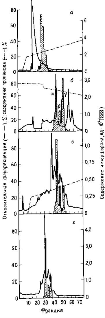
FIG. 6.8. Purification of interferon on Lichrosorb RP-8 using RP- and NP-HPLC. (a) 63 mL of interferon solution in 4 M urea – 0.1 M sodium acetate was applied to the column; the column was washed with 1 M sodium acetate, pH 7.5, and the protein was eluted with an n-propanol gradient (see text); (b) interferon-containing eluate fractions from (a) were pooled, adjusted to 80% n-propanol, applied to the column, and eluted with 0.1 M sodium acetate using an n-propanol gradient (72.5–50%) at a flow rate of 0.25 mL/min [26]; (c) interferon-containing fractions from the NP separation were pooled and subjected to RP-HPLC in pyridine-formate buffers using an n-propanol gradient; (d) chromatography of the product recovered from separation (c). Separation conditions were identical to (c).
Note. Fluorescence measurements were performed on the eluate following post-column derivatization of the fractions.
Last update: 06/08/2026
Editorial and Educational Adaptation: This material has been compiled based on the primary/original source text. The project team performed an editorial review, corrected technical inaccuracies, structured sections, and adapted the content for an educational format.
What was processed:
- elimination of formatting defects (OCR errors, structural breaks, corrupted characters);
- editorial organization of content;
- standardization of terminology in accordance with academic sources;
- verification of factual statements against the original source text.
All mentions of the author, publication year, and origin of the primary text have been preserved in accordance with the source.