Chemistry and Biology of Proteins - F. Haurowitz 1953
Hydrolytic Cleavage of Proteins
Determination of Amino Acids in Proteins and Protein Hydrolysates
Based on the fact that the reactive groups of Certain Amino acids remain free in polypeptide chains, some authors have attempted to determine the quantitative content of these amino acids directly in the protein by the intensity of color reactions. These attempts, however, lacked a sufficient foundation, since the color obtained with Proteins is generally weaker than that obtained with the corresponding protein hydrolysates. This is most likely due to the fact that within the protein molecule, some reactive groups are concealed inside the globule and are consequently inaccessible to the action of the coloring reagent (see Chapter VII). Therefore, to determine the Amino Acid Composition of a protein, it must be subjected to complete Hydrolysis. Most amino acids can be determined in an acid hydrolysate, but certain Amino acids are detected only after Protein Hydrolysis with barium hydroxide (see above). Separating a mixture of amino acids is a difficult task because amino acids are ampholytes, soluble in Water and insoluble in organic Solvents such as alcohol. Only the imino acids Proline and hydroxyproline are soluble in ethyl alcohol. Since amino acids possess similar physicochemical properties, they cannot be separated by fractionation with alcohol or neutral salts. However, Some amino acids can be separated by precipitation under appropriate conditions. For example, the solubility of cystine at a neutral reaction and of Tyrosine at a slightly acidic reaction is so low that upon adjusting the medium's reaction to the corresponding pH value, they precipitate almost completely. Other Amino Acids can be precipitated using specific Reagents. Nevertheless, none of these Methods is completely satisfactory in quantitative terms, since all corresponding precipitates are soluble to a certain degree.
Attempts were made to overcome this difficulty by employing the so-called isotope dilution method [61]. To determine a given amino acid in a hydrolysate, a small amount of this amino acid labeled with an isotope is added to it. The amino acid under investigation is then precipitated by some method, and the isotope content is determined in both the precipitate and the filtrate. Based on the fact that The ratio of the isotope concentration in the filtrate (ci.f) to the isotope concentration in the precipitate (ci.p) is equal to the ratio of the amino acid concentration in the filtrate (ca.f) to the amino acid concentration in the precipitate (ca.p), i.e., from the relation 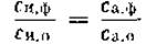 , one can calculate The amount of the corresponding amino acid originally present in the hydrolysate. If C0 designates the isotope content in the added amino acid, and CB — in the isolated amino acid, then the amount (B) of the amino acid originally present in the hydrolysate is determined by the formula
Class="center">B = A(C0/CB — 1),
where A is the added amount of the labeled amino acid. A major advantage of this method is that there is no need to carry out the quantitative Isolation of the target amino acid in a chemically pure form. The accuracy of the method is also unaffected by the co-precipitation of other substances, provided these substances do not contain the isotope used to label the amino acid being determined. This method has been applied for the Quantitative determination of the following amino acids: Glycine, leucine, aspartic and glutamic acids, phenylalanine, tyrosine, Arginine, and Lysine [61]. N15, C14, S35, and deuterium were used for amino acid labeling. When working with deuterium, it must be remembered that upon boiling amino acids with 20% Hydrochloric acid, the deuterium atoms in the a-position slowly exchange with hydrogen atoms [62]. Deuterium atoms in the ß- or y-position are stable under these conditions [62].
If the isolated compound is contaminated with the element used for isotopic labeling, the isotope dilution method may yield a significant error. For instance, in experiments with amino acids labeled with N15, the co-precipitation of other amino acids alters the C0/CB ratio. To circumvent this difficulty, it was proposed to convert all studied amino acids into isotopic derivatives and, by adding a large excess of an unlabeled derivative of the same Structure, subsequently separate them from the isotopic derivatives of other amino acids [63]. For example, a protein hydrolysate is treated with p-iodobenzenesulfonyl chloride containing J131. This reaction yields p-iodobenzenesulfonamide derivatives of the amino acids. Then, a large excess of non-isotopic p-iodobenzenesulfonylglycine is added as a carrier, and the isotope content in the isolated sample of p-iodobenzenesulfonylglycine is measured [63]. This method provides a powerful new tool for the quantitative Determination of amino acids in protein hydrolysates.
Recently, Moore and Stein [27] successfully quantified the amino acid composition of bovine serum albumin and ß-lactoglobulin on a starch Column. The respective hydrolysates were passed through the column, and the amino acids were eluted first with a mixture of n-butyl alcohol, n-propyl alcohol, and 0.1 N hydrochloric acid (1:2:1), and then with a mixture of propyl alcohol and 0.5 N hydrochloric acid (2:1). In this manner, these authors determined over 99% of all amino acids contained in the hydrolysate. This method is based on the fact that individual amino acids are eluted from the column at different rates. The amount of amino acid in small portions of the various extract fractions can be determined using the ninhydrin reaction. Based on the data obtained, a curve is plotted (Fig. 3) from which the amount of each amino acid in the hydrolysate can be calculated. Each point on this curve indicates the content of an individual amino acid in 0.5 ml of eluate, determined colorimetrically. A total of only 2.5 mg of bovine serum albumin was used to prepare the hydrolysate.
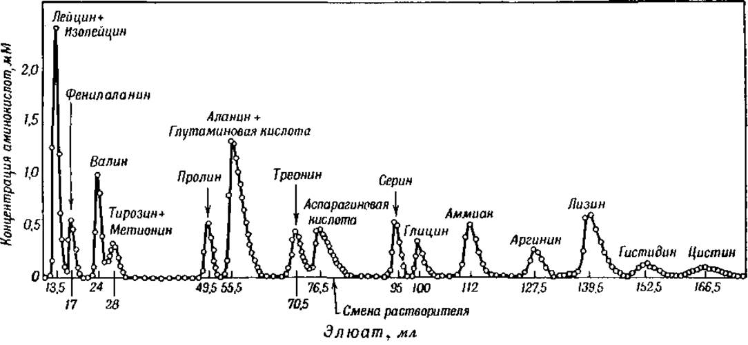
Fig. 3. Chromatographic Separation of bovine serum albumin hydrolysate [27].
Another novel method for the quantitative determination of amino acids is the microbiological method. Various cultures of lactic acid Bacteria, the culture *Leuconostoc mesenteroides*, and certain *Neurospora* strains are used for this purpose. The intensity of culture growth is determined by the turbidity of the bacterial suspension, the amount of lactic acid formed, or by weighing the mycelium [64–66]. One Modification of the microbiological method is the determination of amino acids from the amount of carbon dioxide formed As a result of the enzymatic Decarboxylation of amino Acids by bacterial preparations. In this way, tyrosine, Histidine, lysine, and glutamic acid can be determined [67]1. For the quantitative determination of a given amino acid, microbes are inoculated onto a synthetic medium containing all necessary Amino Acids and growth factors except for the amino acid under investigation.
The growth rate of bacteria on this medium is compared with that on synthetic media of the same composition, but additionally containing varying amounts of the amino acid being determined. By measuring the bacterial growth intensity at different concentrations of the target amino acid, a standard curve is obtained, from which the amino acid content in the analyzed material is calculated [64].
The main advantage of microbiological methods lies in their exceptional sensitivity. Another very important advantage is that microbiological amino acid determination requires a minuscule amount of the test material: less than 1 mg, and sometimes only a few micrograms. It should be noted, however, that There are also several difficulties associated with The Use of these methods. We still know very little about how the requirement of bacteria for a particular1 amino acid changes depending on the presence of any other amino acid. Since the amino acids used as standards for plotting the curve usually contain traces of other amino acids, it is impossible to rule out non-specific stimulation or inhibition of bacterial growth by these impurities. Therefore, the results of microbiological analyses must be treated with a certain degree of caution.
1 In 1947, S. R. Mardashev isolated a bacterial culture possessing the specific ability to decarboxylate aspartic acid (S. R. Mardashev, *Microbiology*, 16, 469, 1947). Dry preparations were successfully prepared from this culture, enabling the quantitative determination of l-aspartic acid content in protein hydrolysates (S. R. Mardashev and V. N. Gladkova, *Biochemistry*, 13, 315, 1948). In subsequent studies, S. R. Mardashev and V. V. Mamaeva (S. R. Mardashev and V. V. Mamaeva, *Biochemistry*, 15, 465, 1950) succeeded in developing a method allowing the determination of Aspartic Acid and Asparagine when simultaneously present in a solution via enzymatic decarboxylation. — Ed. note.
Infrared spectrophotometry is also a new method for the identification and quantitative determination of amino acids. Each Amino Acid and each a-chloro acid (obtained by the action of hydrochloric and nitric acids on amino acids) has a characteristic absorption curve in infrared light [68]. Using infrared spectrophotometry, it has been shown that the microbiological determination of leucine and isoleucine yields excessively high values [69].
The amino acids resulting from the complete hydrolysis of a protein are optically active compounds (unless the hydrolysis is accompanied by racemization). All amino acids isolated from well-characterized proteins possess the same configuration, i.e., the identical spatial arrangement of the four radicals around the a-carbon atom, although some amino acids are dextrorotatory and others are levorotatory. All Naturally Occurring Amino Acids belong to the l-series. Their interrelation is proven by conversion into identical derivatives [70]. Optical stereoisomers of these "natural" amino acids—"unnatural" d-amino acids—have been found in certain ergot Alkaloids and Bacterial toxins (see Chapter XV) [70]. Glycine is optically inactive because it does not contain an asymmetric carbon atom in its molecule.
A multitude of methods serve for the determination of individual amino acids, some of which are absolutely specific. Below we present a description of these methods.
Determination of glycine. Glycine (H2N ∙ CH2∙ COOH) forms an insoluble compound with K3[Cr(C2O4)3] ∙ 3H2O (tripotassium trioxalatochromiate) [71]. Glycine can also be determined microbiologically using *Leuconostoc mesenteroides* as sensitive bacteria [72]. In addition, glycine is determined titrimetrically. The method is as follows: glycine is oxidized with permanganate, yielding oxalic acid (COOH∙COOH), which is then precipitated with calcium and determined by standard titrimetric Procedures [73]. Serine also yields oxalic acid under these conditions, so a preliminary determination of serine must be carried out.
Determination of Alanine. Alanine (I) is converted by deamination with nitrous acid into lactic acid (CH3CHOH ∙ COOH); the latter is oxidized to acetaldehyde, which is determined via a color reaction [74]. Alanine can also be precipitated using nitranilic acid [75]. *Streptococcus faecalis* is used for the microbiological determination of alanine [72]. Upon heating with ninhydrin (see above), alanine is converted into acetaldehyde, which can be determined by titration after steam distillation [76].
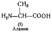
Determination of valine, leucine, and isoleucine. The action of ninhydrin on these amino acids (II), (III), and (IV) also yields volatile aldehydes, which can be determined as described above [76]. The amount of each of these Three amino acids can also be determined microbiologically [64, 77]. Due to its low solubility, leucine can be determined by the isotope dilution method [61].
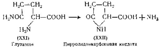
Determination of aspartic and glutamic acids. Aspartic (V) and glutamic (VI) acids can be precipitated as calcium or barium salts from an alcoholic solution [58, 78, 79]. Both of these Amino acids can also be determined microbiologically1 [64, 72], and aspartic acid, in addition, by means of the isotope dilution method [61]. Several new methods have been proposed for the determination of glutamic acid. Upon oxidation with chloramine T (CH3∙ C6H4∙ SO2∙ NHCl), glutamic acid is converted into succinic acid (COOH ∙ CH2∙ CH2∙ COOH); the latter is degraded to carbon dioxide under the action of succinoxidase, which is measured manometrically [81]. The amount of glutamic acid in a protein hydrolysate can be determined from the decrease in amino nitrogen upon heating the hydrolysate under pressure. Under these conditions, glutamic acid is converted into pyrrolidonecarboxylic acid [82], which possesses no free amino groups. Heating glutamic acid with ninhydrin yields 3-formylpropionic acid, which is determined colorimetrically as a dinitrophenylhydrazone [83]. In the protein molecule, aspartic and glutamic acids are present both as dicarboxylic acids with free carboxyl groups and as amides of these acids. The total amount of amides can be determined from the amount of ammonia liberated during acid hydrolysis. There are no reliable Methods for the separate determination of asparagine and glutamine in proteins1.
1 Regarding the determination of aspartic acid, see the note on p. 37. — Ed. note.

In enzymatic hydrolysates, glutamine can be determined by heating the neutralized hydrolysate to 100° [84]. Under these conditions, glutamine is rapidly hydrolyzed to yield an equivalent amount of ammonia, whereas asparagine undergoes no changes whatsoever [85].
Determination of hydroxy amino acids. Upon oxidation of hydroxy amino acids with periodic acid (HJO4), serine (VII) is converted into formaldehyde and Threonine (VIII) into acetaldehyde, with the release of an equivalent amount of ammonia [86]. The sum of hydroxy amino acids can be determined from the amount of cleaved ammonia. Ammonia is also formed upon the action of periodic acid on hydroxylysine, an amino acid found in noticeable quantities only in gelatin [87] (other proteins contain merely traces of hydroxylysine). To determine hydroxylysine, the protein hydrolysate is precipitated with phosphotungstic acid, and the resulting precipitate is treated with periodate [87, 88].
Determination of cystine. Cystine (X) can be determined by precipitation with phosphotungstic acid [89] or by reducing Cysteine with thioglycolic acid [90]. Cysteine (IX) is then determined colorimetrically through the color reaction with Folin's reagent [91], or by titrating sulfhydryl groups in the presence of oxidizing agents, such as iodine [92], porphyrindin [93], or potassium ferricyanide [94]. The resulting potassium ferricyanide is determined colorimetrically based on the color produced by its reaction with ferric chloride (Prussian blue). Cysteine can also be determined by electrometric titration of sulfhydryl groups with silver nitrate [95] or polarographically [96]. When proteins are treated with sodium or potassium cyanide, the Disulfide Bonds of cysteine are cleaved, initially yielding RSH + RSCH compounds, which subsequently form the amino acid lanthionine (XI) [97, 98]. Lanthionine can also be obtained by the action of alkalis on cystine or proteins.
1 S. R. Mardashev and V. V. Mamaeva (S. R. Mardashev and V. V. Mamaeva, Biochemistry, 15, 465, 1950) described a method for the quantitative determination of asparagine which, According to the authors, allows a sufficiently accurate estimation of asparagine content in various biological Tissues. In protein-free Liver extracts, the authors found 3.6 mg% of asparagine relative to the weight of the fresh organ, whereas in the Kidneys the asparagine content was 7.3 mg%. The determination of asparagine in an enzymatic casein hydrolysate showed that about half of the aspartic acid composing this protein exists in the molecule in the form of asparagine. — Ed. note.

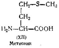
Determination of Methionine. For the quantitative determination of methionine (XII), it is advantageous to exploit its ability to yield methyl iodide upon heating with hydriodic acid. The liberated methyl iodide is determined by titration [99]. Methionine produces a red color in an acidic solution with nitroprusside [100]. Significant amounts of methionine have been found in casein.
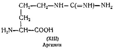
Determination of arginine. Arginine (XIII) is a representative of basic amino acids that can be precipitated from a protein hydrolysate with phosphotungstic acid or isolated by electrolysis [101]. Arginine is degraded upon heating with alkalis, releasing two molecules of ammonia per molecule of arginine. The amount of ammonia can be determined by titration, allowing the calculation of the initial amount of arginine present in the test solution. The enzyme arginase hydrolyzes arginine into Ornithine and urea. The amount of urea can be determined using urease. Arginine is precipitated by flavianic acid (1-naphthol-2,4-dinitro-7-sulfonic acid) [103]. Sakaguchi demonstrated that arginine gives a highly sensitive color reaction with a-naphthol and NaOCl [104]. The red color produced in this reaction is used for the Colorimetric determination of arginine [105].
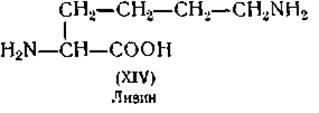
Determination of lysine. Another basic amino acid, lysine (XIV), is separated from the remaining basic acids by precipitation with picric acid. The isotope dilution method [61] and microbiological methods (using Leuconostoc mesenteroides or bacterial decarboxylase) [106] have also been employed for the determination of lysine.
Determination of histidine. The third basic amino acid, histidine (XV), is separated from other bases as a silver salt. The reaction of histidine with diazobenzenesulfonic acid, which produces a red coloration, is used for its colorimetric determination [107].

Determination of proline and hydroxyproline. The imino acids proline (XVI) and hydroxyproline (XVII) differ from other amino acids by their exceptionally high solubility in ethanol. Proline is precipitated by adding Reinecke's salt (NH4Cr(CNS)4(NH3)2) [108] or rhodanilic acid, i.e., tetrathiocyanatodianilidochromic acid [109]. Proline can be determined microbiologically using Neurospora [72]. For the colorimetric determination of hydroxyproline, it is oxidized with sodium peroxide, yielding a substance that forms a red color with isatin in acidic solution [110].

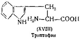
Determination of Tryptophan. Tryptophan (XVIII) is precipitated from its sulfuric acid solution with mercuric sulfate. Tryptophan can also be determined spectrophotometrically, as it exhibits an intense absorption band in the ultraviolet region of the spectrum [72]. Tryptophan yields intense color reactions with many aldehydes, such as formaldehyde, dimethylaminobenzaldehyde, or glyoxylic acid; these reactions can be utilized for its colorimetric determination [111].
Determination of phenylalanine. Upon Treatment with nitric acid, phenylalanine (XIX) is converted into 3,4-dinitrobenzoic acid, the amount of which can be determined colorimetrically [112]. Nitration of phenylalanine can also yield dinitrophenylalanine, which upon reduction with zinc dust to diaminophenylalanine produces a readily colorimetric-active color reaction with naphthoquinonesulfonic acid [113]. Lactobacillus arabinosus is used for the microbiological determination of phenylalanine. Phenylalanine can be precipitated with 2,5-dibromobenzenesulfonic acid [114] and determined by the isotope dilution method [61].
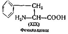
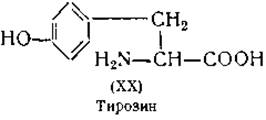
Determination of tyrosine. The solubility of tyrosine (XX) in water is so low that it precipitates upon neutralization of protein hydrolysates. Consequently, it can be determined by the isotope dilution method [61]. Various color reactions of tyrosine are also widely employed for its quantitative determination. The most frequently used for this purpose are: 1) the diazo reaction — a red coloration formed by the reaction of tyrosine with diazobenzenesulfonic acid [115, 116]; 2) Millon's reaction — a red coloration appearing in the presence of tyrosine upon The addition of a mercury-nitric acid solution [117]; 3) a blue coloration resulting from the reducing action of tyrosine on a mixture of phosphotungstic and phosphomolybdic acids [118]. Upon treatment with iodine in a mildly alkaline solution, tyrosine is iodinated. This iodination yields diiodotyrosine and thyroxine [119, 120]. A similar reaction is observed when proteins are treated with iodine [80].
Finally, mention should be made of another substance found among protein hydrolysis products, namely ammonia. It is generally assumed that ammonia is formed exclusively as a result of the Cleavage of asparagine and glutamine (XXI). Both amides have been detected in enzymatic protein hydrolysates [121]. While asparagine is stable in dilute acids and neutral aqueous solutions, glutamine at pH 2–6 and upon heating to 100° forms ammonia and pyrrolidonecarboxylic acid (XXII) [122]. The differing acid Stability of the two amides can be utilized for the separate determination of asparagine and glutamine in enzymatic protein hydrolysates [84, 123].
Last update: 06/08/2026
Editorial and Educational Adaptation: This material has been compiled based on the primary/original source text. The project team performed an editorial review, corrected technical inaccuracies, structured sections, and adapted the content for an educational format.
What was processed:
- elimination of formatting defects (OCR errors, structural breaks, corrupted characters);
- editorial organization of content;
- standardization of terminology in accordance with academic sources;
- verification of factual statements against the original source text.
All mentions of the author, publication year, and origin of the primary text have been preserved in accordance with the source.