FUNDAMENTALS OF ENZYME STRUCTURE AND KINETICS IN BIOLOGICAL SYSTEMS - O. A. Naumenko - 2017
1. General Principles of the Structural Organization of Enzyme Proteins
1.1 Definition of Enzymology as a Science, History of Discovery, Nomenclature, and Classification of Enzymes
1.1.1 Definition of Enzymology as a Science
Enzymology is a branch of biochemistry that studies enzymes. Enzymes are specialized Proteins that act as biological catalysts and possess a native conformation.
Unlike conventional functional proteins, enzymes feature an Active Site (AS) On the surface of the protein globule. This is a region formed by various amino acid residues assembled from different PARTS OF THE polypeptide chain, where substrate binding and conversion take place. A substrate is defined as a chemical compound that undergoes a change during the catalytic process to form a reaction product.
In addition to the active site, some enzymes also possess a regulatory site known as the allosteric site (AlS). This site binds molecules that influence The conversion of the substrate in a chemical enzymatic reaction, while the enzymes themselves remain unchanged.
The Active Site of an enzyme contains amino acid residues with functional groups (-SH, -COOH, -OH, -NH2) that participate in the catalytic process. Functionally, the active site of an enzyme can be divided into two regions: the sorption region, responsible for substrate binding, and the catalytic region, where substrate conversion occurs. The size of an enzyme's active site is determined by the size of the substrate, operating via the induced-fit mechanism. The spatial arrangement of the functional groups in the active site corresponds to The Nature of the substrate, determining the efficiency of its binding and conversion during the enzymatic chemical reaction.
The constant characterizing the efficiency of substrate conversion in the enzyme's active site is called the catalytic constant (Kk), whereas the constant determining the substrate's affinity for the enzyme is the binding constant (Ks). The action of effectors (activators and inhibitors) is determined using the activation constant (Ka) and inhibition constant (Ki).
Enzyme activity depends on the Nature of the enzyme and substrate, their concentrations, intersubunit interactions of protein globules, solution composition, nature of the solvent, Ionic strength of the solution, medium pH, presence of inhibitors and activators, Temperature, pressure, UV irradiation, and other physical factors. Enzyme activity increases with rising ambient temperature; however, at temperatures above 50°C, a decrease in activity is observed due to the disruption of the native Structure OF THE protein globule. Temperature elevation is accompanied by increased mobility of functional groups within the active site region and alterations in the native protein conformation.
The Effect of pH on enzyme activity can manifest through the associative behavior of the ionizing groups of the active site and the Functional groups of the substrate. Enzyme activity can also be influenced by The behavior of groups located on The surface of the protein globule, whose ionization may lead to Changes in the enzyme protein conformation.
1.1.2 History of The Development of Enzymology as a Science
The Water/144.html">Origin of the study of enzymes dates back to the first half of the 19th century. The first scientific concept of enzymes was introduced in 1814 by St. Petersburg scientist K. S. Kirchhoff, who demonstrated that not only germinated barley grains but also malt extracts are capable of saccharifying starch, converting it into maltose. The substance extracted from germinated barley that possesses The ability to convert starch into maltose was named amylase.
J. Liebig and F. Wöhler discovered an agent that cleaves amygdalin found in the essential oil of bitter almonds. This agent was named emulsin. In subsequent years, Other Enzymes were described, notably Pepsin and Trypsin, which cause the breakdown (Hydrolysis) of proteins in the digestive tract. In 1913, Michaelis and Menten proposed The Theory of the METABOLISM/10.html">Mechanism of enzyme Action.
In 1926 (considered the official birth year of enzymology as a science), Sumner isolated crystalline urease and proved its protein nature. Since then, more than 700 enzymes have been discovered and isolated, but many more exist in living organisms. In 1929, another biochemist, J. Northrop, reported the ISOLATION OF A crystalline pepsin preparation, followed by trypsin and other enzymes. In 1946, Northrop and Sumner were awarded the Nobel Prize for the discovery of enzyme proteins.
In 1969, Merrifield (New York) artificially synthesized Ribonuclease and proved that all enzymes are proteins.
1.1.3 Research Areas in Enzymology
Let us outline the main modern research areas in enzymology:
1) investigation of the finer details of the molecular mechanism and Introduction/14.html">Principles of Enzyme action in accordance with the laws of classical organic chemistry and quantum mechanics, as well as the advancement of the theory of Enzymatic Catalysis;
2) study of enzymes at higher levels (supramolecular and cellular) of the Structural Organization of living systems, focusing not just on individual enzymes, but on enzyme complexes within complex systems;
3) investigation of The regulatory mechanisms of enzyme activity and synthesis, and THE CONTRIBUTION OF chemical modification to enzyme action;
4) advancement of research in the creation of artificial low-molecular-weight enzymes—abzymes (synthetic enzyme analogs) endowed with high catalytic Specificity and activity similar to native enzymes, but lacking adverse antigenic properties;
5) research in the field of enzyme engineering (Protein Engineering), the creation of "hybrid" catalysts combining the Properties of Enzymes, Antibodies, and receptors, as well as the development of biotechnological reactors involving individual enzymes or multienzyme complexes to obtain and produce Materials and agents of high value for the national economy and medicine;
6) research in the field of medical enzymology, the main objective of which is to elucidate the Molecular Basis of hereditary and somatic human diseases caused by defects in enzyme synthesis or impaired Regulation of enzyme Activity in The Human Body.
1.1.4 Nomenclature and Classification of Enzymes
The modern Classification and Nomenclature of Enzymes were developed by the Enzyme Commission of the International Union of Biochemistry and adopted at the 5th International Biochemical Congress in Moscow in 1961.
There are three Main principles of Enzyme Classification:
1) by the Chemical Nature of the enzyme;
2) by the chemical nature of the substrate;
3) by the type of catalyzed reaction.
According to the International Classification, enzymes are divided into six main classes, each comprising several subclasses and sub-subclasses, with every enzyme assigned a specific serial number:
1) oxidoreductases;
2) transferases;
3) Hydrolases;
4) lyases;
5) isomerases;
6) ligases (synthetases).
1.1.4.1 Oxidoreductases.
The Class of oxidoreductases comprises enzymes that catalyze oxidation-reduction Reactions Involving Two substrates, which form The basis of Biological Oxidation. Their systematic names are formed using the formula "donor: acceptor oxidoreductase". For example, lactate: NAD+ oxidoreductase for the enzyme Lactate dehydrogenase (LDH).
The following main subclasses of oxidoreductases are distinguished:
- aerobic dehydrogenases or oxidases, which catalyze The transfer of protons (electrons) directly to oxygen;
- anaerobic dehydrogenases, which accelerate the transfer of protons (electrons) to an intermediate substrate rather than to oxygen;
- Cytochromes, which catalyze the transfer of electrons only.
This class also includes the heme-containing enzymes catalase and peroxidase, which catalyze reactions involving hydrogen peroxide.
1.1.4.2 Transferases.
The class of transferases comprises enzymes that catalyze the intermolecular transfer of various atoms, groups of atoms, and radicals. Their names are formed using the formula "donor: transferred group-transferase".
Transferases are known that catalyze the transfer of single-carbon residues, acyl, glycosyl, aldehyde or ketone, and nucleotide residues, nitrogenous groups, as well as phosphoric and sulfuric acid residues, among others. Examples include methyl- and formyltransferases, acetyltransferases, aminotransferases, phosphotransferases, etc.
1.1.4.3 Hydrolases.
The class of hydrolases includes a large group of enzymes that catalyze the Cleavage of intramolecular bonds in Organic compounds with the participation of a water molecule. Their names are formed using the formula "substrate-hydrolase". These include:
1) esterases — enzymes that catalyze the hydrolysis and synthesis of esters;
2) glycosidases, which accelerate the cleavage of glycosidic bonds;
3) Phosphatases and peptide hydrolases, which catalyze the hydrolysis of phosphoanhydride and peptide bonds;
4) amidases, which accelerate the cleavage of non-peptide amide bonds, etc.
1.1.4.4 Lyases.
Lyases are a class of enzymes that catalyze the cleavage of C–O, C–C, C–N, and other bonds, as well as the reversible non-hydrolytic elimination of various groups from substrates. These reactions are accompanied by The formation of a double bond or The addition of groups across a double bond. These enzymes are designated by the term "substrate lyase". For example, the enzyme fumarate hydratase (systematic name "L-malate hydrolase") catalyzes the reversible elimination of a water molecule from malic acid to yield fumaric acid. This group also includes Decarboxylases (carboxy-lyases), amidinolyases, etc.
1.1.4.5 Isomerases.
Isomerases comprise enzymes that catalyze the interconversion of optical and geometric isomers. Their systematic name is formed according to the reaction type: "substrate cis-trans-isomerase". If the isomerization involves the intramolecular transfer of a group, the enzyme is termed a "mutase".
This class also includes racemases and epimerases acting on amino and hydroxy acids, CARBOHYDRATES, and their derivatives; intramolecular oxidoreductases catalyzing the interconversion of aldoses and ketoses; and intramolecular transferases transferring acyl, phosphoryl, and other groups, etc.
1.1.4.6 Ligases (Synthetases).
Ligases are enzymes that catalyze the synthesis of organic substances from two initial molecules using energy derived from The breakdown of ATP (or another nucleoside triphosphate). Their systematic name is structured in the form "X : Y ligase", where X and Y denote the starting substances. An example is L-glutamate:ammonia ligase (commonly recommended as "Glutamine Synthetase"), which facilitates the Synthesis of Glutamine from glutamic acid and ammonia in the presence of ATP.
1.1.5 Monomers and Oligomers
According to their structure, all protein enzymes are divided into:
1) monomers (single-component), which are simple proteins consisting of a single globule;
2) oligomers (two-component), which consist of two or more subunits and contain both a protein moiety (apoenzyme) and a non-protein moiety (cofactor).
1.1.5.1 Monomeric enzymes possess Primary Structure.
This represents the precise sequence of amino acid residues in a polypeptide chain connected by peptide bonds.
The Secondary structure of a monomeric enzyme protein refers to the manner in which the polypeptide chain folds into a specific conformation. The most common Conformations include:
- α-Helix;
- β-pleated sheet;
- random coil (unordered structure);
- loop-like and finger-like structures.
However, the key to the functional activity of a protein enzyme is its tertiary structure. Tertiary, or native, structure is the three-dimensional spatial conformation of a polypeptide chain, or the folding of secondary structure into a compact structure of a specific volume. As secondary structure folds into tertiary structure, domains are formed.
A domain is a region within the protein tertiary structure that exhibits Structural and functional autonomy. Enzyme molecules may contain one or several domains, and their Functions can be either identical or distinct. Domains of the same structural type can be part of different enzyme proteins, imparting similarity to their biological and chemical functions. An example of such a shared domain is the NAD-binding domain of dehydrogenases. Enzymes are engineered on a modular principle, where the domain serves as the module.
1.1.5.2 Structure of oligomeric enzymes.
An oligomer is a molecule structured as a chain composed of a small number of identical constituent units.
This is what distinguishes oligomers from polymers, in which the number of repeating units is theoretically unlimited. The upper molecular weight limit of an oligomer depends on its chemical properties. The properties of oligomers heavily depend on changes in the number of repeating units in the molecule and the nature of the terminal groups; from the point when chemical properties cease to change with increasing chain length, the substance is referred to as a polymer.
Oligomers are also defined as Protein Complexes consisting of two or more subunits. Complexes made of identical subunits are called homooligomers, whereas those made of different subunits are called heterooligomers.
In biochemistry, the term oligomer is also used to denote short fragments of NUCLEIC ACIDS (DNA and RNA). Such oligomers, immobilized on a Glass slide or nylon membrane, are used in DNA Hybridization experiments.
Protein oligomers represent The quaternary structure of proteins and consist of two or more globules (subunits). A subunit is referred to as a protomer.
If the subunits are:
1) identical — homogeneous oligomer;
2) different — heterogeneous oligomer.
As a rule, the number of subunits is even. If the subunits within the oligomer are different, they perform distinct functions: catalytic and regulatory (allosteric).
Isozymes can occur within a single Organism; these are Multiple Forms of an enzyme that catalyze the same reaction yet differ from one another in Physical and Chemical properties, specifically in substrate affinity, maximum velocity of the catalyzed reaction (activity), electrophoretic mobility, or regulatory properties. The nomenclature and numbering of isozymes are based on their electrophoretic activity (anodal mobility) during catalysis. The most mobile isozymes are assigned the first number. The Biological Role of isozymes lies in Metabolic Regulation and adaptation.
For example, the enzyme lactate dehydrogenase exists in two subunit types: Heart (H) and Muscle (M). In the body, the enzyme molecule can be represented by five isozymes with conformations consisting of four subunits: HHHH, HHHM, HHMM, HMMM, MMMM.
Review Questions on the Topic
1. What does enzymology study?
2. Characterize the six main classes of enzymes.
3. Provide the numerical designation for the enzyme Alcohol dehydrogenase. Which class and subclass does this enzyme belong to?
4. What are monomeric and oligomeric enzymes?
5. WHAT IS A homogeneous oligomer?
6. What is a heterogeneous oligomer?
7. What are isozymes?
8. What is a domain?
9. How is the isozyme number determined?
10. What is the tertiary conformation of an enzyme protein?
1.2 Supramolecular Organization of Enzymes
1.2.1 Types of Supramolecular Forms of Enzyme Organization
There are 3 types of supramolecular forms of enzyme organization:
1) multienzyme complexes;
2) multienzyme conjugates;
3) multienzyme assemblies.
Multienzyme complexes are supramolecular structures that comprise multiple Enzymes and Coenzymes. In a multienzyme complex (Figure 1.1), several enzymes are tightly bound together into a single unit to carry out a series of sequential reactions, wherein the reaction product is directly passed on to the next enzyme and serves exclusively as its substrate. The complex is held together by non-covalent interactions. Such complexes significantly increase The rate of molecular conversion.
Figure 1.1 — Structure of a multienzyme complex
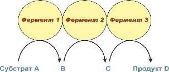
A prime example of multienzyme action is The oxidative decarboxylation of α-keto acids (Pyruvate and α-ketoglutarate) mediated by pyruvate dehydrogenase and α-ketoglutarate dehydrogenase.
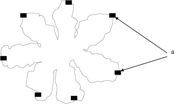
Multienzyme conjugates are complexes in which distinct enzymes are linked within a single polypeptide chain. A multienzyme complex composed of covalently joined enzymes is more stable than one formed through non-covalent bonds. An example of this is the fatty acid synthase (FAS) complex found in bacterial Cells of E. coli and higher plants (Figure 1.2).
Figure 1.2 — Multienzyme conjugate: a — active sites of the enzyme
Multienzyme assemblies (metabolons) represent the most highly organized multienzyme systems associated with large supramolecular structures, such as Ribosomes or membranes. Within a metabolon, each enzyme is in contact with one or more Enzymes of the same metabolic pathway. The structural integrity of the assembly is maintained by the cellular or organellar matrix (scaffold). There are two types of assemblies:
1) adsorption (dynamic);
2) integral (protein-enzyme embedded in the membrane).
1.2.3 THE CONCEPT OF the Prosthetic Group in Complex Enzymes
Complex proteins consist of a protein moiety, termed the apoenzyme, and an additional moiety known as the prosthetic group. Together, the apoenzyme and the prosthetic group constitute the holoenzyme.
The additional group in complex enzymes is called a cofactor; it is a low-molecular-weight, non-protein component essential for the enzymatic reaction to proceed.
Cofactors are divided into two main groups:
1) Metal Ions (as well as certain inorganic anions);
2) coenzymes, which are organic substances.
Roughly one-third of all currently known enzymes are activated by metal ions. Various metal ions can act as enzyme cofactors (Zn2+, Mg2+, Fe2+, Fe3+, Ca2+, u2+, Na+, К). Coenzymes include vitamin derivatives, NUCLEOTIDES (NAD —> NADH, ATP —> ADP), Glutathione, and Lipoic Acid.
The roles of cofactors include:
1) altering the Tertiary Structure of the enzyme protein;
2) ensuring complementarity between the enzyme and the substrate during the enzymatic reaction;
3) participating in the reaction as an additional substrate;
4) modifying The structure of the substrate (acting as an activator).
1.2.3 The Role of Metals as Cofactors
Metal ions may participate in substrate binding at the enzyme active site, directly in catalysis, in stabilizing the optimal conformation of the enzyme molecule, and so forth.
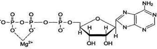
Metal ions function as stabilizers for the substrate molecule, the enzyme active site, and the protein conformation, specifically the Tertiary and Quaternary structures. For certain enzymes, the actual substrate is a complex formed by the reactant and a metal ion. For instance, in most
Kinases, the active substrate is not an ATP molecule itself, but rather the Mg2+ - ATP complex. In this case, the Mg2+ ion does not interact directly with the enzyme; instead, it helps stabilize the ATP molecule and neutralizes the negative charge of the substrate, thereby facilitating its binding to the enzyme's active site (Figure 1.3).
Figure 1.3 - Mg2+ - ATP complex
Schematically, the role of a cofactor in enzyme-substrate interaction can be represented as the E-S-Me complex, where E stands for enzyme, S for substrate, and Me for metal ion.
Coenzymes of oxidation-reduction enzymes (Group 1) are divided into 2 subgroups:
1) redox coenzymes, which transfer hydrogen protons (NAD, NADH, lipoic acid, ubiquinone);
2) nucleoside phosphates, which transfer phosphoric acid residues (ATP, ADP, AMP).
1.2.4. Active Site of Enzymes
The active site is a cluster of specific amino acid side chains arranged in a precise spatial orientation that determines the specificity and catalytic activity of enzymes. It is formed within the tertiary structure of the protein. The active site interacts with either the substrate or the cofactor.
The amino acid residues that make up the active site are referred to as catalytic groups (R-groups).
R-groups are divided into: nucleophilic (negative) and electrophilic (positive) groups (Table 1.1).
Table 1.1 - Electrophilic and nucleophilic groups of the enzyme active site
Nucleophilic groups |
Electrophilic groups |
Histidine imidazole ring |
Positive imidazolium ion |
Unionized carboxyl groups |
|
Lysine NH4-group |
Metal ions |
Ionized carboxyl group of aspartic and glutamic Amino Acids |
Coenzymes |
1.2.5 Structural Features of the Active Site
The active site accounts for only a small fraction of the total volume of the protein molecule. For example, if an entire protein molecule consists of 100,000 amino acid residues, its active site typically comprises merely 10 to 30 amino acids.
The active site may be either bicomponent or monocomponent.
A bicomponent active site occurs in Conjugated Proteins and includes a cofactor or coenzyme.
A monocomponent active site is found in simple protein enzymes and is formed exclusively by amino acids.
The bond between an enzyme and its substrate is relatively weak. Specific enzyme-substrate interaction is determined by the spatial arrangement of atoms within the enzyme's active site. Historically, the correspondence between the enzyme active site and the binding sites of the substrate molecule has often been described using the "lock-and-key" model. However, certain enzyme active sites are not rigid structures; instead, they become complementary only during the Formation of the enzyme-substrate complex (Koshland's theory, or the "induced-fit theory"). In this process, the active site alters its conformation and "tunes" itself to the structure of the substrate-inducer through induced fit (dynamic recognition). Depending on the reactive radicals present within the active site, the enzyme active site exhibits the following properties:
1) acidic;
2) basic;
3) acid-base: exhibiting polyfunctional catalysis if both nucleophilic and electrophilic groups are present in the active site.
X-Ray Diffraction Analysis and Protein Crystallography have established that the active site is localized:
1) in a standard defect, a "dent" located within the architecture of the protein globule. In this position, the substrate is surrounded and simultaneously attacked by multiple protein side chains.
2) at the junction between domains. For example, in Serine proteases, the active site is located at the interface of two domains. This position facilitates the formation of a specialized microenvironment within the active site.
1.2.6 Properties of the Active Site Environment in Enzymes
The environment of the enzyme's active site possesses A number of key properties:
1) microheterogeneity, which is driven by two factors:
- the formation of the enzyme's active site involves not only polar R-groups, but also partially apolar hydrocarbon side chains of amino acid residues. For instance, in alpha-Chymotrypsin, it has been established that 10 out of 23 valines, 10 out of 15 isoleucines, and 6 phenylalanines are exposed on the surface;
- solvent water molecules are firmly integrated into the surface of the side molecule.
2) a low dielectric constant (DC) compared to water. For water, the DC = 80, whereas in the sorption center of chymotrypsin, the DC = 10;
3) reduced polarity (compared to water), as determined by organic compound fluorescence assays relative to water;
4) local high polarity. Adjacent to regions of elevated polarity in the surface areas of the active site, high polarity is registered due to the dipoles of peptide bonds. Their close proximity leads to the formation of zones with high electric field intensity (10 - 100 mV/cm);
5) microviscosity. Within the active site, Amino acids are packed so densely that both translational and rotational mobility of molecules are hindered. All of this contributes to halting diffusion between the enzyme protein molecule and the substrate.
Studying the surface layer of the enzyme helps answer the question, "Why are enzymes able to accelerate reaction rates?":
1) A large number of functional groups are concentrated within the active site, interacting with the substrate simultaneously;
2) the active site environment exhibits microheterogeneity, where hydrophobic patches alternate with hydrophilic regions of local high polarity;
3) the surface layer of the enzyme protein is characterized by microviscosity, which restricts diffusion between the substrate and the enzyme.
All these active site properties facilitate multi-center interactions between the enzyme and substrate molecules.
The factors driving the acceleration of enzyme-catalyzed Chemical Reactions compared to non-enzymatic homogeneous catalytic processes are:
1) sorption interactions between substrate side groups and the enzyme molecule. The substrate undergoes catalytic attack not by a single R-group, but by several. Sorption interactions accelerate the reaction by a factor of 107 or more;
2) the polyfunctional nature of catalysis: nucleophilic or electrophilic catalysis provides an acceleration of enzymatic chemical reactions by a factor of 103;
3) the microheterogeneity of the active site environment provides the highest rate enhancement.
1.2.7 Allosteric Center
Some enzyme proteins possess an allosteric or regulatory center—a region of the enzyme molecule where low-molecular-weight chemical compounds, known as effectors (modifiers), bind and alter enzyme activity. Effectors are divided into:
1) activators (metal ions);
2) inhibitors (heavy metals), which can be either reversible or irreversible.
1.2.8 DETERMINATION OF ENZYME Activity
Enzyme activity is measured in units of activity - katals (kat). 1 katal is The amount of enzyme activity that catalyzes the conversion of 1 molecule of substrate per second at a temperature of 25 degrees.
The rate of an enzymatic reaction is determined by a kinetic parameter representing the number of complete catalytic cycles performed by an enzyme per unit of time (for example, 8 cycles per minute for pepsin).
Specific enzyme activity is the reaction rate referred to 1 milligram of enzyme protein.
Review Questions for the Topic
1. What types of supramolecular organization of enzymes are distinguished?
2. Define the prosthetic group in complex enzymes.
3. What is the role of metals in Enzyme Structure?
4. What is the structure of the active site of enzymes?
5. Where is the active site located?
6. Which nucleophilic residues are part of the enzyme's active site?
7. Which electrophilic residues are part of the enzyme's active site?
8. What is the allosteric site of an enzyme?
9. What is 1 katal?
10. What is the specific activity of an enzyme?
1.3 Principles of Spatial Organization of the Enzyme Molecule
1.3.1 Forces Stabilizing the Tertiary Structure of an Enzyme Protein
The tertiary structure of a protein is its three-dimensional spatial conformation formed through interactions between amino acid residues that may be located at considerable distances along the polypeptide chain.
Covalent bonds (Disulfide Bonds) participate in stabilizing the tertiary structure of a protein molecule, but non-covalent bonds play the primary role:
1) Hydrogen Bonds;
2) Electrostatic Interactions between charged groups;
3) intermolecular Van der Waals forces;
4) Hydrophobic bonds - interactions of nonpolar amino acid residues. The Stability of the tertiary structure depends on the network of non-covalent interactions within the protein globule. Some proteins are additionally stabilized by covalent disulfide bonds between cysteine SH groups.
1.3.2 Thermodynamics of Protein Tertiary Structure Folding
The folding of a protein into its native conformation occurs spontaneously provided There is a decrease in the Gibbs Free energy of the thermodynamic system (∆G <0), in accordance with the Gibbs equation:
∆G = ∆Н - T∆S, (1.1)
where ∆G is the Gibbs free energy; ∆H is the enthalpy of the system; T is the system temperature; ∆S is the Entropy of the thermodynamic system.
For the spontaneous folding process of an enzyme protein molecule to occur, the enthalpy must decrease while the entropy increases. This process is driven by the breaking of certain bonds and the formation of new intramolecular bonds within the protein-enzyme molecule. Hydrogen and hydrophobic bonds play the primary thermodynamic role in protein folding. Disulfide bonds, electrostatic interactions, and van der Waals forces make a significantly smaller contribution.
1.3.3 Importance of the Hydrogen bond
Hydrogen bonds are formed between polar amino acid residues. The energy of a single hydrogen bond is about 3 kcal/mol. Due to the large number of hydrogen bonds formed during the folding of a protein into its tertiary conformation, the enthalpy of the protein molecule increases, which is a thermodynamically unfavorable process. However, the folding involves not only the formation of new hydrogen bonds but also the breaking of pre-existing ones, which overall decreases the system's enthalpy and contributes to a slight increase in entropy, making the process thermodynamically favorable.
During the formation of the tertiary structure, the maximum number of hydrogen bonds is generated within the protein globule. However, while the entire protein globule forms, the entropy of the peptide bonds in the primary structure decreases, whereas the entropy of the solvent (water molecules) increases; this undermines the stability of the protein molecule's structure and likewise facilitates the folding of the enzyme into its native conformation.
The rupture of individual hydrogen bonds within the secondary structure leads to a decrease in enthalpy and an increase in entropy, which also promotes the folding process.
1.3.4 Role of Hydrophobic Bonds
Ranked second in importance for the spontaneous folding of a protein and the stabilization of its tertiary structure are hydrophobic bonds. They form between non-polar Amino Acids and water. The free energy gain results from the creation of cavities within the dynamic structure of water (quasicrystalline structure or "ice-like" water). These dynamic cavities form the interface between the protein and water. Overall, hydrophobic bonds increase the entropy between the protein molecule and the water solvent molecules. The smaller the interface area, the lower the entropy of the protein molecule; therefore, during folding, the protein tends to minimize thermodynamically unfavorable contact with water and forms a spherical Hydrophobic core. In general, hydrophobic bonds increase the entropy of the system.
1.3.5 Significance of van der Waals Bonds
Van der Waals bonds arise when non-polar amino acids come into close proximity, which slightly decreases the entropy of protein folding into the tertiary conformation.
1.3.6 Significance of Electrostatic Bonds
Electrostatic bonds occur between the carboxyl residues (COOH groups) of dicarboxylic amino acids and the amino groups of diaminocarboxylic acids. For instance, aspartic and glutamic acids carry a negative charge, whereas histidine, Arginine, and lysine have positively charged side chains. As a result of differences in electronegativity, electron transfer takes place and salt bridges are formed. Electrostatic bonds cause a slight decrease in entropy during protein folding.
1.3.7 Role of the Disulfide Bond
A disulfide bond forms between cysteine residues. Since cysteine is a minor component in most proteins, the majority of enzymes lack it. Overall, disulfide bonds have a negative effect on the folding process due to a decrease in entropy (entropic penalty).
Significance of disulfide bonds:
1) disulfide bonds facilitate the correct pathway of protein folding into the native conformation, as they are formed among the first;
2) covalent disulfide bonds increase the entropy of the protein molecule;
3) disulfide bonds are preserved in denatured proteins;
4) during protein folding, disulfide bonds restrict the number of possible conformations of the protein-enzyme molecule;
5) the formation of disulfide bonds does not precede the globular folding of the protein, but rather assists and refines this process;
6) incorrect disulfide bonds may form during folding, which are subsequently disrupted after the protein globule is formed.
Thus, the stabilization of the tertiary structure depends not only on the protein's own structure but also on the structure of the water solvent. Consequently, alterations in water structure—such as heating or freezing—destabilize the tertiary structure of the protein and lead to Denaturation, which is the loss of the tertiary conformation and enzymatic activity.
The thermodynamics of protein tertiary structure folding is characterized by a fine balance between folding factors and opposing forces. Therefore, the protein molecule often loses stability even upon minor shifts in system parameters. The energy of a single hydrogen bond is 3 kcal/mol, whereas The change in the free energy of the system during protein folding is ∆G = 10 kcal/mol.
1.3.8 Physical Form of the Enzyme-Protein Tertiary Structure
The native structure of an enzyme features a core composed of non-polar amino acids and a hydrophilic shell formed by polar amino acid residues (Figure 1.4).
Figure 1.4 - Cytology/cytology/92.html">SCHEMATIC STRUCTURE OF an enzyme protein molecule

As a result of folding and the formation of hydrophobic bonds, nonpolar amino acids reside within the interior of the protein molecule. Regions composed of polar amino acid residues within the hydrophobic core form the active site of the enzyme, whereas regions consisting of nonpolar amino acid residues on the enzyme's surface form the allosteric site.
Regions formed by nonpolar amino acid residues can participate in quaternary structure elements and supramolecular structures. Polar residues may also be located within the core, where electrostatic, hydrogen, and van der Waals bonds are formed between them.
Review Questions for the Topic
1. List the forces that stabilize the tertiary structure of an enzyme protein.
2. What energy drives the spontaneous thermodynamic processes involved in the folding of a protein's tertiary structure?
3. What is the role of hydrogen bonding in the folding of the protein tertiary structure?
4. What is the role of hydrophobic interactions in the folding of the protein tertiary structure?
5. What is the role of van der Waals bonds in the folding of the protein tertiary structure?
6. What is the role of electrostatic bonds in the folding of the protein tertiary structure?
7. What is the role of disulfide bonds in the folding of the protein tertiary structure?
8. Describe the physical conformation of the tertiary structure of an enzyme protein.
1.4 Mechanism of Spatial Structure Formation in Enzyme Proteins
1.4.1 Levinthal's Paradox
A polypeptide chain has a vast array of possible conformations (each amino acid residue has about 10 possible conformations, meaning a chain of 100 residues has roughly 10100 possible conformations). Consequently, a protein would theoretically need to search for "its" spatial structure among 10100 potential variants. Given that a transition from one conformation to another takes on average about 10-13 seconds, exhaustively searching all 10100 structures would take approximately 1080 years—a timeframe that dwarfs the lifetime of our Universe, which is roughly 1010 years, making it an infinitesimal fraction. This raises a fundamental question: how does a protein manage to "find" its unique native structural conformation within minutes?
Scientists have established that the native conformation of a protein exhibits several key properties:
1) by all experimental measures, the native Spatial structure of the protein behaves as the most kinetically stable over time;
2) from a thermodynamic perspective, there is no conclusive evidence that this structure is the most thermodynamically stable.
C. Levinthal hypothesized that the native Cell/13.html">Protein Structure is governed not by ultimate thermodynamic stability, but by kinetics; that is, it corresponds not to the global minimum, but to the most rapidly accessible minimum of the molecule's free energy.
The resolution to Levinthal's paradox is as follows:
1) the formation of the enzyme protein's tertiary structure is driven by a cascade of rapid transitions that require no input of external energy, i.e., free energy;
2) the spatial structure of a protein is dictated by its primary structure (encoded within The Genetic Code)—meaning the genetic code itself dictates the spatial structure, rather than the action of extracellular structures during synthesis;
3) The process of protein folding into its native conformation is multistage and proceeds according to a hierarchical principle.
1.4.2 Stages of Protein folding and the Hierarchical Folding Principle
According to current understanding, the protein folding process is hierarchical in nature and proceeds sequentially through four distinct stages:
1) very rapid: the Formation of secondary structure elements that act as "nucleation sites" for more complex architectural motifs (within a fraction of a microsecond, an α-helix spans a peptide of 20–30 residues);
2) very rapid: the specific association of certain secondary structure elements to form Supersecondary structures, such as combinations of several α-helices, several β-strands, or mixed associations of these elements;
3) slow: the formation of a "molten globule" (establishing the main elements of tertiary structure—combinations of α-helices, β-sheets, connecting loops, and the hydrophobic core of the molecule—along with domain formation);
4) formation of the native protein structure—"solidification" and shell formation (taking several minutes).
1.4.3 Thermodynamic CHARACTERISTICS OF THE Folding Process
Protein folding occurs spontaneously due to a decrease in the free energy of the system, driven by an increase in entropy and a decrease in enthalpy. Each stage of protein folding is energetically driven:
Stage 1: driven by the breakage and formation of hydrogen bonds, which lower the enthalpy. Research shows that the more α-helices a protein molecule contains, the lower its enthalpy.
Stage 2: driven by hydrophobic interactions of amino acid residues, which increase entropy.
Stage 3: driven by hydrophobic bonds, which account for 2/3 of the total work required for protein folding.
Stage 4: "solidification" driven by van der Waals, hydrogen, and electrostatic bonds. Hydrogen bonds ensure the precision of interatomic interactions.
1.4.4 Intracellular Regulation of Protein Spatial Structure
Two mechanisms for regulating the folding of native protein structure have currently been identified:
1) Mechanism regulating the folding rate: operates at the stage where the "molten globule" transitions into a rigid protein. This is mediated by specialized enzymes that accelerate the folding process. These include two enzyme proteins:
- peptidyl-prolyl cis-trans isomerase, which converts amino acid residues from the trans-conformation on ribosomes into the cis-conformation required for spatial structure formation.
protein disulfide isomerase, which catalyzes the formation and isomerization of disulfide bonds within the protein molecule.
2) Mechanism protecting partially folded proteins from non-specific aggregation. This is carried out by specialized proteins known as Histones. They enhance the folding efficiency of the polypeptide chain and protect the protein from non-specific aggregation. These proteins are called chaperones.
Chaperones (from the French *chaperon*—an elderly lady accompanying a young woman to balls, etc., or a mentor guiding a group of young people).
Functions of chaperones:
1) Bind to unfolded or partially unfolded conformations of the polypeptide chain, preventing them from tangling;
2) Stabilize the partially unfolded protein;
3) Facilitate the translocation of unfolded proteins across membrane structures into various cell Organelles;
4) Create optimal conditions for efficient protein folding.
Types of histone proteins. Histones belong to the class of "heat Shock" proteins. Under normal conditions, every cell contains a baseline set of chaperones, but their synthesis increases significantly during stress (e.g., elevated temperatures).
Chaperones are classified according to their molecular weight into two main families:
1) chaperonins are large proteins. They form complex structures: hsp 10 and hsp 60;
2) chaperones: hsp 70. They consist of 1 to 2 polypeptide chains and keep the nascent protein emerging from the ribosome in an unfolded state, facilitate its Intracellular Transport through the Cytoplasm upon release from the ribosome, transfer it to another chaperonin to promote further folding, and mediate Transport across mitochondrial and ER membranes. Mitochondria feature their own resident chaperone, hsp 50.
The release of the protein from chaperones is driven by the ability of chaperones to bind ATP or ADP. Cleavage is mediated by the ATPase enzyme.
Review questions on the topic
1. What is the Levinthal paradox?
2. Name the Stages of Protein folding.
3. What is the hierarchical principle of protein folding?
4. Resolution of the Levinthal paradox.
5. Thermodynamic characteristics of the folding process.
6. What intracellular mechanisms regulate the Spatial Structure of Proteins?
7. Which enzymes regulate protein folding?
8. What are chaperones? What function do they perform in cells?
9. What are chaperonins, and what is their function?
1.5 Domains: Structural and Functional Characteristics
1.5.1 Definition of Domains
A domain is a region within the tertiary structure of a protein that exhibits structural and functional autonomy. During protein folding into its native conformation, a hydrophobic core formed by non-polar amino acid residues is established at the Third Stage. X-ray crystallography data reveal that the core contains mosaic regions formed by either hydrophilic or hydrophobic residues, displaying structural and functional autonomy. Domains are formed during the third stage of protein folding.
The process proceeds in the following sequence:
1) independent folding of individual domains;
2) establishment of contacts between individual domains. For monomers, this occurs when an active site is formed between distinct domains. For oligomers, domains are formed at the junction of subunits.
1.5.2 Evidence for the Domain Stage of Protein Folding
In 1969, the first Evidence of the domain stage in protein folding was obtained from Lysozyme crystallography data, which established that the molecule consists of several compact globules.
In 1971, distinct structural entities were isolated from immunoglobulin proteins by Cunningham, who suggested that there is independent Genetic control over domain regions within the protein structure. Such regions were named domains;
In 1972, J. Birktoft and D. Blow identified two domains in alpha-chymotrypsin, each cylindrical in shape and composed of a six-stranded antiparallel beta-sheet. The same structural type was identified in other Proteolytic Enzymes, such as Elastase, trypsin, and aspartic proteinase.
Studies of protein enzymatic activity have shown that only a small part of the protein molecule—the active site—participates in the enzymatic reaction, rather than the entire molecule. The remainder of the protein globule serves as a foundation or scaffold ensuring the proper orientation of amino acid residues within the active site. Consequently, proteins with different primary structures can perform identical chemical functions while belonging to different structural protein classes. Examples include the two-domain protein trypsin and the single-domain protein subtilisin (composed of alpha-helices and beta-sheets);
D. Wetlaufer explains the existence of domains by the specific mechanism of spontaneous self-assembly of the Amino Acid Sequence into the native conformation. He distinguishes Two Types of domains: those with continuous and discontinuous polypeptide chains. The core (nucleation center), consisting of 8–18 residues, is considered the element that cements the domain structure. This core serves as a template for folding the polypeptide chain of a domain, which typically comprises 40–150 amino acid residues. Such domain organization ensures the rapid spontaneous assembly of the entire protein structure;
Parvis et al. established that the domains of complex Oligomeric Proteins are encoded by different exons. It has been found that DNA segments responsible for pausing during Protein Synthesis ensure the correct assembly of multienzyme oligomeric proteins.
1.5.3 Domain Properties
Numerous studies have shown that domains possess the following properties:
1) there is no clear correlation between domain organization and protein size. For instance, the enzyme carboxypeptidase consists of 307 amino acids, a single domain, and one active site, whereas the enzyme lysozyme consists of 164 amino acids and two domains;
2) the structural autonomy of a domain is often complemented by functional autonomy. For example, the nucleotide-binding domain of dehydrogenases is responsible for interacting with one of the reaction substrates—the coenzyme NAD or NADH. The amino-terminal domains (kringles) of Blood Coagulation SYSTEM enzymes mediate binding to Membrane Lipids and other proteins. The amino-terminal domains of IMMUNOGLOBULINS form the Antigen-binding center;
3) incomplete functional autonomy: demonstrated for the protein Papain, whose active site is located in a cleft, with its functional groups situated in different domains; the active site is capable of catalyzing reactions of only a single order;
4) the domain structure of a protein molecule reflects the early evolutionary stages of protein spatial structure, progressing from the simplest life forms to complex ones. It has been established that in the HIV virus, the protease enzyme consists of 99 amino acids and two domains. Structurally, this enzyme resembles pepsin;
5) domain boundaries correspond to exon boundaries within the DNA molecule;
6) domains can independently of other parts of the protein molecule maintain and shape its spatial structure;
7) interdomain interactions during the folding of oligomers have been established;
8) spatial domain swapping has been observed in certain oligomeric proteins, where the tertiary structure formation process occurs via a domain-swapping stage.
1.5.4 Multidomain ORGANIZATION OF THE Enzyme Molecule
Proteins are constructed on a modular principle, where domains serve as the modules. An enzyme protein represents a collection of various domains responsible for specific functions, such as membrane binding (terminal domains of blood coagulation enzymes), substrate binding, or a regulatory domain that governs enzyme activity and inactivity.
Depending on its domain architecture, an enzyme acquires the following properties:
1) absolute specificity;
2) relative specificity, which occurs when:
- the active site is formed at the domain interface;
- the active site is formed following domain closure;
- the active site is formed within a single domain.
Review Questions on the Topic
1. Define Protein domains.
2. What evidence exists for the domain-based stage of protein folding?
3. What are the known properties of domains?
4. What is the nature of the multidomain organization of an enzyme protein molecule?
5. What properties does an enzyme acquire due to identical domains in its structure?
6. What properties does an enzyme acquire if its active site is located at the domain interface?
1.6 Characteristics of the Enzyme-Substrate Complex
The Transformation of a substrate occurs within the enzyme-substrate complex (ES). To understand how an enzyme works, it is necessary to know the structure of the enzyme, the enzyme-substrate complex, as well as reaction intermediates and products. The lifetime of the enzyme-substrate complex is limited. To study the structure of the enzyme-substrate complex, X-ray diffraction analysis of enzyme-product, enzyme-inhibitor, and enzyme-substrate analogue complexes is performed.
1.6.1 Mechanism of Enzyme-Substrate Complex Formation in Solution
During the formation of the enzyme-substrate complex, new intermolecular bonds are established between the enzyme and the substrate. Hydrogen, covalent disulfide, hydrophobic, and electrostatic bonds are formed.
Covalent bonds form between the R-groups of cysteine and the corresponding residues of the substrate.
A hydrogen bond arises between two hydrogen residues located adjacent to oxygen atoms. The formation of such bonds occurs between the substrate and the active site of the enzyme lysozyme during polysaccharide hydrolysis. It has been established that in this case, the hydrogen bond energy is 4 to 8 kcal/mol.
Hydrophobic bonds arise between nonpolar hydrocarbon fragments of the substrate and hydrophobic (at least partially) Regions of the enzyme's active site. This process effectively results in the extraction of the enzyme's R-groups into an organic solvent. For example, the active site of a protein enzyme—containing a region that contacts water via hydrophobic side chains—loses its association with water, which is energetically more favorable. This contributes to
a 105-fold acceleration of the reaction. This type of interaction is quite common.
Electrostatic interactions occur between charged groups of the substrate and amino acid residues of the enzyme's active site. The energy gain ranges from 6 to 9 kcal/mol, and the reaction is accelerated by 104-fold, as seen, for example, in the catalysis of proteins by serine proteases.
The formation of the enzyme-substrate complex is also accompanied by the creation of chelate complexes. Specifically, when an enzyme and a substrate are linked by two or more points of interaction, such complexes are referred to as chelates.
The formation of the enzyme-substrate complex also induces conformational changes within the enzyme molecule. The enzyme can spatially "adjust" its structure, since the R-groups of the active site amino acid residues on the surface of the protein globule are not rigidly fixed and possess a certain degree of mobility. Consequently, this allows for the spatial alignment of the sorption sites on the enzyme globule with the corresponding sorption sites of the substrate molecule.
The enzyme is capable of adopting a conformation distinct from its equilibrium state prior to complex formation. These conformational changes help to minimize entropy losses.
The microviscosity of the environment within the enzyme's active site has a profound effect on the formation of the enzyme-substrate complex. This leads to an almost complete loss of mobility of the substrate molecules, meaning they lose translational and rotational mobility as well as the ability to diffuse.
Conclusion: Thus, the thermodynamically unfavorable process of enzyme-substrate complex formation in solution is compensated by multipoint interactions, the conformational flexibility of the active site's R-groups, the microviscosity of the active site environment, and the formation of various types of bonds between the enzyme and the substrate.
1.6.2 Estimation of the Free Energy of Sorption of the Enzyme-Ligand Complex
From a thermodynamic perspective, the process of enzyme-ligand complex formation can be viewed as a sequential, two-step process:
1) the approach and orientation of the ligand (substrate) in front of the enzyme's sorption center, which requires an energy input (approaching ∆G) and is a thermodynamically unfavorable process;
2) the formation of a chelate enzyme-ligand complex in solution through the creation of intermolecular bonds. The energy cost of complex formation
∆GE • L is compensated by the formation of a chelate complex between the enzyme and the substrate: the greater the number of interaction points, the higher the binding energy ∆G (a thermodynamically favorable process):
 (1.2), 40
∆Gapproach is the energy expended on bringing the enzyme and substrate together;
∆Gassociation is the association energy that lowers the activation energy of the substrate in the subsequent enzymatic reaction converting the substrate into product.
1.6.3 Reasons for Reaction Acceleration by Enzymes
The catalytic properties of enzymes are driven by a sophisticated MECHANISM OF ACTION. From the standpoint of modern enzymology, enzymatic catalysis is attributed to three main factors:
1) sorption of the substrate on the enzyme proceeds in a way that facilitates the subsequent Conversion of the substrate into a product. The activation energy of the substrate is lowered due to the release of the association free energy ∆GE • L;
2) the polyfunctional nature of chemical interactions between the enzyme and the substrate. The active site of the enzyme may contain both nucleophilic and electrophilic groups that provide chemical catalysis of the substrate via both acid and base mechanisms;
3) the microenvironment effects of the enzyme's active site.
Review Questions for the Topic
1. What mechanisms of complex formation exist in a solvent?
2. What intermolecular bonds are formed between the enzyme and the substrate in a solvent?
3. Define a chelate complex.
4. Evaluate the free energy of enzyme-ligand complex sorption.
5. What are the reasons for the acceleration of reactions by enzymes?
6. Through what mechanism does the lowering of the activation energy of the substrate molecule occur?
1.7 Complementarity Between Enzyme and Substrate
1.7.1 The Concept of Complementarity
During the formation of the enzyme-substrate complex, when each R-group of the substrate binds to the corresponding binding sites of the enzyme and the binding energy reaches its maximum value, the enzyme and the substrate are said to be structurally complementary.
The structure of the substrate changes during the course of the enzymatic chemical reaction, but the undeformed enzyme molecule can be complementary to only one form of the substrate. It is catalytically advantageous for the enzyme to be complementary not to the initial state, but to the Transition State of the substrate, when its internal energy reaches its maximum value. In this case, the increase in binding energy as the substrate approaches the transition state structure reduces the activation energy of the chemical transformation step of the substrate into the product.
1.7.2 Catalysis by Serine Proteases
Proteases containing a serine residue in their active center belong to the group of serine proteases: alpha-chymotrypsin, elastase, subtilisin.
Protease enzymes cleave peptide bonds in protein substrates. The Protein Hydrolysis reaction takes place in the presence of a solvent, water (Figure 1.5).
Figure 1.5 - Scheme of protein hydrolysis

The entire mechanism of enzyme action comes down to the sorption of the alpha-acylamide fragment of the substrate onto the carboxyl group of the enzyme's active site (Figure 1.6).
Figure 1.6 - Fragment of the sorbed substrate molecule
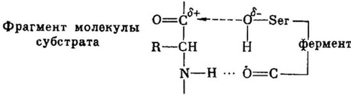
The formation of hydrogen bonds between the carboxyl groups of the enzyme and the alpha-acylamide fragment of the substrate molecule leads to the generation of two polarized nucleophilic and electrophilic serine groupings. As a result of sorption, a transition state of the substrate molecule is formed, which facilitates the nucleophilic attack by the enzyme and leads to reaction acceleration at the enzyme's active site.
The formation of a hydrogen bond imposes significant entropic constraints on the substrate molecule. This should reduce the overall contribution of enzyme-substrate complexation to reaction acceleration.
The stabilization of the substrate transition state has been experimentally proven in numerous chemical enzymatic reactions (CERs) using oligomeric substrates, which demonstrated that the enzymatic process proceeds via The intermediate formation of an acyl-enzyme, wherein a bond is formed between the OH group of serine and the C-peptide bond of the substrate.
The rate of Chemical Reactions Involving enzymes naturally increases with a rise in the number of hydrogen bonds formed between the chemically inert moiety of the substrate molecule and the substrate-binding site of the enzyme.
In enzymatic catalysis, the free energy of substrate sorption on the enzyme acts as the driving force. The thermodynamic prerequisite for approximation and
orientation is that the freezing of the substrate molecule is achieved at the expense of the free energy of sorption.
In the case of serine proteases, these are hydrogen bonds that increase entropy, which leads to a decrease in $\Delta G$ and causes the process to proceed spontaneously.
1.7.3 Models of Enzyme-Substrate Interaction in Catalysis
Currently, there are three theories regarding the formation of the enzyme-substrate complex during enzymatic reactions:
- the lock-and-key mechanism;
- the induced-fit theory;
- the strain theory.
1.7.3.1. The Lock-and-Key Mechanism
In 1894, Emil Fischer proposed the "lock-and-key" model to explain the Mechanism of Enzymatic catalysis. The enzyme binds to the substrate to form a short-lived enzyme-substrate complex. The active site of the enzyme is pre-structured in such a way that the substrate fits into it like a key in a lock. During the formation of the enzyme-substrate complex, neither the structure of the substrate nor that of the active site undergoes any changes. In this case, the enzyme exhibits absolute specificity toward the substrate.
Disadvantages: for many enzymes, "the key does not fit the lock well," in which case additional force must be applied to turn the "key".
1.7.3.2. The Induced-Fit Theory Proposed by Koshland
In the absence of the substrate, the catalytically active groups of the enzyme X and X' are positioned in such a way that they cannot simultaneously interact with the substrate fragment Y (Figure 1.7 a).
Figure 1.7 - Schematic representation of Conformational Changes in the enzyme induced by the substrate
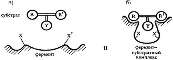
As a result of approximation and orientation, a conformation of the enzyme's active site takes place, leading to the formation of the enzyme-substrate complex (Figure 1.7 b). Part of the free energy of sorption is expended on the formation of the complex, which is directed toward altering the conformation of the active site.
For example, hexokinase is the enzyme, and ATP is the substrate (S). Hexokinase transfers phosphate groups from ATP to glucose. The phosphate group can also be transferred to water.
Koshland put forward three postulates for the induced-fit mechanism:
1) prior to substrate binding, the enzyme exists in an open conformation. It can capture the substrate from water, but does not carry out phosphorylation;
2) upon substrate binding, the domains rotate, the cleft closes, water is excluded, and all Components of the chemical enzymatic reaction are brought together. The enzyme transitions into a closed, catalytically active conformation, and since water is excluded, it does not compete with the substrate for the phosphoric acid residue;
3) following the catalytic act, the enzyme opens up, and the product is released.
Thus, induced fit is achieved through the displacement of either large blocks or entire protein domains, rather than by a simple rearrangement of the active site's side chains. By analogy with Muscle contraction, the fingers (domains) close into a fist.
1.7.3.4 The Strain Theory
The strain theory was developed by Lumry, Eyring, and Jencks. The active site is organized in such a way that, as a result, the deformed substrate molecule becomes activated—that is, it acquires certain properties essential for the formation of the transition state of the reaction. The transition state of the substrate is thus formed.
In the substrate molecule, bonds are compressed or stretched, and the Bond Angles change. Such a "strained" structure of the substrate helps lower the activation energy required for the transformation of the substrate molecule.
Review Questions for the Topic
1. Define the complementarity between the enzyme and the substrate.
2. Which enzymes belong to serine proteases?
3. What are the structural and thermodynamic Prerequisites for the Proximity and Orientation mechanism in enzymatic catalysis?
4. What models of enzyme-substrate interaction exist in catalysis?
5. What is the underlying basis of the "strain" theory?
6. Who proposed the "lock-and-key" theory? What is the core concept of this theory?
7. What mechanisms are involved in catalysis by serine proteases?
8. What postulates did Koshland put forward regarding the induced-fit mechanism?
9. How does the conformation of the enzyme change as a result of approximation and orientation?
1.8 Polyfunctional Nature of the Enzymatic Catalysis Mechanism. The Essence of Acid-Base Catalysis
Enzyme-catalyzed reactions are not only physical but also chemical processes. The active site of an enzyme concentrates R-groups that can be electrophilic, nucleophilic, or both simultaneously. In an enzymatic reaction, these groups can act as:
- acids;
- bases.
Due to this structure of the enzyme's active site, chemical attack on the sorbed substrate molecule within the enzyme-substrate complex can occur via two main chemical mechanisms:
1) general acid catalysis;
2) general base catalysis.
1.8.1 Mechanism of General Acid Electrophilic Catalysis
The active site of the enzyme acts as a hydrogen proton donor and functions as an acid. Let us consider this using the dehydration reaction of the amide group in protein-based substrates as an example (Figure 1.8).
Figure 1.8 - Dehydration reaction of the amide group in protein-based substrates
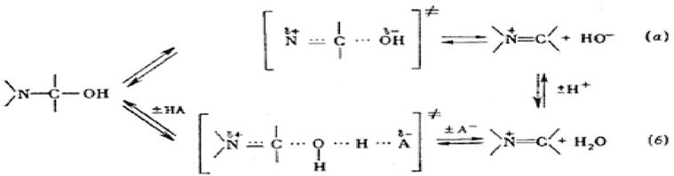
The non-catalytic pathway yields a thermodynamically unstable product. The catalytic pathway involves the active site of the enzyme, which
contains a free hydrogen proton - H+. The electrophilic active site acts as a hydrogen proton donor. In the forward reaction, a hydrogen proton is cleaved from the enzyme's active site and combines with a hydroxyl radical OH to form a water molecule. In the reverse reaction, the nucleophilic radical of the enzyme's active site is restored by the conjugate Base of the active site R2: the OH group and the hydrogen proton return to their original positions in the active site R1, and the enzyme recovers its electrophilic properties.
1.8.2 Mechanism of General Base Nucleophilic Catalysis
An example of general base catalysis is the hydrolysis reaction, which involves the addition of water to the acylimidazole substrate.
The active site of the enzyme acts as an electron acceptor. The reaction can proceed via catalytic and non-catalytic pathways.
In the non-catalytic non-enzymatic pathway, a kinetically unstable hydronium ion compound is formed.
Catalytic pathway: within the active site of the enzyme, the OH group can accept a positive hydrogen proton. In the forward direction, the active site abstracts a hydrogen proton and binds it (acting as a base).
In the reverse reaction, the active site is restored by the conjugate acid of another R-group of the enzyme, regaining its negative nucleophilic properties.
The acceleration of chemical enzymatic reactions via general base or general acid mechanisms is driven by intramolecular bonds. The reduction of entropy losses is ensured by the formation of chelate complexes, the microenvironment properties of the active site, and the proximity and orientation effect.
1.8.3 Efficiency of Polyfunctional Catalysis
The rate of a chemical enzymatic reaction increases by hundreds of thousands of times when catalysis is polyfunctional in nature. This is facilitated by three factors:
1) positive and negative R-groups can simultaneously reside in the active site;
2) the R-groups within the active site are closer together and more precisely oriented in the initial state of the reaction than in most non-enzymatic reactions;
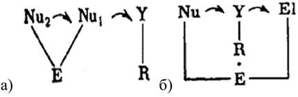
3) the dehydrated hydrophobic microenvironment of the enzyme active site enhances the interaction between the electrophilic and nucleophilic centers of the enzyme. Therefore, only two MECHANISMS OF ENZYME-substrate complex formation or two mechanisms of polyfunctional catalysis are possible for all enzymes (Figure 1.10 a, b):
Figure 1.10 - Two mechanisms of polyfunctional catalysis
Review Questions for the Topic
1. What is The Mechanism of general acid electrophilic catalysis?
2. What are the mechanisms of general base and nucleophilic catalysis?
3. What mechanisms of polyfunctional catalysis exist?
1.9 Mechanisms of Enzyme Regulation
1.9.1 Types of Regulatory Mechanisms. Definition of Metabolism. The Concept of Constitutive and Adaptive Enzymes
Metabolism encompasses the sum total of all biochemical processes within an organism. There are two primary directions of metabolism in living systems:
- anabolism — the synthesis of compounds, which requires an energy input;
- Catabolism, or Energy Metabolism — the breakdown of compounds with the release of energy, which supplies The Cell with high-energy molecules.
These processes occur concurrently and simultaneously in living systems, and the REGULATION OF METABOLISM at all Levels of organization—from the cell to the whole organism—is carried out by enzyme proteins.
Depending on how Metabolic pathways are regulated, all Enzymes can be divided into 3 major groups:
1) anabolic (regulating biosynthetic pathways);
2) catabolic (catalyzing energy metabolism reactions);
3) amphibolic (regulating both processes).
Conclusion: thus, The regulation of Metabolism in the Body ultimately comes down to the regulation of enzyme activity.
There are Two main mechanisms for regulating enzyme activity:
1) extensive (slow), implemented by regulating the rate of enzyme synthesis and proceeding slowly, as hours are required for synthesis;
2) intensive (fast) — the Regulation of the activity of already existing enzymes present in the cell.
The first, extensive mechanism. The reaction rate in the cell is determined by the amount of enzyme: the higher the Enzyme Concentration, the higher the reaction rate. An increase in the rate of enzyme synthesis regulates the reaction rate. A decrease in the reaction rate occurs through the hydrolysis of the enzyme, which reduces its amount in the cell.
The Rate of protein-enzyme synthesis depends on cellular conditions. There are protein-enzymes that are always present in the cell in a specific amount known as constitutive protein-enzymes.
Others appear under certain conditions in response to the presence of a specific substrate, known as adaptive protein-enzymes.
1.9.2 Classification of Mechanisms Regulating Enzyme Activity via the Intensive Pathway
The second, much faster intensive pathway of activation, aimed at regulating The activity of pre-existing enzymes, is represented by the following mechanisms:
1) with covalent modification:
- irreversible covalent modification — Limited proteolysis;
reversible covalent modification — regulation via covalent binding — direct action on the active site of the enzyme using an inhibitor or activator;
2) without covalent modification — without direct action on the active site of the enzyme:
- conformational (allosteric) mechanism;
- dissociative mechanism (enzyme oligomerization);
- adsorption mechanism (due to the binding of enzymes to Cellular Membrane Structures).
1.9.3 The Concept of Activators and Inhibitors
Chemical reaction rates are influenced by various substances: activators and inhibitors, collectively referred to as effectors.
Enzyme activators are substances that increase the rate of an enzymatic reaction. Metal ions such as iron, copper, cobalt, magnesium, and others most commonly act as activators. A distinction should be made between metals that are part of metalloenzymes—the so-called cofactors—and those acting as enzyme activators. Cofactors can bind tightly to the protein portion of the enzyme, whereas activators easily dissociate from the apoenzyme. Cofactors are mandatory participants in the catalytic act; in their absence, the enzyme is inactive. Activators enhance the catalytic effect, but their absence does not prevent the enzymatic reaction from occurring. As a rule, a cofactor metal interacts with negatively charged groups of the substrate. A metal with variable valence participates in electron exchange between the substrate and the enzyme.
Activator metals participate in the formation of a stable transitional conformation of the enzyme, which facilitates the more rapid formation of the enzyme-substrate complex. For example, magnesium ions stabilize enzymes of NUCLEIC ACID METABOLISM, while Calcium Ions stabilize alpha-amylase.
The Rate of Enzymatic reactions can be partially reduced or completely blocked by certain substances known as Enzyme Inhibitors. Some enzyme inhibitors serve as effective drugs for animals and humans, while others are lethal poisons.
Based on their mode of action, inhibitors are divided into irreversible and reversible.
In irreversible inhibition, the enzyme completely loses its activity.
Reversible inhibitors interact with enzymes without forming covalent bonds. Following incubation with a reversible inhibitor, enzyme activity is restored upon removal of the free inhibitor via dialysis.
The degree of inhibition characteristic of a given system is generally reached relatively quickly and remains time-independent thereafter, indicating that an equilibrium is established during the formation of the complex between the inhibitor (I) and the enzyme (E): E + I —> E I.
There are three MAIN TYPES OF reversible Enzyme Inhibition: competitive, non-competitive, and uncompetitive.
Competitive inhibition occurs when an inhibitor reversibly binds to the active site of an enzyme. Typically, Competitive Inhibitors structurally resemble the substrate; they compete for binding to the enzyme and can be displaced from the enzyme-inhibitor complex by an excess of substrate. A hallmark of competitive inhibition is that its effectiveness depends on the concentration ratio of the substrate to the inhibitor (rather than the absolute concentration of the inhibitor). Interaction with a competitive inhibitor does not cause denaturation or inactivation of the enzyme; therefore, replacing the inhibitor with the substrate does not reduce the rate of the enzymatic reaction.
The interaction between an enzyme and a competitive inhibitor alters the $K_m$ value of the corresponding enzymatic reaction.
The structural similarity between the substrate and the competitive inhibitor is sufficient for binding and the formation of the enzyme-inhibitor complex, but insufficient for the enzymatic reaction to proceed.
Many medicinal drugs inhibit Human and Animal enzymes through a competitive mechanism.
Non-competitive inhibitors interact with enzymes outside the active site rather than at it, and they cannot be displaced from the complex by any excess of substrate. Typically, they bear no structural similarity to the substrate. The effectiveness of non-competitive inhibition is determined by the inhibitor concentration and is independent of The ratio of inhibitor to substrate concentrations. When the inhibitor binds to the enzyme, it induces a conformational change followed by a partial disruption of the active site. The interaction of an enzyme with a non-competitive inhibitor alters the $V_{max}$ of the enzymatic reaction. They can reversibly bind to both the free enzyme and the ES complex without competing with the substrate, meaning they do not displace it from the enzyme-substrate complex.
Uncompetitive inhibition occurs when the inhibitor binds exclusively to the enzyme-substrate complex, thereby preventing its breakdown.
The activity of many enzymes is inhibited by an excess of substrate, and several mechanisms account for this phenomenon.
If multiple functional groups of the enzyme participate in forming the enzyme-substrate complex, two or more substrate molecules may attach to the active site simultaneously, inevitably leading to the formation of an inactive complex.
In the presence of excess substrate, it may bind not only to the active site but also to other chemical groups functionally linked to it. Such interactions can interfere with the enzymatic reaction.
1.9.4 Irreversible Covalent Modification and Limited Proteolysis
This belongs to intensive regulation mechanisms. In humans and mammals, the activity of blood coagulation enzymes is regulated via limited proteolysis. The mechanism proceeds in 3 stages:
E + S ⇄ ES* ⇄ E + R, (1.3)
S stands for substrate, ES for the enzyme-substrate complex, and R for the reaction product.
If the reaction proceeds in the forward direction yielding R, the reaction product R then binds to an inhibitor of the proteolytic enzyme. In living organisms, this is how the activity of intracellular proteolytic enzymes, such as ASAT or ALAT, is regulated.
R + Inhibitor*ALAT ⇄ R*Inhibitor*ALAT, (1.4)
where Inhibitor*ALAT represents the inactive form of the enzyme, and R*Inhibitor*ALAT is the ternary complex.
During the Second Stage of the mechanism, a ternary complex R*Inhibitor*ASAT is formed, consisting of the product, the inhibitor, and the proteolytic enzyme. This complex is active toward enzyme E (e.g., ASAT) and induces its degradation—proteolysis—into inactive fragments $E_1$ and $E_2$. Notably, the proteolytic enzyme (ASAT) is most sensitive to the action of the ternary complex:
3) E + R*Inhibitor*Proteolytic enzyme (ASAT) —> $E_1$ + $E_2$ + R + Inhibitor*Proteolytic enzyme (ASAT), (1.5)
where the Inhibitor*Proteolytic enzyme ASAT complex is the inactive form of the enzyme, and $E_1$ and $E_2$ are the inactive fragments of the ASAT enzyme resulting from proteolysis.
If the product concentration decreases, the formation of the ternary complex ceases. Consequently, the proteolytic enzyme (ASAT) stops degrading, the enzyme becomes active, and the forward biochemical reaction resumes (Stage 1).
1.9.5 Reversible Covalent Modification: Regulation by Covalent Binding
Modification of enzyme activity can involve its inhibition through the formation of covalent bonds between the enzyme and another low-molecular-weight substance within the cell, operating on THE PRINCIPLE OF reversible competitive inhibition. In eukaryotes, phosphorylation reactions represent the most widespread mechanism of enzyme regulation.
Example: Synthesis and degradation of Glycogen in Liver and muscle cells.
Glycogen —> glucose-1-phosphate ⇄ glucose
The processes of synthesis and degradation are regulated by enzymes: Glycogen phosphorylase (A and B), which catalyzes the breakdown of glycogen into glucose, and glycogen synthase (A and B), which catalyzes glycogen synthesis.
For glycogen phosphorylase (GP), cyclic AMP (cAMP) acts as a regulatory substance. Therefore, GPA is the active form of the enzyme, formed by the covalent attachment of cyclic AMP. GPB is the inactive form of glycogen phosphorylase.
If Cyclic AMP is present in the cell, GPA is activated via phosphorylation and the addition of a phosphate group, initiating the breakdown of glycogen into glucose-1-phosphate and glucose. If cAMP levels are low, GP is not phosphorylated, the enzyme becomes inactive, and the breakdown of glycogen into glucose is halted.
For glycogen synthase, glucose-6-phosphate serves as such a regulatory substance.
Principle of covalent binding: a low-molecular-weight molecule attaches to a specific site on the enzyme via a covalent bond, leading to a decrease in the enzyme's activity.
In living systems, covalent modification is also represented by adenylylation and uridylylation reactions, where nucleotides bind to enzymes
via covalent bonds and regulate the rate of Nitrogen metabolism. Enzyme activity will depend on the nitrogen content within the cell.
1.9.6 Mechanisms of ENZYME ACTIVITY REGULATION via Non-Covalent Modification
1.9.6.1 Allosteric mechanism: occurs through conformational changes in the enzyme.
The allosteric mechanism is a process by which enzyme activity is controlled through alterations in its conformation.
Conformational changes occur due to the binding of a regulatory metabolite to the allosteric site of the enzyme, which is spatially distinct from the active site.
A change in the enzyme's conformation leads to an alteration of its catalytic activity by modifying the structure of the active site, resulting in either its activation or inhibition.
The regulatory metabolite that modifies the activity of the active site is called an allosteric regulator.
An oligomeric enzyme molecule consisting of multiple subunits may contain several active and allosteric sites, each sensitive to specific effectors. In such an oligomer, two types of interaction between the sites are possible:
1. All.S —> AS1 and AS2
2. AllS1 —> AS1; AllS2 —> AS2.
The first type of interaction between the enzyme's active site and the substrate is called cooperative. The curve representing the reaction rate as a function of Substrate Concentration for allosteric enzymes regulated by a cooperative mechanism assumes a sigmoidal shape.
types:
1) concerted allosteric mechanism (developed by Changeux, Monod, and Wyman);
2) sequential allosteric mechanism (developed by Koshland).
1.9.6.2 Concerted Allosteric Mechanism.
The enzyme exists in two interconvertible states. If the enzyme consists of R subunits, it is in the R-form; if it consists of T subunits, it is in the T-form. The enzyme can exist in two forms: RR and TT.
Figure 1.11 - R and T forms of the enzyme
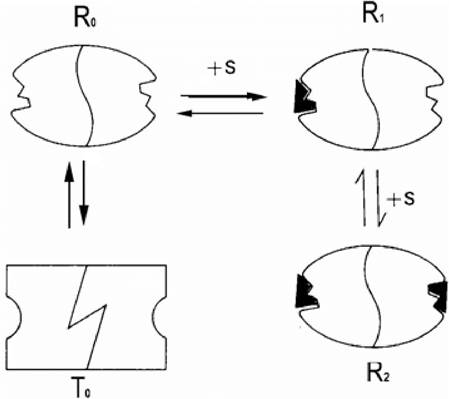
Substrate binding leads to a conformational change in one subunit. When another substrate molecule binds, the conformation of a second subunit changes, which exemplifies a concerted mechanism.
1.9.6.3 Sequential Allosteric Mechanism.
Substrate binding alters the conformation of the specific subunit it attaches to, shifting it from an inactive to an active form, while the conformation of the other subunit remains unchanged.
Conformational changes induced by the substrate in one subunit can either increase or decrease the substrate affinity of another subunit.
Allosteric interactions between an enzyme and a ligand can be of two types:
1) homotropic (interaction of enzyme subunits with identical ligands);
2) heterotropic (interaction of enzyme subunits with different ligands).
In the Concerted mechanism of allosteric interactions, homotropic effects are always positive and cooperative, whereas heterotropic effects can be either positive or negative.
Review Questions
1. What types of mechanisms regulate enzyme activity?
2. Define metabolism.
3. What are the Main Pathways of metabolism?
4. Define constitutive and adaptive enzymes.
5. Classify the mechanisms of intensive regulation of enzyme activity.
6. Describe the mechanisms of extensive regulation of enzyme activity.
7. Define effectors, activators, and inhibitors.
8. What is the mechanism of irreversible covalent modification?
9. What are the stages of limited proteolysis?
10. Which mechanisms are related to regulation by covalent binding?
1.10 Associative mechanism of enzyme activity regulation
Oligomers are enzymes composed of two or more subunits. These enzymes represent a transition into a new multimeric
structure known as the Quaternary Structure of the enzyme protein. If an oligomer consists of identical subunits, it is a homogeneous oligomer; if of different ones, a heterogeneous oligomer. The individual subunits are called protomers.
A multimeric structure containing three or more subunits is referred to as a chimera. The formation of the quaternary structure alters the Molecular Weight of the enzyme protein and, most importantly, its functional properties. In a living organism, the same enzyme can exist in various forms.
Each of these forms exhibits different enzymatic activity. Under certain conditions, the enzyme has a greater tendency to form associates (tetramers, dimers, etc.), whereas under other conditions, it exists as monomers. The activity of monomeric and oligomeric forms varies for each enzyme. For some enzymes, tetramers are active, while for others, dimers are active.
If the associative form of the enzyme is active, it means that the active site of the enzyme is formed at the contact site of the subunits. If the active site is located within a single subunit, only its monomeric form is active.
1.10.1 Mechanisms of Enzyme Association
Two types of oligomeric enzyme associations are distinguished:
1) associations resulting in closed oligomeric structures;
2) linear association of protomers of unlimited length.
1. Associations resulting in closed oligomeric structures: these are formed through the interaction of two or more identical association centers located on different subunits. The dimer has a closed structure, which is why the association centers are closed. This mechanism of association formation is referred to as isologous. An example of this formation is the enzyme glycogen phosphorylase B. In this case, the dimer is the active form, and the tetramer is inactive. Shielding of the enzyme's active site (AS), located at the center of the closed association from the substrate, takes place.
2. Linear association of protomers of unlimited length: this is formed through the interaction of two or more different association centers. An example is the enzyme lysozyme: M <=> M2 <=> M3 <=> M4... Only one form of the enzyme, which possesses a specific molecular weight, is active.
From a practical standpoint, the presence of Various Forms of oligomeric enzymes in a cell can be determined using association constants.
1.10.2 Dependence of Enzyme Activity on its Oligomeric Association
The pattern of distribution of enzymatic activity among various forms of enzyme associations indicates that the specific activity of associative enzymes depends on enzyme concentration.
The dependence can be:
1) linear
2) non-linear.
Non-linear dependence:
1. Directly proportional. The higher the concentration, the higher the specific reaction rate. This is characteristic of enzymes in which the AS is formed at the subunit contact site, i.e., the associative form is the active one.
For example, the enzyme NAD-dependent isocitrate dehydrogenase.
2. Inversely proportional. The higher the enzyme concentration, the lower the specific enzyme activity and the lower the reaction rate. Upon formation of the associative form, the AS becomes inaccessible to the substrate, meaning the oligomeric form will be inactive.
Non-linear dependence. For certain enzymes, only specific associative forms of optimal size are active.
For example, the enzyme Phosphofructokinase exists as a monomer, dimer, tetramer, etc.: M <=> M2 <=> M4, etc. Here, M2 is the only active form. Oligomerization of this enzyme proceeds via the formation of associations of unlimited length. The chimera is inactive.
1.10.3 Regulation of Oligomeric Enzyme Activity by Specific Ligands
The state of equilibrium between various forms of oligomeric enzymes can be regulated by specific ligands, which can be a substrate or an allosteric effector (activator or inhibitor):
1) it is mediated by specific allosteric ligands that act either as activators or inhibitors and bind covalently to the allosteric site of the enzyme. This leads to conformational changes in the subunit structure and, consequently, to alterations in the structure of association centers responsible for Protein-Protein Interactions.
For example, in glucuronyltransferase B, the dimer is inactive, whereas the tetramer is active;
2) the binding sites of the oligomeric enzyme coincide with the enzyme's active site, in which case, upon formation of the oligomeric form, the active site is blocked from the substrate. In a living organism, the transition from one form to another is reversible.
Review Questions on the Topic Studied
1 What are oligomeric enzymes?
2 What are the structural Features of oligomeric enzymes?
3 What is the mechanism of heterologous enzyme association?
4 How does enzyme activation depend on its oligomeric association?
5 How is the activity of oligomeric enzymes regulated by specific ligands?
6 What is the mechanism of association referred to as isologous?
1.11 Adsorption Mechanism of Enzyme Activity Regulation
1.11.1 Physiological Significance of Enzyme Adsorption on Subcellular Structures
Enzymes can reversibly bind to subcellular structures such as cell and organelle membranes (mitochondria, Lysosomes, ribosomes, inclusions, the Cytoskeleton, and myofibrils).
The Physiological Role of such binding includes:
1) reversible adsorption of an enzyme leads to changes in its catalytic and regulatory properties, i.e., it acts as a factor regulating enzyme activity;
2) adsorption of the enzyme on the membrane ensures the compartmentalization of metabolites at the membrane. This is a state in which the metabolic process proceeds in an isolated manner, without the release of intermediates (participants of the enzymatic reaction: enzyme, substrate, reaction product, effector, etc.) into the surrounding volume;
3) adsorbed enzymes can form metabolons—multienzyme structures that regulate major metabolic processes;
4) enzymes can adsorb onto membrane pores and participate in The Active Transport of metabolites across the membrane;
5) adsorbed forms of enzymes are more stable than free forms. Thus, adsorption can serve as a factor that reduces the rate of enzyme degradation within the cell.
1.11.2 Adsorption Mechanism of Regulation
Key signs of the existence of the adsorption mechanism:
1) the existence of a reversible equilibrium between the free form of the enzyme and the adsorbed enzyme;
2) changes in the catalytic characteristics of the enzyme upon adsorption;
The plot showing the dependence of the enzymatic reaction rate on substrate concentration is presented in Figure 1.18.
Figure 1.18 - Dependence of the enzymatic reaction rate on substrate concentration: 1) free form of the enzyme; 2) adsorbed form of the enzyme.
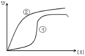
The higher the content of the enzyme In the second (adsorbed) form, the greater the increase in the enzymatic reaction rate: (1) the plot has a sigmoidal shape, which indicates the presence of a cooperative effect with respect to the substrate and low enzyme activity. As can be seen from the graph, the adsorbed form (2) is active.
3) sensitivity of the mobile equilibrium between the free and bound forms of the enzyme to the presence of cellular regulatory metabolites, also known as effectors (inhibitors and activators):
Let us consider the adsorption mechanism using 6-phosphofructokinase as an example. The enzyme exists in two forms: free and membrane-adsorbed.
human erythrocytes. An equilibrium exists between these forms. The metabolite-effector is ATP, high concentrations of which act as inhibitors. Enzyme activity changes upon adsorption onto the human membrane. The transition of the enzyme to the adsorbed state is accompanied by the disappearance of kinetic cooperativity for the substrate, fructose-6-phosphate.
For this enzyme, all three signs of an adsorption-based mechanism of enzyme activity regulation are present, namely:
1) the transition from the free form to the bound form occurs on The erythrocyte membrane;
2) the Effect of Substrate cooperativity disappears for the bound form of the enzyme, confirming the alteration of its catalytic properties;
3) in the presence of ATP, the adsorbed form of the enzyme becomes less sensitive to the inhibitor compared to the free form, i.e., it begins to exhibit its activity.
Example 2: the enzyme lactate dehydrogenase (LDH) can exist in two forms: free and bound. The substrate is NADH, an increase in the concentration of which promotes the transition of LDH into the free, active form.
1.11.3 Relay model of enzyme action
The adsorption mechanism of enzyme activity regulation ensures the compartmentalization of metabolites near the surface of subcellular structures onto which the enzyme is adsorbed.
Consider the relay model of hydrolytic enzyme action through which this mechanism operates:
S0+ E1 —> E1S’ + Е2 ⇄ E1S’E2⇄ E1 + S»E2+ Е3 ⇄ E2S»E3, (1.6)
where S0 is the initial form of the substrate; E1 is the first enzyme; Е2 is the second enzyme; ЕЗ is the third enzyme; E1S’ is the adsorbed form of the first enzyme with the substrate.
1) direct transfer of the intermediate from the active site of one enzyme to the active site of another;
2) reversible adsorption of the enzyme on subcellular structures;
a) the first enzyme E1 is released from the intermediate, adsorbs onto the surface, and transitions into an inactive form;
b) the second enzyme Е2, along with its product, is released into the bulk solution, acquiring active properties.
Review questions on the topic studied
1 What is the physiological significance of enzyme adsorption on subcellular structures?
2 What are intermediates?
3 What is metabolite compartmentalization?
4 What is the adsorption mechanism of enzymatic activity regulation?
5 What are the hallmarks of the adsorption mechanism of enzyme activity regulation?
6 Provide examples of such regulation.
7 What is the relay model of enzyme action?
8 How is this mechanism implemented?
9 Provide examples of the relay model.
Last update: 06/08/2026
Editorial and Educational Adaptation: This material has been compiled based on the primary/original source text. The project team performed an editorial review, corrected technical inaccuracies, structured sections, and adapted the content for an educational format.
What was processed:
- elimination of formatting defects (OCR errors, structural breaks, corrupted characters);
- editorial organization of content;
- standardization of terminology in accordance with academic sources;
- verification of factual statements against the original source text.
All mentions of the author, publication year, and origin of the primary text have been preserved in accordance with the source.