Principles of Biochemistry, Volume 1 - A. Lehninger 1985
Biomolecules
Globular Proteins: Structure and Function of Hemoglobin
Forces Stabilizing the Tertiary Structure of Globular Proteins
We already know how the Introduction/11.html">Secondary Structure of Polypeptides is formed and maintained. Intrachain Hydrogen Bonds hold the α-Helix together, while interchain hydrogen bonds stabilize the β-conformation of the chains forming pleated sheets (Sec. 7.11). Given the appropriate Amino Acid Sequence, the α-helix and β-conformation are the most stable structures that a given region of the chain can adopt; in other words, they represent states of lowest Free energy. What forces stabilize the tertiary Structure of Globular Proteins? There are four types of interactions whose combination ensures the correct spatial arrangement of all coils and loops in Globular proteins (Fig. 8-9) under normal biological conditions (Temperature, pH, ionic concentration).
1. Hydrogen bonds between the R-groups of residues located in adjacent loops of the polypeptide chain. For example, the hydroxyl group of a Serine residue in one segment of the polypeptide chain can form a Hydrogen bond with a nitrogen atom in the ring of a Histidine residue located in an adjacent loop of the same chain.
Class="center">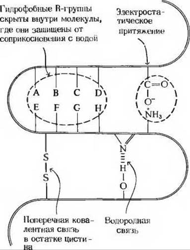
Fig. 8-9. Forces stabilizing the Tertiary Structure of globular proteins.
2. Electrostatic attraction between oppositely charged R-groups. For example, the negatively charged carboxyl group (—СОО—) of a glutamic acid residue can attract the positively charged ε-amino group (—NH3) of a Lysine residue located in an adjacent loop.
3. Hydrophobic interactions. The hydrophobic R-groups of Certain amino acid residues (see Table 8-1) avoid contact with the aqueous environment and tend to cluster together inside the globular structure, where they are shielded from contact with Water.
4. Covalent cross-links. Adjacent loops of the polypeptide chain in some proteins, such as Ribonuclease, contain cystine residues that form intrachain covalent cross-links between adjacent loops. Such covalent cross-links are, of course, much stronger than the non-covalent interactions listed above, but they do not occur in all proteins. Consequently, proteins that lack disulfide cross-links maintain their native tertiary structure through a multitude of weak non-covalent interactions and various contacts, which collectively provide the structure with sufficient stability.
Although the native tertiary structure of each globular protein corresponds to a minimum of Free Energy and is therefore the most stable conformation that a given polypeptide chain can adopt, the tertiary structure of globular proteins should not be considered completely rigid and static. Many globular proteins normally undergo conformational changes while performing their biological Functions. For example, the Hemoglobin molecule, which we will discuss later, changes its conformation upon binding oxygen and returns to its original conformation after releasing it. In addition, the molecules of many Enzymes undergo conformational changes upon binding substrates—this is part of their catalytic action. The polypeptide backbone of globular proteins is characterized by a certain degree of flexibility, making these proteins subject to local internal fluctuations, meaning they undergo a kind of "breathing".
Last update: 06/08/2026
Editorial and Educational Adaptation: This material has been compiled based on the primary/original source text. The project team performed an editorial review, corrected technical inaccuracies, structured sections, and adapted the content for an educational format.
What was processed:
- elimination of formatting defects (OCR errors, structural breaks, corrupted characters);
- editorial organization of content;
- standardization of terminology in accordance with academic sources;
- verification of factual statements against the original source text.
All mentions of the author, publication year, and origin of the primary text have been preserved in accordance with the source.