Fundamentals of Biochemistry - Filippovich, Y. B. 1999
Enzymes
Classification of enzymes and characteristics of certain groups
According to the earliest Classification in The history of enzymology, Enzymes were divided into two main groups: Hydrolases, which accelerate hydrolytic reactions, and desmolases, which catalyze non-hydrolytic Cleavage reactions. Later, an attempt was made to classify enzymes based on the number of substrates involved in the reaction. Accordingly, enzymes were categorized into three groups: 1. Those catalyzing The conversion of two substrates simultaneously in both directions: A + B ⇄ C + D. 2. Those accelerating the conversion of two substrates in the forward reaction and one in the reverse reaction: A + B ⇄ C. 3. Those facilitating the catalytic modification of a single substrate in both forward and reverse reactions: A ⇄ B.
Simultaneously, another approach was developing, which based Enzyme Classification on the type of reaction undergoing catalytic transformation. Along with enzymes that accelerate Hydrolysis reactions (hydrolases), researchers studied enzymes involved in The transfer of atoms and atomic groups (pherases), isomerization (isomerases), cleavage (liases/lyases), various syntheses (synthetases), and so on. This approach proved to be the most fruitful because it grouped enzymes not by artificial or formal criteria, but according to the type of fundamental biochemical processes underlying the vital activity of any living Organism. Based on this principle, all enzymes are divided into 6 classes.
1. Oxidoreductases — catalyze oxidation-reduction reactions. 2. Transferases — catalyze the transfer of functional groups and molecular residues. 3. Hydrolases — catalyze hydrolytic cleavage reactions. 4. Lyases — catalyze the non-hydrolytic removal of specific groups of atoms from substrates with The formation of a double bond (or add groups of atoms across a double bond). 5. Isomerases — catalyze spatial or structural rearrangements within a single molecule. 6. Ligases — catalyze synthetic reactions coupled with The breakdown of energy-rich bonds. These classes form The basis of the modern scientific Classification of Enzymes.
1. Oxidoreductases. The Class of oxidoreductases includes enzymes that catalyze oxidation-reduction reactions. Their general reaction scheme can be represented as follows:

Oxidation proceeds as the removal of H atoms (electrons) from a substrate, whereas reduction proceeds as The addition of H atoms (electrons) to an acceptor. If we denote the acceptor as A and the substrate as B, the equation for an oxidation-reduction reaction involving oxidoreductases takes the following form:

A characteristic feature of oxidoreductase activity in living Cells is their ability to form systems (so-called chains of redox enzymes) that carry out multi-step transfer of hydrogen atoms or electrons from the primary substrate to the final acceptor—typically oxygen—resulting in the formation of Water.
Those oxidoreductases that transfer H atoms or electrons directly to oxygen atoms are called aerobic dehydrogenases or oxidases. In contrast, oxidoreductases that transfer H atoms and electrons from one component of the Electron Transport Chain to another without passing them to oxygen atoms are called anaerobic dehydrogenases or reductases.
If an enzyme catalyzes the removal of H directly from the substance being oxidized (the primary substrate), it is called a primary dehydrogenase. If the enzyme accelerates the removal of hydrogen atoms from a secondary substrate that received H atoms via a primary dehydrogenase (the secondary substrate may be the coenzyme of the primary oxidoreductase itself), it is called a secondary dehydrogenase (see Chapter X).
Another characteristic feature of oxidoreductases is that, being two-component enzymes with a rather limited set of active groups (Coenzymes), they are capable of accelerating A wide variety of redox reactions. This is achieved because the same coenzyme can bind to many different apoenzymes, each time forming an oxidoreductase specific to a given substrate or acceptor.
Yet another, perhaps primary, feature of oxidoreductases is that they catalyze chemical processes associated with the release of energy. This energy is utilized both to drive synthetic processes within the organism and for other cellular needs.
About five hundred individual oxidoreductases have been discovered in natural sources. The most widespread are oxidoreductases containing nicotinamide adenine dinucleotide, or NAD+, as their active group (on the Structure of NUCLEOTIDES, see Chapter VI):

More than half of all currently known oxidoreductases contain NAD+ as a coenzyme. By combining with a specific protein and thus forming a two-component enzyme—briefly referred to as a pyridine protein—NAD+ drastically enhances its ability to undergo reduction at the nicotinamide ring. As a result, pyridine Proteins are able to Abstract H atoms in the form of hydride ions (H-) and protons (H+) from substrates (such as alcohols, aldehydes, dicarboxylic and keto acids, amines, etc.), thereby oxidizing these compounds. All pyridine proteins function as anaerobic dehydrogenases, meaning they do not transfer the hydrogen atoms removed from the substrate to oxygen, but pass them on to the next enzyme in the Respiratory Chain.
Let us examine the structure and MECHANISM OF ACTION of one such pyridine protein: Alcohol dehydrogenase from animal Liver. It is a protein with M = 73,000, consisting of two subunits, each carrying an NAD+ molecule and a Zn atom. During the abstraction of H atoms from an alcohol, a ternary apoenzyme-coenzyme-substrate complex is formed, held together by Zn2+. The structure of this complex and The Mechanism of alcohol oxidation to an aldehyde are illustrated in Fig. 53. One hydrogen atom is transferred directly from the alcohol molecule to nicotinamide adenine dinucleotide in the form of a hydride ion (H-)—i.e., a hydrogen atom carrying an extra electron. Conversely, the second hydrogen atom removed from the alcohol molecule loses an electron, turning into a proton (H+), and is released into the reaction medium. Therefore, the equation for The oxidation of alcohol in the presence of NAD+ is written as follows:


Fig. 53. Mechanism of action of alcohol dehydrogenase
NAD+ is held On the surface of the protein molecule by bonds formed between the positively charged nitrogen atom of the pyridine ring and the negatively charged sulfur atom (from the HS group), as well as between the nitrogen atoms of the purine ring and the sulfur atom mediated by Zn2+. The alcohol molecule binds to the Active Site of the enzyme via a coordination bond between the oxygen atom and Zn2+. The catalytic function in transferring hydrogen atoms from the alcohol molecule to NAD+ is carried out by the imidazole radical of Histidine. A hydrogen atom, previously bonded to the carbon atom bearing the alcohol group, adds to the pyridine ring of NAD+. The hydrogen atom of the alcohol group becomes protonated (I). The binding of NAD+ to the apoenzyme occurs via the nucleotide-binding domain (II), which is composed of α-helices and β-sheets. The STRUCTURE OF THE nucleotide-binding domain is similar in all NAD-dependent dehydrogenases; the AMP moiety attaches to the lower part of the domain (β-strands A, B, and C), while the nicotinamide ribose phosphate moiety of the NAD+ molecule binds to the upper part (β-strands D, E, and F).
In any case, NAD+ receives two electrons through the addition of a hydride ion (H-).
In addition to NAD+, pyridine enzymes contain nicotinamide adenine dinucleotide phosphate (NADP+) as a coenzyme. This coenzyme is a derivative of NAD+ in which the hydrogen of the 2'-OH group of the adenosine ribose is replaced by a phosphoric acid residue.
NADP+, upon combining with specific proteins, forms a large group of pyridine proteins characterized by their own distinct substrate Specificity. The mechanism of oxidation involving NADP+ as a coenzyme is analogous to that mediated by NAD+. Furthermore, NADH and NADP+, as well as NADPH and NAD+, can exchange hydrogen atoms and electrons via the catalytic action of a specific enzyme, transhydrogenase:

The partners of the reduced forms of pyridine proteins in the oxidoreductase chain are typically flavoproteins (FP). One such flavoprotein, for instance, is an enzyme that carries phosphorylated vitamin B2 as its active group. The oxidized form of this flavoprotein (M = 52,000) is colored. Each enzyme molecule carries a molecule of riboflavin phosphate (or flavin mononucleotide, FMN), which is capable of accepting and donating two H atoms at the nitrogen atoms of the isoalloxazine ring:

The second coenzyme found in flavoproteins is flavin adenine dinucleotide (FAD):

FMN and FAD combine with various apoenzymes to give rise to approximately thirty flavoproteins, which differ in their substrate specificity.
The primary function of flavoproteins is the transfer of electrons (H atoms) from reduced pyridine proteins to Other components of the redox chain, meaning that in most cases, FPs act as secondary dehydrogenases. However, certain flavoproteins, particularly those containing FAD as a coenzyme, are capable of directly removing an H atom from the substrate.
Quinones also serve as coenzymes for oxidoreductases. For instance, by binding with proteins, ubiquinones form ubiquinone-Protein Complexes, which are a vital component of oxidoreductase assemblies that mediate the transfer of H atoms and electrons.
Ubiquinones are derivatives of benzoquinone and feature a side chain composed of A large number of isoprenoid residues:

The number of isoprenoid fragments in the side chain (n) ranges from 6 to 10. It has been established that ubiquinones participate in the body's redox processes by facilitating the transfer of H atoms:

In plants, this function is carried out by plastoquinone, a compound structurally similar to ubiquinone:

The most complex yet widespread type of cellular redox process involves the oxidation of H atoms—removed from the substrate—via the cytochrome system.
The cytochrome system consists of several oxidoreductases that contain iron-Porphyrins as their prosthetic groups (Fig. 54). As early as 1915, two decades before O. Warburg, A. Ya. Danilevsky and B. P. Solovtsov drew attention to the potential role of iron-containing proteins in Biological Oxidation. By binding with various structural proteins, four types of iron-porphyrins (A, B, C, and D) give rise to a family of Chromoproteins collectively known as Cytochromes. Dozens of cytochromes are currently known, and the list continues to grow. Each individual cytochrome is designated by a lowercase Latin letter (a, b, c, and d) with a corresponding subscript index, such as b1, b2, b3, etc., while the cytochrome class is indicated by an uppercase Latin letter (A, B, C, or D). A cytochrome's class is determined by the structure of its prosthetic group (iron-porphyrin), whereas its specific identity is defined by the structure of the apoenzyme (protein). Recently, along with the numerical index, it has become preferred to indicate the characteristic wavelength at which absorption occurs in the visible spectrum (e.g., cytochrome b6 or b563, found in METABOLISM/14.html">Chloroplasts, etc.).
The Introduction/19.html">Primary Structure of several cytochromes has been elucidated, revealing that their species specificity is linked to minor variations in Amino acid sequences. These findings are fundamentally important for understanding The Nature of species and Other types of Enzyme Specificity: it appears to be determined primarily by differences in the primary structure of the apoenzymes. Cytochromes b1, b2, b3, etc., which contain the exact same prosthetic group, differ from one another precisely due to this characteristic.

Fig. 54. Structure of cytochrome c from horse Heart Muscle
The prosthetic group of cytochrome c is represented by an iron-porphyrin (with an Fe2+ ion at the center). Its attachment to the protein occurs through the interaction of the heme vinyl radicals with the HS groups of the 14th and 17th Cysteine residues of the polypeptide chain. The amino group of the N-terminal Glycine is acetylated. In the three-dimensional model of cytochrome c, numbers indicate the positions of The amino acid residues, each of which is also represented by its Spatial Structure
Figure 54 illustrates the structure of cytochrome c from horse heart. The Molecular Weight of cytochrome c is relatively small, around 13,000. Under a 1,300,000-fold magnification (via Electron Cell/15.html">Microscopy), it can be seen that the polypeptide chain of cytochrome c is folded into an α-Helix 12–15 nm in length, which is further coiled into a secondary helix with a diameter of 4–5 nm, effectively isolating the heme group inside the surrounding polypeptide chain. These ring-like molecules readily form larger aggregates. The structures of other cytochromes have been studied in less detail; however, it is known that the molecular weights of some are significantly higher. The ability to aggregate with one another appears to be an intrinsic property of cytochrome molecules.
This is precisely why cytochromes form the cytochrome system—an ordered association of various cytochromes, such as b, c, and a, within a single complex.
The cytochrome system is capable of accepting electrons stripped from the H atoms of reduced ubiquinone (UQ). It then transfers these electrons down the cytochrome chain and ultimately to an oxygen molecule; the latter, upon combining with ionized H atoms, forms an H2O molecule.
Within the cytochrome chain, each individual cytochrome occupies a strictly defined position. The simplest configuration of the cytochrome system is shown in the following diagram:

As seen from the diagram, electron transfer along the cytochrome chain is driven by Changes in the valence state of the iron atom within the porphyrin ring. Among all the cytochromes, only Cytochromes c and a3 transfer electrons to oxygen. Therefore, they complete the cytochrome chain and are referred to as cytochrome oxidase. In addition to iron atoms (as part of the heme group), cytochromes a and a3 also contain copper (Cu) atoms, which are associated with their oxidative properties (see Ch. X, Fig. 132).
Such are the Characteristic Features of the Action of Certain key oxidoreductases and redox systems that drive various metabolic transformations within The Cell.
2. Transferases. This class includes enzymes that accelerate the transfer reactions of functional groups and molecular residues from one compound to another. It is one of the most extensive classes, comprising about 500 individual enzymes. Depending on the Nature of the transferred groups, a distinction is made between phosphotransferases, aminotransferases, Glycosyltransferases, Acyltransferases, transferases transferring single-carbon residues (methyltransferases, formyltransferases), and others.
Phosphotransferases. These include enzymes that accelerate the transfer reaction of a phosphoric acid residue. This reaction is of exceptional importance for the vital activity of the organism, ensuring the conversion of A number of Organic compounds into phosphoric esters, which possess increased chemical reactivity and enter subsequent reactions more easily. The Transfer of phosphate groups proceeds to alcoholic, carboxyl, nitrogen-containing, phosphorus-containing, and other groups of various organic compounds. Accordingly, several subclasses are distinguished among phosphotransferases.
In most cases, adenosine triphosphate (ATP) serves as the donor of phosphate residues, although other sources are also possible. Phosphotransferases include, for example, hexokinase, an enzyme that accelerates the transfer of a phosphoric acid residue from an ATP molecule to glucose (glucose transformation usually begins with this reaction):

Hexokinase is ubiquitous. Yeast hexokinase has been studied particularly well. Its molecule (M = 96,000) is composed of 4 subunits. The multimer is stable at pH 5. Upon a decrease or increase in the pH of the solution, the hexokinase molecule dissociates into 4 protomers (M = 24,000) devoid of phosphotransferase activity. In accordance with the dual nature of the subunits in the multimers, 5 hexokinase isozymes have been discovered.
The transformation of many other Monosaccharides also begins with their phosphorylation mediated by phosphotransferases: this is the case with ß-D-fructose, ß-D-ribose (see p. 338), and a number of other sugars.
Special attention has recently been paid to The Study of phosphotransferases that ensure the transfer of a phosphate residue from ATP to proteins—namely, protein Kinases. They transfer phosphate to the ser, thr, tyr, lys, and his residues of a number of proteins, resulting in a dramatic change in the biological activity of the latter. This, in turn, affects the intensity of chemical processes in the organism, i.e., the REGULATION OF METABOLISM (see Ch. XIII).
Aminotransferases. These enzymes accelerate the Transamination reaction of Amino Acids with keto acids and are very important for ensuring the Biosynthesis OF AMINO Acids. Aminotransferases are two-component systems: their prosthetic group in all cases is Pyridoxal phosphate, covalently attached to the apoenzyme through its aldehyde group (see Fig. 55, B) and by an ionic bond through the phosphoric acid residue:

The mechanism of the transamination reaction is now well elucidated, and the reaction itself was discovered back in 1937 by A. E. Braunshtein and M. G. Kritsman.
For convenience, we will designate the pyridoxal enzyme using the following structure:  retaining only the functionally significant aldehyde group of its coenzyme.
At The First stage of Enzymatic Catalysis, the prosthetic group of the enzyme (for simplicity, it is assumed to be free rather than connected by an aldimine bond to the apoenzyme via the ε-amino group of the lys residue) interacts with the amino acid undergoing transamination. The reaction proceeds via the amino group of the Amino Acid and the aldehyde group of the pyridoxal phosphate residue:
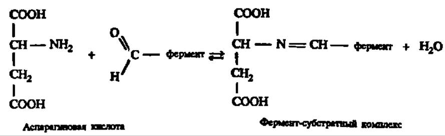
At the Second Stage of catalysis, the transformation of the substrate takes place, which in this case consists of a tautomeric rearrangement:

This rearrangement is carried out with the participation of imidazole-containing residues of his, which are part of the catalytic center of the enzyme:

As a result of subsequent hydrolysis, the keto acid and the enzyme in the form of the pyridoxamine enzyme are released:

Next, an enzyme-substrate complex is formed again between the pyridoxamine enzyme and another keto acid:

The substrate within it undergoes transformation once more due to a tautomeric conversion:
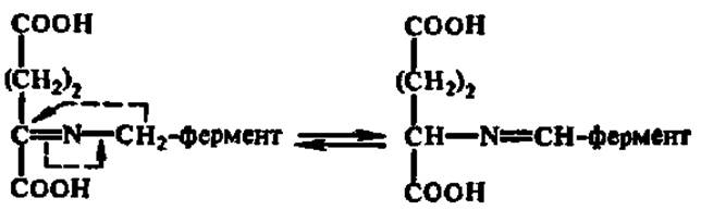
The resulting compound is hydrolyzed, and a new amino acid is formed:

Consequently, as a result of a series of Reactions Involving the alternating formation of enzyme-substrate complexes, aspartic acid is converted into oxaloacetic acid, while α-ketoglutaric acid is converted into glutamic acid. This can be represented by the following overall equation:
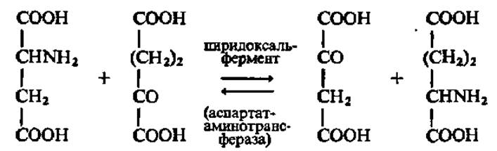
A central role in pyridoxal catalysis is played by the shift of electron density within the enzyme-substrate complex:

As a result, the bonds between the α-carbon atom of the amino acid residue and its substituents (nitrogen, the COOH group, etc.) are weakened, facilitating the Cleavage of the respective bonds.
Aspartate aminotransferase has a molecular weight of 93,000 and consists of two identical subunits (M = 46,500), each bound to a pyridoxal phosphate molecule. Upon dilution of aspartate aminotransferase solutions, its dimers dissociate into catalytically active monomers. Thanks to research conducted primarily by Soviet scientists (A. E. Braunstein et al., Yu. A. Ovchinnikov et al., and B. K. Vainshtein et al.), the primary and tertiary structures of this enzyme, as well as the structure and detailed functioning mechanism of its active center, have been elucidated. The subunit of the cytosolic isoenzyme of aspartate aminotransferase from pig heart (the other isoenzyme is localized in the Mitochondria) is represented by a polypeptide chain of 412 amino acid residues. A significant portion of it adopts an α-helical conformation, while the coenzyme-binding domain—where the active center is located (Fig. 55)—resides in a distinct region of the globule. Notably, the coenzyme-binding domain of pyridoxal enzymes bears a striking resemblance to the nucleotide-binding domain of NAD+- and NADP+-dependent dehydrogenases (see Fig. 53, II), which is presumably due to the presence of a pyridine ring in both coenzymes.
Since aspartate aminotransferase consists of two subunits and consequently bears two pyridoxal phosphate residues, the subunits function in a coordinated manner during the transamination reaction, with a phase shift in the utilization of the energy required to drive chemical transformations. As a result, the dimeric structure of the enzyme provides a significant advantage in executing the catalytic process.

Fig. 55. One of the possible tertiary structures of aspartate aminotransferase (A) and the structure of its active center (B)
As can be seen in Fig. B, pyridoxal phosphate is linked via an aldimine bond to the ε-amino group of a Lysine residue within the apoenzyme; it is precisely at this aldimine bond that the amino acid's amino group attaches, displacing the lysine ε-amino group
Glycosyltransferases. These enzymes accelerate the transfer reactions of glycosyl residues from phosphoric ester molecules or Other Compounds to molecules of monosaccharides, Polysaccharides, or other substances, playing a key role in the Synthesis and degradation of oligo- and polysaccharides in both the animal and plant kingdoms. Below is the equation for the breakdown of sucrose facilitated by sucrose: orthophosphate α-glucosyltransferase, also known as sucrose phosphorylase:

Starch phosphorylase, Glycogen phosphorylase, and other glycosyltransferases act in a similar manner. When glycosyl residues are transferred to H3PO4, this process is referred to as phosphorolysis, because it is formally analogous to hydrolysis, except that hydrogen and the phosphate group of phosphoric acid attach at the site of the oxygen bridge cleavage instead of water elements (for more details on glycogen phosphorylase and its mechanism of action, see Chapter VIII).
It has recently been established that the transfer of glycosyl residues is particularly readily accomplished by enzymes of this group when a nucleoside diphosphate monosaccharide serves as the substrate. This reaction apparently represents the primary pathway for the natural synthesis of oligo- and polysaccharides and will be discussed in detail in Chapter VIII. Nucleoside diphosphate sugars function as coenzymes for glycosyltransferases.
Acyltransferases. These enzymes accelerate the transfer of acyl groups (carboxylic acid residues) to amino acids, amines, alcohols, and other compounds. The universal source of acyl groups in all these reactions is acyl-coenzyme A, which can rightfully be regarded as the active group of acyltransferases.
The acyl group most frequently transferred in biological systems is the acetic acid acyl radical—acetyl 
Coenzyme A (see formula on p. 163), by binding to the acetyl residue—which takes THE PLACE OF hydrogen in its HS group—forms acetyl-coenzyme A. The latter serves as a cofactor in the corresponding transfer reaction. One example of a transacylation reaction is the synthesis of acetylcholine:
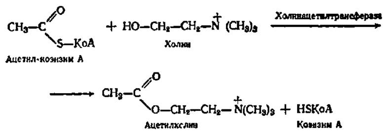
Of major importance among transferases are enzymes that accelerate the transfer of single-carbon fragments (methyl, oxymethyl, formyl, etc.), as well as nucleotidyltransferases, which catalyze the transfer of nucleotide residues during nucleic acid synthesis. Their mechanism of action will be described below.
3. Hydrolases. The class of hydrolases comprises enzymes that accelerate the cleavage (and occasionally the synthesis) of organic compounds involving water: R'R"+HOH ⇄ R'H+R"OH. Depending on the nature of the substrate undergoing hydrolysis, hydrolases are divided into a number of subclasses, of which the most important are the following:
1) esterases, which accelerate the hydrolysis of esters;
2) glycosidases, which accelerate the hydrolysis of Glycosides, including CARBOHYDRATES;
3) peptidyl hydrolases, which accelerate the hydrolysis (and in special cases, the synthesis) of proteins, Peptides, and other compounds containing peptide bonds;
4) hydrolases acting on C—N bonds other than peptide bonds (e.g., amidases, etc.). In total, there are nearly 500 different enzymes classified as hydrolases.
Esterases. These enzymes catalyze the hydrolysis of esters formed by alcohols with organic and inorganic acids. The most important sub-subclasses of esterases are carboxylic ester hydrolases and Phosphatases. Lipase will be considered as a representative of the first sub-subclass.
Lipases accelerate the hydrolysis of outer, i.e., α-ester bonds in triacylglycerol (fat) molecules:

The mechanism of action of several esterases has been studied in detail. One example is discussed in this chapter (see the section on the Mechanism of enzyme Action). The characteristics of lipases are given in Chapter IX.
Phosphatases catalyze the hydrolysis of phosphate esters. Phosphatases acting on sugar phosphates, such as glucose-1-phosphatase, are particularly widespread:

Phosphatases function across a broad pH range from 3 to 9, and most of them exhibit broad substrate specificity. Protein phosphatases are especially important for Metabolic Regulation; they remove phosphate groups from phosphorylated proteins, thereby altering their biological activity, notably their enzymatic activity.
Glycosidases. These enzymes accelerate the hydrolysis of glycosides. Depending on which spatial isomer (α or β) the enzyme acts upon, it is classified as either an α- or β-glycosidase. Thus, glycosidases display pronounced stereospecificity. In addition to glycosides containing monohydric alcohol residues as aglycones, the substrates for various glycosidases include oligo- and polysaccharides. Among the glycosidases acting on Oligosaccharides are maltase (α-glycosidase) and sucrase (β-glycosidase), which accelerate the hydrolysis of maltose and sucrose, respectively:

Among the glycosidases that act on polysaccharides, amylases are the best known. Several types of amylases occur in nature, accelerating the hydrolysis of glycosidic bonds in starch to yield glucose, maltose, or oligosaccharides. Their characteristics and mechanism of action are discussed in Chapter VIII.
The hydrolysis of other natural polyglycosides, such as Cellulose, inulin, and Xylan, is likewise accelerated by the corresponding glycosidases. Furthermore, certain glycosidases catalyze the transfer of glycosyl residues and thus function as transglycosidases.
Peptide hydrolases. Enzymes of this subclass accelerate the hydrolysis of peptide bonds in PROTEINS AND PEPTIDES and, under certain conditions, can also promote peptide bond formation, although this is not the physiological pathway of Protein Synthesis. The chemistry of protein and peptide hydrolysis mediated by peptide hydrolases can be represented by the following scheme:

Peptide hydrolases are subdivided into proteinases, or peptidyl amino acid hydrolases, which catalyze the hydrolysis of a small number of internal peptide bonds within a protein molecule, resulting in its breakdown into smaller peptides. Consequently, they act as Endopeptidases. In contrast, peptide hydrolases known as peptidases function as exopeptidases, cleaving free amino acids from the peptide chain.
Depending on their mechanism of action on internal peptide bonds within a protein molecule, proteinases are divided into four subclasses: 1) Serine proteinases, which feature serine and histidine residues in their active site that drive the catalytic act—Examples include Chymotrypsin and Trypsin secreted by the Pancreas, and subtilisin produced by Bacteria; 2) thiol (cysteine) proteinases, containing a cysteine residue in the active site, which include Papain from the latex of the papaya tree Carica papaya, ficin from fig latex, bromelain from pineapple stem juice, and cathepsin B, an intracellular enzyme found in vertebrates; 3) acidic (carboxyl) proteinases, which have an optimal pH below 5 and contain dicarboxylic amino acid residues in their active site—this group encompasses Pepsin secreted by the gastric mucosa, intracellular cathepsin D, and a number of acidic proteinases produced by various microorganisms; and 4) metalloproteinases, whose catalytic activity depends on the presence of Metal Ions (Ca2+, Zn2+) in the active site, with examples including collagenase and several proteinases of microbial origin (such as Thermolysin and the pronase component).
Pepsin, trypsin, and chymotrypsin are secreted by glandular cells as inactive proenzymes, or zymogens—pepsinogen, trypsinogen, and chymotrypsinogen, respectively—because their active sites are blocked by polypeptide chain fragments. Once these fragments are hydrolytically cleaved, the enzyme acquires activity. This phenomenon was first discovered in the laboratory of I. P. Pavlov.
A crucial feature of proteinases is the selective nature of their action on peptide bonds within a protein molecule. For instance, pepsin selectively accelerates the hydrolysis of peptide bonds formed by phe and leu; trypsin acts on arg and lys; chymotrypsin on aromatic amino acids; and papain on arg, lys, and phe, among others. Consequently, an individual protein is always cleaved by a specific peptidyl peptide hydrolase into a strictly limited number of peptides. This property is practically applied in determining the Primary Structure of Proteins and plays a vital role in metabolic regulation. Many products of selective Protein Hydrolysis exhibit exceptionally high biological activity: this is precisely how enzymes are generated from proenzymes, and Hormones and releasing factors from their precursors. The selectivity of peptide peptide hydrolases is due to the fact that the amino acid residue adjacent to the cleaved peptide bond is involved in the Formation of the enzyme-substrate complex.
Peptide hydrolases that catalyze the hydrolysis of peptides into free Amino acids can cleave the latter starting from either the amino acid with a free NH2 group or the one with a free COOH group. In the first case, they are referred to as aminopeptidases (alpha-aminoacylpeptide hydrolases), and In the second, as Carboxypeptidases (peptidylamino acid hydrolases).
A diagram illustrating the action of amino- and carboxypeptidases, as well as certain endopeptidases, is shown in Fig. 56.
Some amino- and carboxypeptidases exhibit high specificity, meaning they cleave strictly defined N- or C-terminal amino acids. The third subclass of peptidases is represented by dipeptide hydrolases, or dipeptidases, of which about ten are known. They complete protein hydrolysis. Recently, two additional subclasses have been distinguished among peptidases: dipeptidylpeptide hydrolases, which cleave a dipeptide from the N-terminus of a polypeptide, and peptidyldipeptidase hydrolases, which cleave a dipeptide from the C-terminus.
Amidases. These enzymes accelerate the hydrolysis of acid amides. Among them, urease, asparaginase, and glutaminase play a pivotal role in the biochemical processes of the organism.

Fig. 56. Sites of action of Proteolytic Enzymes on peptide bonds within a protein molecule
Urease was one of the first enzyme proteins to be obtained in crystalline form (J. Sumner, 1926). It is a single-component enzyme (M = 480,000) with a globular molecule composed of 8 identical subunits. Urease accelerates the hydrolysis of urea into NH3 and CO2.
Asparaginase and glutaminase accelerate the hydrolysis of amides of dicarboxylic amino acids, namely aspartic and glutamic acids, for example:

Hydrolases acting on C—N bonds other than peptide bonds include, alongside amidases, enzymes that catalyze the hydrolysis of C—N bonds in linear amidines. Arginase is a prime example of this group. Mediated by arginase, the amino acid Arginine is hydrolyzed into Ornithine and urea:
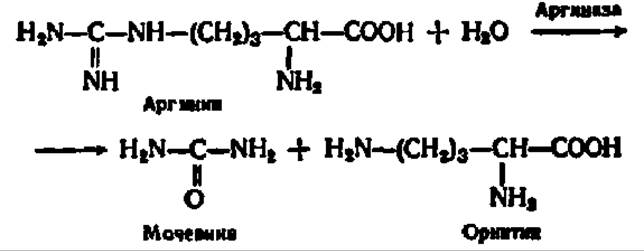
Since urea is released during the hydrolysis of arginine, the systematic name for arginase is L-arginine ureohydrolase. This reaction is widespread in nature, serving as The final stage of urea biosynthesis—one of the End products of nitrogenous compound degradation. Like urease, arginase exhibits absolute reaction specificity.
4. Lyases. The class of lyases comprises enzymes that accelerate non-hydrolytic cleavage reactions of organic compounds across C—C, C—N, and C—O bonds, among others. These processes involve the formation of double bonds and the release of simple end products such as CO2, H2O, NH3, and the like. Some of these reactions are reversible, meaning that under appropriate conditions, the corresponding enzymes catalyze not only degradation but also synthesis. Consequently, the name of this enzyme class does not always reflect the full scope of the processes they accelerate.
One of the most important groups of enzymes in this class is the carbon-carbon lyases (C—C lyases). Among them, carboxylyases (Decarboxylases) and aldehyde lyases are of particular significance.
Decarboxylases of keto acids and Amino acids are widespread in nature, catalyzing reactions according to the following schemes:

These enzymes are two-component systems; in many cases, their prosthetic groups are phosphoric esters of Water-Soluble Vitamins: thiamine (B1) in keto acid carboxylyases and pyridoxal (B6) in amino acid carboxylyases.
The mechanism of keto acid decarboxylation mediated by decarboxylases with thiamine pyrophosphate as a coenzyme will be discussed in Chapter IV. The mechanism of AMINO ACID DECARBOXYLATION by carboxylyases using Pyridoxal phosphate as a coenzyme is closely analogous to that operating during AMINO ACID TRANSAMINATION. Here, a Schiff base is also formed, in which the electron density at the a-carbon atom of the amino acid is sharply attenuated. As a result, the bond between this carbon atom and the carboxyl group is significantly weakened, facilitating the ready cleavage of the latter.
A characteristic representative of aldehyde lyases is aldolase, which catalyzes the reversible cleavage of fructose-1,6-diphosphate into phosphotrioses:

This reaction holds a central place in Carbohydrate Metabolism. Other aldehyde lyases function in a similar manner.
Another important group of lyases comprises carbon-oxygen lyases (hydro-lyases), which accelerate the Hydration and dehydration reactions of organic compounds. As an example of hydro-lyases, we can cite fumarate hydratase:

Hydration and dehydration reactions occur continuously during the breakdown and synthesis of carbohydrates and Higher Fatty acids; therefore, hydratases play a major role in the vital activity of organisms.
An example of a carbon-nitrogen lyase is aspartate ammonia-lyase, which accelerates the direct deamination of aspartic acid:

This enzyme is characteristic of bacteria and a number of plants.
Certain lyases accelerate not only degradation but also synthesis reactions. For instance, L-serine hydro-lyase has been isolated from yeast; it cleaves water from serine and adds hydrogen sulfide, thereby synthesizing the amino acid cysteine:

To distinguish such lyases from Enzymes of the ligase class (which accelerate exclusively synthetic reactions and are termed synthetases accordingly), they are also referred to as synthases.
5. Isomerases. The enzymes belonging to this rather small class (comprising about 90 individual enzymes) accelerate geometric or structural rearrangements within a single molecule. These alterations may involve the intramolecular transfer of hydrogen, phosphate, or acyl groups, changes in the spatial arrangement of atomic groupings, or the relocation of double bonds, among others.
The most important isomerases include Triosephosphate isomerase, phosphoglycerate phosphomutase, aldose mutarotase, and isopentenyl pyrophosphate isomerase.
Triosephosphate isomerase accelerates the transfer of H atoms during the conversion of 3-phosphoglyceraldehyde into phosphodihydroxyacetone and vice versa:

The transformation likely proceeds via a common enediol intermediate. Phosphoglycerate phosphomutase ensures an adequate rate of conversion of 2-phosphoglyceric acid to 3-phosphoglyceric acid and vice versa:

Both processes are of paramount importance in the living world, as they represent the most critical stages in the breakdown and synthesis of carbohydrates.
Mutarotase is a representative of stereoisomerases; it accelerates the conversion of a-D-glucopyranose into ß-D-glucopyranose:

Enzymes of this type (stereoisomerases) facilitate, in particular, the interconversion of numerous spatial isomers of monosaccharides, and this pathway is sometimes the only one available for the synthesis of certain monosaccharides in nature. Stereoisomerases also include cis-trans isomerases, such as retinol isomerase, which converts trans-retinol to cis-retinol (see Chapter IV).
Isopentenyl pyrophosphate isomerase catalyzes the rearrangement of isopentenyl pyrophosphate into dimethylallyl pyrophosphate, which involves the shift of the double bond from the 3rd to the 2nd position:

Isopentenyl pyrophosphate isomerase contains free sulfhydryl groups, likely in the form of cysteine residues within the protein molecule. It is precisely these groups that drive the aforementioned reaction, which is of utmost importance for the synthesis of polyisoprenoids and sterols.
6. Ligases (synthetases). The characteristic features of this class of enzymes have been elucidated only recently, owing to significant breakthroughs in understanding the mechanisms of fat, protein, and carbohydrate synthesis. It turned out that the traditional views regarding the formation of these compounds—according to which they arise via the reversal of hydrolysis reactions—are incorrect. The pathways of their synthesis are fundamentally different.
Their primary characteristic is the coupling of synthesis with the breakdown of substances capable of supplying energy to drive the biosynthetic process. One such naturally occurring compound is ATP. When one or two terminal phosphate residues are cleaved from its molecule in the presence of ligases, a large amount of energy is released, which is utilized to activate the reacting substances. Ligases, in turn, catalytically accelerate the synthesis of organic compounds from starting Materials activated at the expense of ATP cleavage. Thus, ligases comprise enzymes that catalyze the joining of two molecules together, coupled with the hydrolysis of a pyrophosphate bond in an ATP molecule or another nucleoside triphosphate.
The mechanism of action of ligases is not yet fully understood, but it is undoubtedly very complex. In a number of cases, it has been proven that one of the substances participating in the primary reaction first forms an intermediate compound with a fragment of the degrading ATP molecule, after which this intermediate product interacts with the second partner of the main chemical reaction to yield the final product.
An example of ligase action is the synthesis of pantothenic acid from a,y-dihydroxy-ß,ß-dimethylbutyric acid and ß-Alanine:
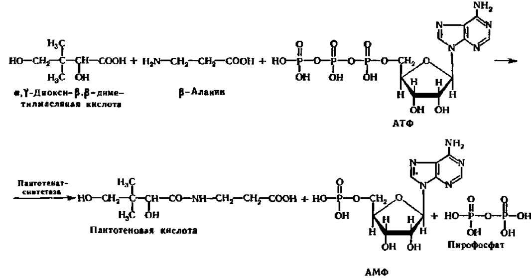
Pantothenate synthetase, as follows from the given equation, belongs to the group of ligases that accelerate the synthesis of C—N bonds. This group of ligases includes about 40 representatives. Currently, more than 75 different ligases responsible for vital synthetic processes in animal and plant cells have been studied.
In addition to accelerating the synthesis of C—N bonds, particularly peptide bonds, ligases catalyze the formation of C—C, C—O, and C—S bonds. The group of ligases that form C—C bonds includes carboxylases. They mediate the carboxylation of various compounds, resulting in the elongation of carbon chains. One of the most important carboxylases is Pyruvate carboxylase, which accelerates the formation of oxaloacetic acid from pyruvic acid and carbon dioxide (IV):

This reaction is of exceptional importance in metabolism, providing The Link Between carbohydrate and Protein metabolism as well as the fixation of CO2.
Ligases that catalyze the synthesis of C—O bonds play a crucial role in Protein Biosynthesis, as they accelerate the activation of amino acids prior to their incorporation into a peptide bond. One of the simplest reactions of this type is the formation of aminoacyl adenylates:
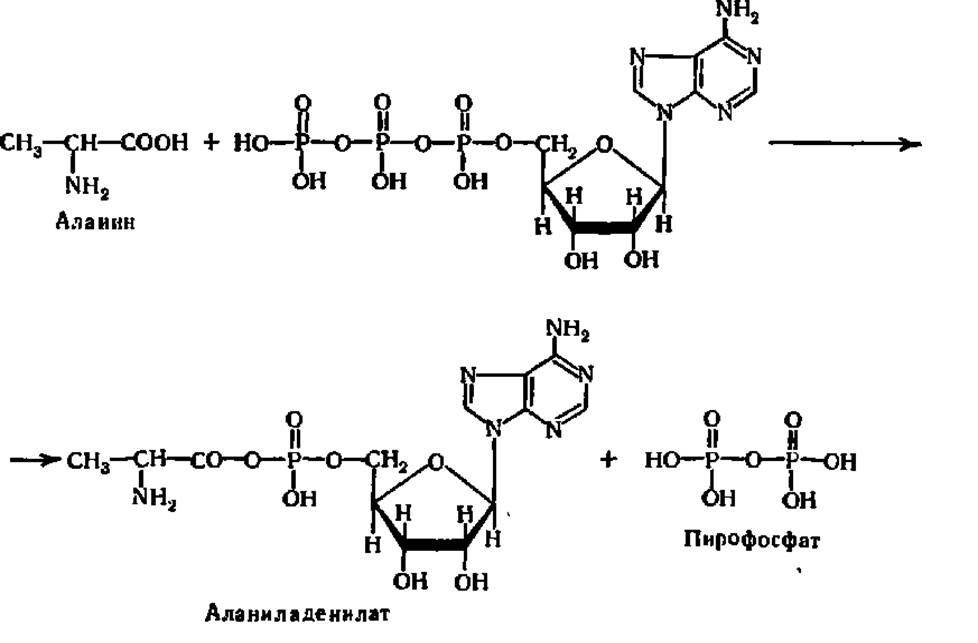
From aminoacyl adenylates, amino acids are transferred to tRNA to form aminoacyl-tRNA, which is utilized directly in polypeptide synthesis. These processes will be discussed in detail in Chapter VII.
Furthermore, ligases facilitate the formation of C—S bonds by acting as acyl-CoA synthetases. An example of acyl-CoA synthetase action is the Formation of Acetyl-CoA from acetic acid and coenzyme A (see the formula on page 163), which proceeds coupled with ATP hydrolysis:

Acetyl-CoA serves as a coenzyme in transacylation reactions; therefore, the action of these two groups of enzymes—ligases and acyltransferases—is tightly coordinated in living systems. A similar interdependence is characteristic of many Other Enzymes.
Last update: 06/08/2026
Editorial and Educational Adaptation: This material has been compiled based on the primary/original source text. The project team performed an editorial review, corrected technical inaccuracies, structured sections, and adapted the content for an educational format.
What was processed:
- elimination of formatting defects (OCR errors, structural breaks, corrupted characters);
- editorial organization of content;
- standardization of terminology in accordance with academic sources;
- verification of factual statements against the original source text.
All mentions of the author, publication year, and origin of the primary text have been preserved in accordance with the source.