FLAVONOIDS: BIOCHEMISTRY, BIOPHYSICS, MEDICINE - Tarakhovsky Yu. S. - 2013
CHAPTER 2. MECHANISMS OF FLAVONOID ACTION
2.5. Effect of flavonoids on cellular signaling systems
Cellular signaling systems are involved in transmitting chemical signals from The Cell surface to the Cytoplasm, enabling Cells to respond to environmental changes. To achieve this, cells feature specialized receptors on their Plasma Membrane capable of recognizing specific molecules in their environment, known as extracellular signaling molecules. Other molecules of various types, known as second messengers—including Proteins, Peptides, Lipids, NUCLEOTIDES, and metal cations—Relay this signal into the interior of the cell. This process often involves "signal Amplification," which is an increase in the number of Molecules Participating in the pathway. As a consequence, a single signaling molecule can trigger A wide variety of diverse responses.
Plant polyphenol compounds are capable of influencing the function of cytokine receptors, receptor Tyrosine Kinases (RTKs), G protein-coupled receptors (GPCRs), and a broad Class of transmembrane signal-transducing proteins known as Integrins (Fig. 71). The molecular mechanisms underlying these effects and the exact signal Transduction pathways remain insufficiently understood. In recent years, only fragmented data have emerged indicating Changes in the activity or expression of proteins within specific signaling systems in the presence of certain polyphenols.
Fig. 71. Simplified diagram of cellular signaling illustrating several signal transduction pathways from the cell surface to The Nucleus that are modulated by Flavonoids. In addition to moving from the surface to the nucleus, signals can propagate "horizontally," engaging other signaling systems in the process. The outcome of signal transduction may include alterations in Gene Expression, cell proliferation or apoptosis, and changes in cellular motility and adhesive properties. JAK - Janus kinase, STAT - signal transducer and activator of METABOLISM/31.html">Transcription, RTK - receptor tyrosine kinase, PKB - protein kinase B, IkB - inhibitor of nuclear factor kappa-B, PKA - protein kinase A, GPCR - G protein-coupled receptor, GRB2 - growth factor receptor-bound protein 2, SOS1 - son of sevenless homolog 1 (a membrane protein involved in cell growth and differentiation signaling), Ras - small GTPase superfamily proteins, Raf - proto-oncogene, MAPK and MAP2K - mitogen-activated protein kinases, PTK - protein tyrosine kinase, SHC1 - Src Homology 2 domain-containing-transforming protein C1.

2.5.1. Cytokine receptors
Cytokines are small proteins, peptides, or Glycoproteins that mediate Intercellular Communication, determining the growth, differentiation, functional activity, and apoptosis of cells across various tissues. Over 30 different cytokines (including interleukins, lymphokines, chemokines, and interferons) are known to act as immunomodulators of inflammatory processes. Under normal conditions, their tissue concentration may be around 10-12 M, but during inflammatory responses associated with trauma or infection, cytokine levels can surge up to 10-9 M. Specific cytokine receptors located on the cell surface initiate cascades of Intracellular Signaling processes that regulate the expression of target genes.
For instance, the protein cytokine known as tumor necrosis factor alpha (TNF-α) is produced by macrophages and certain other cells, transported via the Blood AND Lymph to various Organs, and acts as a trigger for inflammation and apoptosis. By binding to its receptor On the surface of various cells, TNF-α can participate in The regulation of numerous physiological processes (Fig. 72).
Fig. 72. Involvement of the TNF-α cytokine receptor in various physiological processes. NO - nitric oxide, AP1 - activator protein 1 (transcription factor), TF - transcription factor, PAF - platelet-activating factor, MCP-1 - monocyte chemoattractant protein 1, NF-kB - nuclear factor kappa-B, p38 - mitogen-activated protein kinase, JAK - Janus kinase, STAT-3 - signal transducer and activator of transcription 3, VCAM1 - vascular Cell Adhesion molecule 1, ICAM1 - intercellular adhesion molecule 1, IL-8 - interleukin 8, MCP-1 - monocyte chemoattractant protein 1, MMP-9 - matrix metallopeptidase 9, ROS - reactive oxygen species.

It is currently known that plant polyphenol compounds can influence the functioning of receptors for cytokines such as tumor necrosis factor (TNF) or certain interleukins (IL), which holds promise for the Treatment of Cancer, immune disorders, and other diseases. For instance, quercetin and naringenin are capable of protecting pancreatic beta cells from cytokine-induced apoptosis. This protective effect may be associated with the activation of Akt and Bad, which belong to the PI3-K kinase signaling pathway. Quercetin can also prevent The Development of contact dermatitis by blocking the release of inflammatory cytokines IL-8 and TNF by mast cells, an effect achieved by increasing intracellular calcium concentration and activating NF-kB. Furthermore, quercetin suppresses The production of the inflammatory cytokines TNF-α and IL-1β in the bronchoalveolar lavage fluid of experimental animals. In protecting cells against ultraviolet radiation, the action of quercetin goes beyond simply shielding against penetrating rays. To a large extent, cell protection is ensured by suppressing the production of inflammatory cytokines, which leads to a decrease in the binding of the nuclear factor NF-kB to DNA and a reduction in the production of cytokines such as IL-1β, IL-6, IL-8, and TNF-α.
EGCG is one of the most active green tea flavonoids, possessing The ability to normalize numerous cellular processes by neutralizing the damaging effects of high cytokine concentrations that arise during inflammation. For example, when acting on Insulin-producing pancreatic β-cells, EGCG protected against the effects of IL-1β and TNF-α and restored the cells' ability to produce insulin in response to glucose. In doing so, it decreased the levels of oxidation products and reactive oxygen species in the cytoplasm, restored mitochondrial Membrane Potential, halted the release of cytochrome c from Mitochondria into the cytoplasm, and lowered the cytoplasmic concentration of nitric oxide by suppressing cytokine-Induced Expression of nitric oxide synthase genes. Model experiments on rats demonstrated that EGCG normalized TNF-α levels and exerted a vasodilatory effect in response to acetylcholine. A decrease in fractalkine levels was also observed due to the downregulation of the nuclear factor NF-kB, alongside the inhibition of the monocyte chemoattractant MCP-1 and an increase in AP-1 protein activity in vascular endothelial cells.
Apigenin, luteolin, kaempferol, quercetin, and certain other flavonoids are capable of reducing monocyte adhesion to human umbilical vein endothelial cells (HUVECs) by normalizing the levels of adhesion molecule 1 and E-selectin expression induced by elevated TNF-α concentrations. A phenolic extract from olive oil (Olea europaea L.) also exerts a beneficial effect on pancreatic β-cells by mitigating the toxic action of the cytokines IL-1β and TNF-α. This results in a decreased concentration of reactive oxygen species in the cytoplasm and the restoration of insulin production. EGCG can also influence T-lymphocyte function by suppressing the expression of interleukin receptors IL-7 and IL-15, which are responsible for T-cell Homeostasis. The combination of EGCG and the cytostatic drug methotrexate exhibits anti-inflammatory properties and prevents the development of Arthritis by suppressing the expression of the cytokines IL-6 and TNF-α. At the same time, a significant antioxidant effect is manifested through an increase in the concentrations of catalase, superoxide dismutase, and Glutathione reductase. In a study investigating the protective effects of green tea EGCG and grape resveratrol—both abundant in red wines—it was found that a decrease in the concentration of the cytokine TNF-α is accompanied by a sharp reduction in lipid oxidation products, as indicated by the malondialdehyde content in the medium. Flavonoids extracted from wormwood (Artemisia herba alba) can positively influence the course of a multisystem inflammatory disorder (Behçet's Disease) by regulating cytokine production by Th1 and Th2 helper T cells. Additionally, they counteract the damaging effects of nitric oxide.
2.5.2. Receptor Tyrosine Kinases
Receptor tyrosine kinases (RTKs) play a vital role in regulating processes associated with cell proliferation or cell death.
Moreover, these receptors serve as molecular targets for numerous therapeutic agents used in cancer treatment. An RTK is a transmembrane protein that interacts with various growth factors, Cell Division signals, and certain Hormones (Fig. 73). Accordingly, about 20 Different types of RTKs are distinguished. These include the insulin receptor, receptors for epidermal growth factor, fibroblast growth factor, vascular endothelial growth factor, and hepatocyte growth factor, as well as the receptor for ephrin (a protein that regulates cell-cell interactions and cell migration), the angiopoietin receptor (responsible for angiogenesis), and others.
Fig. 73. Regulation of various processes via receptor tyrosine kinases (RTKs). Upon interacting with a Ligand, the receptor forms a dimer and transmits a signal to the nucleus. Two signal transduction pathways are shown: 1) Via RAS GTPases, the Serine/Threonine-protein kinase RAF, the mitogen-activated protein kinase MEK, and the protein kinase Erk. 2) Via phosphatidylinositol 3-kinase, the serine/threonine-protein kinase AKT, and the rapamycin-sensitive protein mTOR.

Green tea polyphenol compounds, and catechins in particular, can exert therapeutic effects on many types of cancer cells as well as on animal tumor development in model experiments by suppressing RTK signaling. Located in The Plasma Membrane, this receptor is sensitive to changes in lipid physical properties, which can be modulated by flavonoids. Among them, tea catechins are arguably some of the most effective anticarcinogenic agents found in plant polyphenols. One potential explanation for their activity suggests that flavonoids act as mimetics of the adenine moiety of the ATP molecule and are capable of blocking the ATP-binding sites of protein kinase receptors. Furthermore, attention is drawn to their ability to influence the lateral segregation of plasma Membrane Lipids and The formation of lipid rafts, thereby disrupting the function of Membrane Receptors such as RTKs or the epidermal growth factor receptor (EGFR). For instance, the flavonoid EGCG prevents the binding of epidermal growth factor to its corresponding receptor and inhibits the functioning of other RTKs, which dictates the anticarcinogenic effect of these polyphenols. The flavonoid silibinin likely exerts a similar action on RTKs.
Green tea EGCG also affects other cytoplasmic Components of the RTK signaling cascade, including the mitogen-activated protein kinase (MAPK) signaling pathway. EGCG blocks the DNA-binding activity of NF-kB, the cytoplasmic accumulation of A number of interleukins, and the expression of pro-inflammatory genes induced by the presence of bacterial lipopolysaccharides. Notably, it is the phosphorylation of proteins within the MAPK signaling system, including MEK, Raf, and others, that underlies the inhibitory action of tea catechins. This phosphorylation is suppressed As a result of blocking the action of Insulin-like Growth Factors IGF-I and IGF-II. Other polyphenols, such as quercetin, resveratrol, and ferulic, vanillic, and tannic acids, also act upon the MAPK signaling system.
2.5.3. Integrins
Integrins are cell-surface receptors that transmit signals into the cytoplasm regarding changes in The chemical composition of the Extracellular matrix surrounding cells. Found on The surface of most Multicellular Organisms—from Sponges to mammals—integrins typically consist of two subunits, α and β, which combine to form 24 different dimeric molecules. Each subunit features a transmembrane segment, as well as extracellular and cytoplasmic domains. Integrins can facilitate the attachment of pathogens to cell surfaces (e.g., Viruses) and are involved in bacterial invasion. They are of great importance in regulating cell-cell interactions, cell adhesion, and cell migration. Integrins are implicated in various pathologies, including tumor development and metastatic processes. Consequently, integrins serve as targets for the therapeutic action of various pharmacological agents.
It has been discovered that green tea catechins, and above all EGCG—the most active among them—can reduce the motility and adhesive properties of blood macrophages (monocytes), which play a crucial role in the Development of the Immune Response and inflammatory processes. This is attributed to the ability of EGCG to inhibit The activity of integrin β1, making this flavonoid a promising anti-inflammatory agent. Quercetin and catechins can influence the expression of cyclooxygenase-2 (COX-2) and integrin β2, thereby diminishing the pro-inflammatory response of monocytes. EGCG can modulate the adhesive properties and migration of mast cells, as well as their ability to activate monocytes, by downregulating the expression of integrins α5β3, which is essential for regulating tumor growth and metastasis. Moreover, EGCG can inhibit the migration and adhesive capacity of B lymphocytes—which also participate in the immune response—by blocking the expression of the integrin CD11b. EGCG is likewise capable of suppressing the expression of the epidermal growth factor receptor through its action on integrin α5β1, which is of great significance in the development of human carcinoma. There is also evidence indicating that EGCG affects fibroblast motility and migration by suppressing the expression of integrin α2β1, a mechanism that may contribute to the antitumor activity of this catechin.
Other flavonoids can also influence integrin expression, thereby preventing tumor growth and metastasis. For instance, apigenin, found in many medicinal herbs (such as chamomile, violet, Adonis, and lemon balm), can block integrin $\beta$5 in breast cancer cells. Kaempferol, a flavonoid derived from caraway, tea, and Guelder rose, suppresses TNF-$\alpha$-induced expression of eosinophil integrin $\beta$2, which prevents their infiltration into the respiratory epithelium in mice with allergic asthma. Glabridin, a licorice flavonoid, inhibits the expression of integrin $\alpha$v$\beta$3, which—along with suppressing the activity of other signaling components (FAK/Src, Akt, RhoA)—prevents the migration, invasion, and angiogenesis of lung tumor cells.
2.5.4. Peroxisomes and PPAR
Polyunsaturated Fatty acids, as well as their oxidation products such as Prostaglandins and Leukotrienes formed via lipoxygenases, can participate in the Introduction/30.html">Regulation of Gene Expression. One of the most thoroughly studied pathways of this regulation begins with the interaction of these substances with peroxisome proliferator-activated receptors (PPARs) located in the nucleus. These receptors are capable of functioning as transcription factors, participating in cell development and differentiation, and influencing protein, lipid, and Carbohydrate Metabolism (Fig. 74).
Fig. 74. Gene regulation by fatty acids, prostaglandins, and leukotrienes via the PPAR receptor. Fatty acids dissolved in the blood enter the cell and bind to the PPAR (peroxisome proliferator-activated receptor) located in the nucleus. Products of enzymatic lipid oxidation involving lipoxygenase (LOX) or cyclooxygenase (COX) also interact with PPAR. Upon activation of PPAR by the corresponding cytokine, a PPAR/RXR (retinoid X receptor) heterodimer is formed, which binds to the HRE (hormone response element) sequence of DNA, thereby driving the transcription of specific genes.

Following the binding of a fatty acid molecule via the PPAR-$\alpha$ receptor or a prostaglandin via the PPAR-$\beta$ receptor, the receptor forms a heterodimer with RXR (retinoid X receptor) and subsequently interacts with a specific DNA sequence responsible for hormonal regulation, known as the HRE (hormone response element). The HRE acts as a gene promoter that binds specific Hormone Receptors and similar regulators, leading to the expression of corresponding genes.
It has been discovered that isoflavones act as PPAR agonists. Due to this property, they exhibit anti-inflammatory activity and help prevent metabolic syndrome, atherosclerosis, and other inflammation-related conditions. The ability to activate PPAR was demonstrated in a Study of the total methanolic fraction of soybean seed isoflavones, which contained daidzin, daidzein, glycitin, genistin, malonyldaidzin, malonylglycitin, and genistein. This fraction promoted an increase in PPAR transcription. Notably, in in vitro experiments, the PPAR activity of these flavonoids exceeded that of bezafibrate, a pharmaceutical drug used in the treatment of metabolic syndrome. An alcoholic extract of isoflavones from Pterocarpus marsupium, a plant used in traditional Indian medicine to treat diabetes, also exhibited PPAR activity. One of the fractions from this extract lowered blood glucose levels comparably to insulin or rosiglitazone, an antidiabetic drug and selective PPAR agonist. Earlier experiments on mice showed that soy isoflavones also exhibit antidiabetic effects through PPAR receptor activation. Furthermore, the action of isoflavones, such as genistein, on PPAR receptors may exert beneficial effects in the treatment of Alzheimer's disease, with genistein's anti-inflammatory effect being comparable to that of estradiol. The protective effect of baicalein on Nerve Cells under ischemia-reperfusion conditions is attributed to its inhibition of PPAR protein expression. Upregulation of PPAR expression is likewise observed upon treatment with flavonoid extracts from the roots of Sophora flavescens or the flowers of black elderberry (Sambucus nigra), both of which possess anti-inflammatory and antidiabetic properties.
However, not all flavonoids effective against obesity and insulin resistance act through the PPAR signaling pathway. For example, quercetin can increase adiponectin secretion without affecting PPAR. Conversely, another study on Lung Cancer cells demonstrated that quercetin suppresses cell division by acting on the PPAR signaling system.
Naringenin from grapes is also capable of activating PPAR, which disrupts the assembly of hepatitis C virus particles. In cell culture experiments, The Effect of naringenin was comparable to that of interferon.
2.5.5. Nuclear Factor NF-$\kappa$B and Inflammatory Mediators
Inflammation is a primary mechanism of tissue repair following injury or stress initiated by pathogens. This mechanism involves a cascade of cellular responses to the presence of external or internal agents recognized by the host Organism as "foreign." The inflammatory cascade includes increased capillary permeability, migration of immune system cells to the site of injury, release of inflammatory mediators, cellular apoptosis, clearance of apoptotic cells via phagocytosis, and, ultimately, the growth of new tissue and Blood Vessels.
Several distinct signaling pathways are involved in the development of inflammatory processes, with the nuclear factor NF-$\kappa$B molecule occupying a central regulatory position. This pathway originates from proinflammatory cytokines—such as tumor necrosis factor alpha (TNF-$\alpha$) and interleukin-6 (IL-6)—passes through NF-$\kappa$B, and extends further along the arachidonic acid signaling pathway (Fig. 75). Consequently, NF-$\kappa$B serves as a crucial target for anti-inflammatory therapeutics.
Fig. 75. The signaling pathway of the nuclear transcription factor NF-$\kappa$B, a protein complex regulating the development of inflammatory processes through the release of Eicosanoids. TNF-$\alpha$ – tumor necrosis factor alpha; IL-6 – interleukin-6; Akt – protein kinase B (phosphatidylinositol-3-kinase); Stat3 – signal transducer and activator of transcription 3; IKK – I$\kappa$B kinase; iNOS – inducible nitric oxide synthase; PLA2 – phospholipase A2; COX – cyclooxygenase; LOX – lipoxygenase.

Next comes the arachidonic acid pathway, which is regulated by proinflammatory cytokines such as Akt, TNF-$\alpha$, and IL-6. Arachidonic acid (AA) is a lipid mediator generated through the action of phospholipase A2 on membrane lipids. Subsequently, AA undergoes oxidation by cyclooxygenase and lipoxygenase Enzymes, yielding substances belonging to the eicosanoid group, which function as inflammatory mediators (Fig. 76).
Fig. 76. Biosynthetic pathways of eicosanoids from arachidonic acid (AA), which is cleaved from phospholipid molecules by the action of phospholipase A2 (PLA2). The diagram illustrates two eicosanoid synthesis pathways: (I) – formation of prostaglandins (PG) and thromboxanes (TX) involving cyclooxygenases (COX); (II) – formation of HPETE (hydroperoxyeicosatetraenoic acid) and subsequent leukotrienes (LT) involving lipoxygenases (LOX).

Eicosanoids are signaling molecules generated during the enzymatic oxidation of polyunsaturated Essential Fatty Acids with a C20 hydrocarbon chain belonging to the omega-3 ($\omega$-3) and omega-6 ($\omega$-6) groups. Fatty acids serving as precursors for eicosanoids include eicosapentaenoic acid (EPA), an $\omega$-3 acid with five double bonds; arachidonic acid (AA), an $\omega$-6 acid with four double bonds; and dihomo-gamma-
linolenic acid, an $\omega$-6 acid with three double bonds. Eicosanoids belonging to the $\omega$-6 group are more potent inflammation Inducers than those of the $\omega$-3 group. There are four main groups of eicosanoids: prostaglandins, prostacyclins, thromboxanes, and leukotrienes. These groups are further subdivided into two or three subgroups derived from either $\omega$-3 or $\omega$-6 fatty acids.
Eicosanoid synthesis is triggered by various stimuli, including mechanical trauma, cytokines, and certain hormones. These stimuli activate phospholipase A2, which is capable of cleaving fatty acids from the phospholipid MOLECULES OF CELL membranes (Fig. 77).
Fig. 77. Examples of eicosanoids. A phosphatidylcholine molecule containing an arachidonic acid residue is shown, which is cleaved by phospholipase A2 (PLA2). Free arachidonic acid is oxidized by respective oxidases, resulting in the formation of leukotrienes, prostaglandins, thromboxanes, and prostacyclins.

Plant polyphenols include A large number of substances capable of inhibiting the activity of phospholipase A2, as well as the LOX and COX enzymes involved in eicosanoid synthesis. Certain polyphenolic COX-1 inhibitors act at concentrations below 1 $\mu$M and may be more effective than acetylsalicylic acid (aspirin), which also targets COX-1, given that therapeutic daily doses of aspirin range from tens of milligrams to several grams and the drug carries undesirable side effects.
Table 10. Examples of polyphenolic (predominantly flavonoid) inhibitors of enzymes in the arachidonic acid signaling cascade.
Phospholipase A2 |
IC50 |
Cyclooxygenase 2 |
IC50 |
12-Lipoxygenase |
IC50 |
Kaempferol |
7.50 |
Gambogic acid |
13.60 |
Anadanthoflavone |
13.00 |
Morelloflavone |
0.60 |
Genistein |
15.00 |
Artonin E |
2.30 |
Quercetin |
6.50 |
Glycitein |
2.50 |
Baicalein |
0.64 |
Cyclooxygenase 1 |
Tectorigenin |
3.00 |
Cirsiliol |
1.00 |
|
Artonin E |
2.50 |
Phenethyl ferulate |
4.35 |
Fisetin |
0.25 |
Chrysin |
5.00 |
Rosmarol |
2.50 |
5-Lipoxygenase |
|
Isoliquiritigenin |
8.00 |
Baicalein |
1.00 |
Artonin E |
0.36 |
Kurarinone |
0.60 |
Resveratrol |
50.00 |
Cirsiliol |
0.10 |
Kuraridin |
0.60 |
Ginkgetin |
0.33 |
||
Morusin |
1.60 |
Kenusanone A |
0.50 |
||
Quercetin |
8.00 |
15-Lipoxygenase |
Luteolin |
0.10 |
|
Sophoraflavanone G |
0.10 |
Apigenin |
4.00 |
Quercetin |
0.30 |
Sophoraflavanone A |
5.00 |
Baicalein |
1.60 |
Gingerol |
0.00 |
Catechin |
0.11 |
Fisetin |
1.50 |
Ginkgetin |
0.33 |
Baicalein |
0.67 |
Luteolin |
0.60 |
Baicalein |
7.00 |
Resveratrol |
15.00 |
Morusin |
3.30 |
Resveratrol |
1.37 |
Note. The most effective inhibitors are presented. Polyphenol concentrations sufficient to inhibit the indicated enzymes by 50% (IC50) are given in µM. More detailed information can be found in the literature.
Furthermore, as shown in the table (Table 10), plants also contain a large number of highly effective inflammatory signal inhibitors acting on various targets, the efficacy of which remains to be investigated. For instance, the efficacy of the most thoroughly studied flavonoids, such as quercetin, in suppressing allergic processes appears quite modest compared to the activity of resveratrol and, especially, baicalein, which are capable of acting simultaneously on COX- and LOX-dependent signaling pathways.
2.5.6. G Protein-Coupled Receptors
Eicosanoids act on G protein-coupled receptors (GPCRs), also known as seven-transmembrane domain receptors, which are involved in signal transduction across the membrane of Eukaryotic cells—including mammalian and human cells—from the surface to the cytoplasm. The ligands binding to these receptors are extremely diverse: odorant molecules, hormones, Neurotransmitters, and even quanta of light in visual receptors. These receptors participate in signaling pathways such as the cyclic adenosine monophosphate and glutamate pathways. However, the largest number of proteins belongs to rhodopsin-like receptors. Collectively, these receptors are encoded by approximately 800 genes, accounting for 4% of The Human Genome or about 10% of Membrane Proteins. GPCR family proteins are crucial therapeutic targets in the treatment of conditions such as cancer, inflammatory processes, and Disorders of the immune, nervous, cardiovascular, and excretory systems. These proteins serve as targets for more than 40% of pharmaceutical drugs.
The quantity and ratio of these groups of eicosanoids are directly related to the development of various diseases, such as rheumatoid arthritis, allergies, asthma, and cancer. For example, rheumatoid arthritis is associated with the infiltration of immune system cells (such as lymphocytes, neutrophils, and macrophages) into the synovial fluid of joints, where these cells release inflammatory mediators, resulting in Cartilage destruction. Type 1 T helper cells (Th1) play a dominant role in these processes, induced by the pro-inflammatory cytokines TNF-α, IL-1, IL-6, and IL-17. Conversely, type 2 T helper cells (Th2), induced by the cytokines IL-4 and IL-13, are involved in the development of allergies and asthma. Accordingly, the Th1/Th2 ratio is of great importance in determining The Nature of the immune response.
One of the most promising approaches in treating immune disorders is controlling the levels of the cytokine IL-23, which plays a pivotal role in the development of the immune response. It has been shown that IL-23 is involved in the activation of type 17 T helpers (Th-17). A number of other interleukins, as well as TNF-α, are also implicated in these processes. The Use of Antibodies against these interleukins, particularly IL-23, successfully treats certain types of allergic diseases, such as psoriatic arthritis. In addition, suppressing the immune response heavily relies on the use of phospholipase A2 inhibitors, which consequently reduce the concentration of free arachidonic acid.
The NF-κB protein plays an essential role in the final stages of inflammatory processes by promoting the expression of anti-inflammatory genes and regulating leukocyte apoptosis. Present in the cells of all tissues, this protein is capable of interacting with DNA molecules and serves as the most universal transcription-regulating factor. Its activation is the cell's primary "rapid" response to damaging stimuli. Inducers of NF-κB activity include reactive oxygen species, nitric oxide, ionizing radiation, TNF-α, certain interleukins, Bacterial Cell wall lipopolysaccharides, and many other toxic agents. Suppressing NF-κB activity can be an effective strategy for treating numerous immune disorders. Furthermore, blocking signaling pathways at specific stages can be crucial for disease management. For instance, suppressing the production of prostaglandins, thromboxanes, and leukotrienes can exert a beneficial effect in alleviating pain associated with osteoarthritis. Controlling prostanoid production (Prostaglandins and thromboxanes) or blocking receptors for these agents is vital in treating various allergic and immune diseases, including different types of dermatitis, asthma, rheumatoid arthritis, encephalomyelitis, and multiple sclerosis.
2.5.7. Adipokines and Metabolic Syndrome
Metabolic syndrome encompasses a cluster of abnormalities that increase the risk of cardiovascular disease and type 2 Diabetes Mellitus. The most prominent sign of metabolic syndrome is central adiposity (abdominal fat accumulation) and an increased waist circumference. In addition, glucose and Lipid Metabolism are impaired, and blood pressure rises. Although current understanding of the Causes and Mechanisms underlying metabolic syndrome remains somewhat contradictory, most researchers believe that visceral fat deposits, particularly hepatic fat accumulation, may drive the Impairment of the insulin-dependent regulatory system, which fuels all subsequent metabolic disturbances. Thus, dysregulation of carbohydrate metabolism associated with increased insulin resistance—termed prediabetic syndrome—directly follows metabolic syndrome and acts as a risk factor for cardiovascular diseases. Their development begins with endothelial cell damage and leads to disruptions throughout the Arterial System, accompanied by erosion and thrombosis. This is linked to the activation of leukocytes causing oxidative stress, as well as the upregulated expression of metalloproteinases that degrade extracellular matrix proteins such as Collagen, Fibronectin, and Laminin. Furthermore, inflammatory processes, allergic reactions, and tissue destruction occur due to the activation of Connective Tissue immune cells (mast cells), leading to the release of histamine, cytokines, chemokines, and proteases (chymases and tryptases) into the bloodstream. Metabolic syndrome can trigger not only cardiovascular diseases but also a multitude of other dysfunctions associated with inflammation, tissue accumulation of reactive oxygen species, and allergic reactions. Consequently, the risk of neurological disorders—including stroke, Alzheimer's disease, depression, and sexual dysfunction—is elevated, alongside a potential development of prostatitis, nephrological dysfunctions, and periodontal disease.
To fully understand the Pathogenesis of metabolic syndrome, it is necessary to examine the anatomical and PHYSIOLOGICAL CHARACTERISTICS OF adipose tissue. Adipose tissue is a loose connective tissue containing adipocytes, whose cytoplasm features lipid droplets. Additionally, adipose tissue harbors fibroblasts, macrophages, and endothelial cells, and is richly vascularized by small blood vessels. In humans, adipose tissue is distributed as subcutaneous fat beneath the Skin, visceral fat surrounding Internal Organs, intramuscular fat within Muscle tissue, and within Bone Marrow and breast tissue. The biochemical composition, physiological characteristics, and localization of adipose tissue vary across different organs.
In young women, fat is predominantly deposited in the buttocks and thighs, a pattern determined by hormones. With the onset of menopause and declining estrogen levels, fat redistributes toward the waist and accumulates primarily in the abdomen. A similar abdominal fat distribution is characteristic of men. Abdominal fat can be superficial, but typically the majority is stored as visceral fat deposits (mesenteric, perirenal, and perihepatic adipose tissue). Abdominal obesity correlates with cardiovascular disease, type 2 diabetes, insulin resistance, and certain types of cancer and arthritis. Significant fat deposits also lie directly beneath the skin surface. Subcutaneous fat is believed to be less prone to triggering cardiovascular disease compared to visceral fat and may potentially exert a protective effect on the organism.
Adipocytes are designed to maintain required blood concentrations of free Fatty Acids and triglycerides, as well as to regulate insulin resistance. Abdominal fat can exacerbate insulin resistance and contribute to Hypertension. The accumulation of abdominal fat increases during stress and can provoke hormonal shifts detrimental to health. This is because adipose tissue functions as a major endocrine organ. It produces cytokines known as adipokines, which participate in glucose homeostasis, the regulation of inflammatory and immune responses, blood clotting, lipid metabolism, angiogenesis, carcinogenesis, and many other processes.
Table 11. Adipokines and their effects on metabolic processes.
Adipokine |
Processes |
Diseases and risk factors |
Adiponectin |
Improves insulin sensitivity, anti-atherogenic effect |
|
Apelin |
Hypotensive effect, cardiac contractility stimulation, suppression of vasopressin secretion, increased insulin resistance |
Cardiovascular diseases, type 2 diabetes mellitus, and obesity |
Visfatin |
B-lymphocyte maturation, neutrophil apoptosis, promotion of normal cell proliferation, Prevention of cancer cell apoptosis, hyperglycemic effect |
Type 2 diabetes, rheumatoid arthritis, cardiovascular diseases, oncogenesis, inflammation and Immunity, Alzheimer's disease |
Plasminogen activator inhibitor-1 (PAI-1) |
Reduction of Fibrinolysis, inhibition of matrix metalloproteinase activity |
Coronary heart disease, hemorrhagic diathesis, cancer, metabolic syndrome, fibrosis |
Interleukin-6 (IL-6) |
Inflammation, immune response, Cell Differentiation |
Oncological diseases, cardiovascular diseases, diabetes |
Leptin |
Appetite suppression, reduction of thyroid, pancreatic, and gonadal activity |
Anorexia, hemorrhagic vasculitis, vascular thrombosis, obesity, cancer |
Tumor necrosis factor alpha (TNF-α) |
Suppression of certain tumors, cell apoptosis, systemic inflammation, Regulation of Immune response |
Infectious diseases, cancer therapy, rheumatoid arthritis, systemic lupus erythematosus |
Resistin (ADSF) |
Inflammatory processes, energy homeostasis, endocrine function, insulin resistance |
Obesity, type 2 diabetes mellitus, cardiovascular diseases |
To date, over 600 adipokines have been identified as protein biomarkers of metabolic syndrome, exerting diverse effects on the body, although the functions of only a few are well-studied. It has been found that aberrant adipokine secretion observed in obesity can trigger various diseases (Table 11).
Overall, obesity is linked to excessive caloric intake and insufficient Energy Expenditure. Genetic and hormonal factors also heavily contribute to the development of obesity. Reducing food intake is not always effective against obesity if energy expenditure is low and the body strives to maintain its weight. Therefore, various pharmacological agents may be utilized for weight loss. Plant polyphenol compounds can also be effective in combating obesity and preventing various obesity-related disorders. Studies indicate that their Mechanisms of action may vary.
For instance, investigating the effects of quercetin and resveratrol on adipokine secretion in animals demonstrated that these flavonoids can reduce visfatin secretion, whereas neither flavonoid affected leptin or adiponectin secretion. Among more than 20 evaluated flavonoids, quercetin and resveratrol proved to be the most effective in normalizing blood TNF-α levels, which in turn normalized levels of inflammation-related factors regulated by TNF-α, such as certain interleukins, monocyte chemoattractant protein-1 (MCP-1), c-Jun N-terminal kinase (JNK), and PPAR-γ. Thus, quercetin and resveratrol are potent inhibitors of obesity-associated inflammation. Studies of rutin in rats showed that this flavonoid can restore normal physiological parameters in many chronic processes associated with metabolic syndrome, such as glucose tolerance, hepatic and vascular dysfunctions, oxidative stress, and inflammation. Onion peel extract, rich in quercetin and its derivatives, can also be utilized in treating metabolic syndrome. Notably, onion peel extract exerted a more pronounced beneficial effect on animal models than purified quercetin.
A distinct MECHANISM OF ACTION against metabolic syndrome was uncovered in studies examining naringin in mice fed a high-calorie diet. Naringin was shown to activate AMP-activated protein kinase (AMPK-α) by initiating the phosphorylation of this protein, leading to reduced insulin resistance and the suppression of oxidative stress. Alongside naringin (or its aglycone naringenin), the ability to lower blood glucose levels was also observed in studies involving daidzein.
In the prevention of metabolic syndrome, genistein proved effective in preventing the development of non-alcoholic steatohepatitis. Metabolic syndrome is known to be accompanied by hepatic triglyceride accumulation, which triggers free radical oxidation, stimulates inflammatory processes, and promotes fibrosis ranging up to Liver cirrhosis and hepatocellular carcinoma. Treatment with genistein led to decreased concentrations of TNF-α and IL-6 in liver tissue and Blood Plasma, inhibition of IκB-α phosphorylation, and activation of JNK kinase. It is possible that the primary target of genistein is the mitochondrial membrane of adipocytes, the disruption of which leads to lowered cellular ATP levels. Mitochondrial metabolic dysfunction is also observed upon treatment with resveratrol. Consequently, this suppresses adipocyte growth and differentiation, reduces adipose tissue mass, and lowers tissue adipokine concentrations, accompanied by a decrease in blood glucose levels. Impaired adipocyte differentiation was also noted with daidzein treatment, although the mechanisms of action of these flavonoids differ slightly.
Green tea catechin EGCG exhibits pronounced anti-obesity activity. This flavonoid suppresses adipogenesis by arresting adipocyte cell division. An inhibition of genes responsible for the fibroblast-to-adipocyte transition—namely C/EBP-α and PPAR-γ—is observed. Concurrently, dietary Lipid Absorption decreases while fecal lipid excretion increases. Genes responsible for Fatty acid oxidation are expressed in Skeletal Muscle mitochondria, meaning fat-burning processes are activated within the body, although other researchers attribute this fat-oxidation effect primarily to caffeine rather than EGCG. Green tea polyphenols can be effective in normalizing blood glucose, triglyceride, and Cholesterol levels. They lower plasma Lipid Peroxidation products and enhance plasma antioxidant status by increasing the concentrations of superoxide dismutase and other antioxidant enzymes.
A substantial body of research is dedicated to investigating The Role of grape polyphenols and wine products in preventing metabolic syndrome. For instance, grape seed proanthocyanidins were found to reduce inflammatory marker levels in animals fed a fat- and carbohydrate-rich diet. The levels of TNF-α, IL-6, and macrophage markers decrease, while adiponectin expression increases. Concurrently, hepatic Lipogenesis is suppressed, and a series of genes involved in hepatic Glycogenesis, Glycolysis, and lipid metabolism are upregulated.
Polyphenol extracts, particularly abundant in red grape skins, can also modulate LIPID METABOLISM IN humans and animals consuming high-fat, high-sugar diets. A reduction in muscle triglyceride content is observed, along with an upregulation of the insulin-dependent glucose transporter GLUT4—which alleviates insulin resistance and lowers blood glucose concentration—and the expression of mitochondrial fatty acid oxidation enzymes, such as the mitochondrial hydroxyacyl-CoA dehydrogenase gene. Furthermore, fatty acid transport and lipid accumulation in Muscles are diminished.
Table 12. Effects of plant polyphenols and polyphenol-rich plant products on metabolic syndrome.
Polyphenols or their source |
Mechanism of action |
|
Apple juice (unclarified) |
Reduction in body weight of subjects after 4 weeks of intake at 750 mL/day. Clarified juice lacks this effect. |
|
Citrus flavonoids |
Suppression of stearoyl-CoA desaturase-1 gene expression, reduction of blood lipid and sugar levels (regulation via PPAR-α and PGC-1α). Potential application in atherosclerosis prevention |
|
Mandarin juice |
Protection against oxidative stress |
|
Cinnamon polyphenols |
Expression of TTP family proteins, exerting anti-inflammatory effects |
|
Pomegranate peel polyphenols |
Stimulation of intestinal bifidobacteria growth, reduction of blood cholesterol levels |
|
Black rice anthocyanins |
Reduction of platelet aggregation, lowering of blood triglyceride concentrations, body weight reduction |
|
Rapeseed oil |
Expression of antioxidant system proteins, prevention of atherosclerosis |
|
Flavonoids of spikemoss (Selaginella tamariscina) |
Antidiabetic activity: reduction of blood glucose, triglycerides, cholesterol, and fatty acids. Upregulation of antioxidant system proteins |
|
Sweet potato extract (Ipomoea batatas) |
Reduction of leptin secretion, suppression of inflammatory factor expression and lipid synthesis, activation of lipolysis factors |
|
Blueberry polyphenols |
Improvement of insulin sensitivity, PPAR expression, reduction of liver weight, body weight, and fat mass percentage, lowering of blood triglycerides |
|
Various PARTS OF THE pomegranate plant (Púnica granatum) |
Anti-inflammatory, antioxidant, and antitumor activity |
|
Cocoa polyphenols |
Suppression of adipogenesis and obesity. Acts on the insulin receptor, inhibiting IR kinase and related signaling pathway components |
|
Bean polyphenols (Vigna angularis) |
Normalization of lipid metabolism, reduction of hepatic lipids, elimination of excess lipids via feces, lowering of blood triglycerides, activation of glycerol-3-phosphate dehydrogenase |
|
Nut extract (Semecarpus anacardium) |
Significant reduction (normalization) of blood glucose concentration, increase in antioxidant enzyme levels |
|
Abacopterin A from Abacopteris penangiana |
Hypolipidemic and anti-inflammatory action via inhibition of NF-κB expression |
|
Flavonoids from Litsea coreana |
Enhanced insulin sensitivity, increased superoxide dismutase concentration, reduction of blood free fatty acids and triglycerides, normalization of liver function. |
|
Carrot juice anthocyanins |
Antioxidant and anti-inflammatory effects. Improvement of glucose tolerance, normalization of liver function |
|
Strawberry |
Reduction of blood triglyceride and cholesterol concentrations, decreased oxidized product content in low-density Lipoproteins |
|
Flavonoids from Potentilla discolor |
Hypoglycemic and hypolipidemic activity, elevation of antioxidant enzyme levels, protection of pancreatic β-cells |
|
Dodonaea viscosa extract |
Reduction of insulin resistance, suppression of oxidative stress, normalization of blood lipid profiles |
|
Pineapple fruit, meal, and oil |
Clinical trials indicate a significant reduction in plasma cholesterol and high-density lipoproteins |
|
Soy isoflavones |
Prevention of obesity through the regulation of hypothalamic function |
|
Lotus flavonoids (Nelumbo nucifera) |
Reduction of body weight and fat mass. Regulation of hepatic lipid metabolism enzymes |
|
Cissus quadrangularis stem extract |
Improved insulin sensitivity, restoration of liver function. Effects comparable to the drug metformin |
|
Olive oil extract |
Normalization of cardiovascular and hepatic functions. No effect on hypertension |
|
Flavonoids from licorice oil (Glycyrrhiza glabra) |
Reduction of abdominal obesity, regulation of hepatic PPAR-α and SREBP-1c enzymes |
|
Flavonoids from fragrant solomon's seal (Polygonatum odoratum) |
Increased adipocyte insulin sensitivity |
|
Sea buckthorn flavonoids (Hippophae rhamnoides) |
Significant reduction in body weight, hepatic fat, and visceral fat deposits, lowering of blood cholesterol, triglycerides, and glucose. |
|
Aqueous extract of mulberry fruit (Morus alba) |
Reduction of body weight and hepatic fat content, normalization of lipid metabolism, elevation of enzymes protecting against oxidative stress |
|
Coffee bean polyphenols |
Reduction of hepatic fat accumulation and normalization of lipogenic enzyme activity |
|
Flavonoids of Pilea microphylla |
Antidiabetic effect: prevention of obesity, protection of pancreatic islet cells, prevention of hepatocyte hypertrophy, elevation of blood antioxidant levels. |
|
Alcoholic extract of Hibiscus (Abelmoschus manihot) |
Reduction of triglyceride accumulation in adipocytes, lowering of blood triglyceride and free fatty acid levels, Regulation of transcription factors PPAR-γ, CEBP-α, ap2 |
Note. The table omits the most thoroughly studied polyphenols whose effects are described in detail above. Most data were obtained from animal experiments unless otherwise indicated.
Furthermore, grape polyphenols suppress inflammatory processes by acting as antioxidants and activating the expression of antioxidant proteins. They also block the expression of pro-inflammatory cytokines, kinases, and transcription factors involved in the progression of inflammation, while conversely upregulating genes that inhibit inflammatory pathways. Enriching grape seed extracts with catechins, or using combined extracts of red grape, soy isoflavones, and L-carnitine, may further enhance the prophylactic efficacy of these preparations.
Polyphenols found in various dietary and medicinal plant products can exert beneficial effects on metabolic syndrome and type 2 diabetes manifestations. However, their mechanisms of action remain poorly understood and are likely highly diverse (Table 12). Most studies to date have been conducted on animal models (mice and rats), with a severe lack of robust clinical trials evaluating the effects of these compounds in humans. Consequently, the widespread use of such botanical Materials as dietary supplements should be approached with caution.
It is also worth noting that the binding affinity of polyphenol compounds to proteins may be reduced during sustained hyperglycemia. This is driven by the covalent cross-linking of glucose with proteins through a non-enzymatic process known as glycation. Research has shown that the binding of polyphenols to glycated Plasma Proteins can decrease tenfold or more compared to non-glycated proteins. If this phenomenon is broadly applicable to the interaction of polyphenols with various cell-surface receptors, a decline in polyphenol efficacy might be expected under chronic Metabolic Disorders.
2.5.8. TRAIL Apoptosis Factor and Anticarcinogenic Activity
Apoptosis, or programmed cell death, is the primary target of most therapeutic strategies in cancer treatment. Consequently, the apoptotic cytokine TRAIL (TNF-related apoptosis-inducing ligand), a member of the TNF protein superfamily, has garnered intense research interest since its discovery in 1995. In literature, TRAIL is also referred to as cluster of differentiation 253 (CD253), APO2-L, or TRAIL/Apo2L.
While The Physiological Role of TRAIL is not yet fully elucidated, this protein has been shown to be involved in T-cell memory formation, hematopoiesis, the development of autoimmune diseases, and numerous other biological processes. TRAIL plays a significant role in the antitumor activity of T-lymphocytes and natural killer (NK) cells. Thus, TRAIL-mediated regulation of tumor growth and metastasis constitutes a crucial component of the body's immune defense against carcinogenesis.
This protein consists of 281 Amino Acids and forms a homotrimer composed of three identical subunits. TRAIL is expressed on the surface of certain immune cells (T cells, NK cells), and a soluble form of TRAIL also exists. The soluble form exhibits lower hepatotoxicity compared to the membrane-bound form and can be utilized to induce apoptosis in tumor cells. Circulating TRAIL molecules bind to transmembrane death receptors DR4 (TRAIL-R1) or DR5 (TRAIL-R2) located on the plasma membrane of cancer cells (Fig. 78), thereby triggering a cascade of biochemical events that leads to apoptosis.
The apoptosis factor TRAIL is produced by immune cells (T AND NK lymphocytes) and binds to death receptors DR4/DR5 on the surface of cancer cells, resulting in the Formation of the DISC complex, which also incorporates the adaptor protein FADD and procaspase-8 or -10. Subsequently, activated caspase-8 or -10 activates caspase-3 (and potentially caspase-6 or -7), which acts as the executioner of apoptosis. This pathway is designated as the extrinsic pathway. It can be modulated by the apoptosis regulator protein c-FLIP (also known as CASP8). Alternatively, caspase-3 can be activated via the mitochondria (MTC). In this pathway, caspase-8 or -10 activates the pro-apoptotic protein Bid (also designated as BH3), which acts on mitochondrial membranes via Bax and/or Bak proteins. This induces pore formation in the outer mitochondrial membrane, releasing cytochrome c (Cyt C), which in turn can initiate apoptosis via caspases. The activity of the Bax protein is regulated by associated anti-apoptotic Bcl-2 family proteins, such as Bcl-2, Bcl-XL, and the induced myeloid leukemia cell differentiation protein Mcl-1. The activities of caspase-9 and caspase-3 can be modulated by inhibitors of apoptosis (XIAP, cIAP, and Survivin), which are themselves regulated by the mitochondrial second activator of caspases, SMAC (also known as Diablo). Mitochondrial damage may also be triggered by the tumor suppressor protein p53 in the presence of reactive oxygen species (ROS) or protein kinase Akt, the latter being activated by phosphoinositide 3-kinase (PI3K). As illustrated in the diagram, Akt is also involved in regulating cell division through cyclin-dependent kinase inhibitors p21 and p27, or promoting cell survival via the IKK kinase and the nuclear factor NF-kB.
Table 13. Effects of plant polyphenols on molecular targets of TRAIL-induced apoptotic pathways (modified and supplemented from the review).
Molecular target |
Polyphenol |
Cancer type |
Upregulation of death receptors DR4/DR5 |
Quercetin, kaempferol, apigenin, luteolin, baicalein, EGCG, silibinin |
Colorectal, pancreatic, prostate, cervical, lung cancer, leukemia, melanoma, glioma |
Downregulation of c-FLIP |
Resveratrol, quercetin, myricetin, genistein |
Melanoma, glioma |
Activation of caspase-8 |
Quercetin, kaempferol, myricetin, apigenin, luteolin, EGCG, genistein, silibinin, resveratrol |
Colorectal, pancreatic, liver, prostate, Cervical cancer, leukemia, glioma |
Expression and activation of Bid |
Quercetin, apigenin, luteolin, genistein, silibinin |
Colorectal, liver, prostate, cervical cancer, leukemia, glioma |
Upregulation of Bak |
Quercetin, resveratrol |
Prostate cancer, neuroblastoma, lymphoma |
Upregulation of Bax |
EGCG, resveratrol |
Gastric and prostate cancer |
Downregulation of Bcl-2 |
Kaempferol, myricetin, EGCG, genistein, daidzein, resveratrol |
Liver, prostate, cervical cancer, glioma |
Downregulation of Bcl-XL |
Kaempferol, EGCG, resveratrol |
Liver, prostate, cervical cancer, melanoma |
Release of cytochrome c |
Quercetin, resveratrol |
Colorectal and prostate cancer |
Downregulation of Akt |
Quercetin, kaempferol, EGCG, genistein |
Prostate, lung cancer, glioma |
Downregulation of survivin |
Quercetin, kaempferol, EGCG, silibinin, resveratrol |
Prostate, lung cancer, glioma, neuroblastoma |
Downregulation of c-IAP |
Flavopiridol |
Leukemia |
Downregulation of XIAP |
Luteolin, resveratrol, flavokawain, luteolin |
Prostate and cervical cancer |
Activation of caspase-8, -3 |
Quercetin, EGCG, genistein, silibinin, resveratrol |
Colorectal, pancreatic, liver cancer |
TRAIL induces apoptosis in tumor cells not only in vitro, but also in whole-organism models, as demonstrated in preclinical cancer studies using laboratory animals. For reasons not yet fully understood, activation of the TRAIL signaling pathway does not exhibit toxicity toward normal cells, distinguishing this factor from TNF or FasL. While the latter can also trigger apoptotic pathways, their medical application is highly problematic due to severe toxicity toward healthy cells in various organs, particularly hepatocytes.
Fig. 78. Effect of polyphenols on apoptotic signaling pathways. Adapted with modifications from reviews. Plus (+) and minus (-) signs denote pathway components positively or negatively regulated by polyphenols. DISC – death-inducing signaling complex, c-FLIP – apoptosis regulator, Bid – pro-apoptotic protein, Mcl-1 – induced myeloid leukemia cell differentiation protein, Cit C – cytochrome c, PI3K – phosphoinositide 3-kinase, Akt – protein kinase B, p21 and p27 – G1-phase cell division inhibitors, IKK – IkappaB kinase.

Clinical trials utilizing recombinant human TRAIL combined with conventional Chemotherapy have shown promising results. However, certain cancer cells exhibit resistance to TRAIL pathway activation. Overcoming this resistance and enhancing cellular apoptotic susceptibility could significantly improve the treatment of various malignancies.
Many polyphenol compounds, predominantly flavonoids, demonstrate synergistic effects with TRAIL by modulating various proteins involved in the regulation of apoptosis, cell survival, or tumor proliferation rates. For instance, Nishikawa was the first to discover that green tea epigallocatechin-3-gallate (EGCG) can potentiate TRAIL-mediated cytotoxicity in human hepatocarcinoma cells through the downregulation of Bcl-2α and Bcl-XL proteins. A similar mechanism of action via Bcl-2, Bcl-XL, and other proteins (not shown in the diagram) was observed when EGCG and TRAIL were applied to prostate carcinoma cells. Subsequently, the efficacy of kaempferol combined with TRAIL was demonstrated in glioblastomas, where this flavonoid induced survivin degradation (survivin, Fig. 78) and Akt inhibition, ultimately leading to carcinoma cell death.
Quercetin can enhance TRAIL-induced effects through Akt deactivation (dephosphorylation) and caspase activation in human adenocarcinoma cells, without exhibiting cytotoxicity toward normal cells. The same authors demonstrated that quercetin can activate caspase-3, -8, and -9. Furthermore, quercetin was found to interact with the survivin promoter and repress the expression of this protein [1171]. Data concerning the Effects of Flavonoids and related compounds on various components of the TRAIL-dependent apoptotic signaling system are summarized in Table 13.
2.5.9. Prevention of Neurodegenerative Diseases
For centuries, traditional medicine has utilized plant materials to treat various neurological disorders. Certain Alkaloids are well known for their ability to interact with various Central Nervous system receptors. However, recent studies have revealed that plant polyphenols can also exert diverse effects on The Nervous System, engaging with specific receptors on the surface of Neurons and microglial cells, and protecting neural cells against oxidative stress.
2.5.9.1. Blood-Brain Barrier Permeability.
To evaluate the potential of various polyphenol compounds to influence the central nervous system (CNS), it is first necessary to examine the ability of these substances to cross the blood-brain barrier (Fig. 79).
Brain capillaries exhibit several distinct features compared to capillaries in other organs. One such feature is the formation of tight junctions between endothelial cells, which significantly reduces Capillary Wall permeability to certain molecules. This functional property is termed the blood-brain barrier (BBB).
Fig. 79. Simplified diagram of substance Transport Across the blood-brain barrier (BBB). The endothelial cell layer lining the brain capillaries is shown. Unlike the endothelium in other organs, brain endothelial cells are interconnected by tight junctions, primarily composed of occludin and claudin proteins. These tight junctions restrict paracellular transport that typically occurs in capillaries of other tissues. Although this limits unrestricted transendothelial flux, substances are still transported from the blood into brain parenchyma via Passive Diffusion Across cell membranes (1) and through tight junctions (2), via specialized protein receptors and transporters (3), or through transcytosis, which involves sequential Endocytosis and Exocytosis (4).

The presence of the barrier does not imply a restricted nutrient supply across capillary walls. On the contrary, the brain is the most metabolically demanding organ in The Human Body, requiring a constant delivery of energy substrates and the removal of Metabolic waste products. The BBB is essential for tightly regulating these fluxes and preventing The entry of compounds that could disrupt or alter brain function. Similar barriers exist not only in the brain, but also in the Spinal Cord and retinal capillaries.
Due to the presence of endothelial tight junctions, most substances cannot freely diffuse paracellularly, establishing the conditions for highly selective transport across endothelial cell membranes, which is likewise heavily restricted by the hydrophobic barrier of the phospholipid bilayer. Lipophilic substances that are poorly Water-soluble can cross this hydrophobic barrier primarily by diffusion. Many compounds are transported via specialized carrier proteins. Assessing blood-brain barrier permeability for lipophilic substances remains a complex task and is typically determined experimentally using both cellular models and animal subjects.
Table 14. Effects of flavonoid consumption (mostly oral) and flavonoid-rich plant products on cognitive functions and intracellular brain signaling systems.
Substance |
Effect |
|
Cocoa decoction, cocoa epicatechin |
Improved memory and learning, reduced risk of Alzheimer's disease and stroke, increased neuronal viability under intoxication, enhanced synaptic plasticity |
|
Flavonoid extracts from Ginkgo leaves (Ginkgo biloba) |
Increased extracellular dopamine and acetylcholine levels |
|
Juices or flavonoids from blueberries, strawberries, blackberries, grapes, and plums |
Reduced risk of cognitive decline in the elderly, positive effects on rodent cognitive functions, increased microglia activity. Activation of NF-kB and MAPK |
|
Green tea EGCG or green tea decoction |
Neuroprotective activity, cognitive enhancement, improved attention, tranquilizing and anxiolytic effects, modulation of the cholinergic system, glutathione system, CREB and Bcl-2 systems, protection against oxidative stress |
|
Grape proanthocyanidins |
Improved memory, synaptic plasticity, and learning capacity, reduced risk of Alzheimer's disease |
|
Açaí palm anthocyanidins |
Protective effects on microglial cells, reduction of COX-2, p38, TNF-α, NF-kB |
|
Purple sweet potato polyphenols |
Cognitive enhancement, promotion of mitochondrial biogenesis in hippocampal neurons |
|
Naringenin, naringin |
Tranquilizing and anxiolytic effects, improved tolerance to immobilization stress, neuroprotective, anti-inflammatory, and antioxidant effects, interaction with the diazepam binding site on the GABA receptor, mitochondrial protection, increased brain TNF-α levels |
|
Pycnogenol from maritime pine |
Improved attention, memory, executive function, and mood in a study on students. Alleviation of menopause symptoms in older women |
|
Alcoholic extract of noni fruit (Morinda citrifolia L.) |
Memory improvement, increased cerebral blood flow, inhibition of oxidative stress and acetylcholinesterase activity |
|
Genistein |
Improved memory and learning, long-term cognitive improvement in Sanfilippo syndrome |
|
Silymarin |
Protection against oxidative stress, Mn chelation, acetylcholinesterase activation, amelioration of Alzheimer's disease pathology |
|
Silibinin |
Memory improvement, reduction of oxidative stress in the brains of diabetic mice, effects on the cholinergic system, improvement of brain Energy Metabolism, inhibition of beta-amyloid aggregation |
|
Extra virgin olive oil |
Improved memory and learning in aged mice, reduction of Alzheimer's disease manifestations |
|
Walnuts |
After 8 weeks of consumption, college students showed an 11.2% improvement in verbal logic test scores. No changes were observed in non-verbal logic, memory, or mood tests |
|
7,8-dihydroxyflavone |
Tropomyosin receptor kinase B (TrkB) agonist involved in the pathogenesis of Alzheimer's disease. The molecule crosses the blood-brain barrier |
|
Luteolin |
Antidepressant. At concentrations of 1–10 µM, prevents neuronal death and affects stress protein expression in the hippocampus |
|
Liquiritigenin |
Improved memory and learning capacity, inhibition of hippocampal astrocytes and the Notch-2 signaling pathway associated with Alzheimer's disease |
|
2'-methoxy-6-methylflavone |
Sedative and anxiolytic effects. GABA(A) receptor activator and modulator |
|
Morin |
Treatment of Alzheimer's disease, reduction of tau-protein phosphorylation and neurofibrillary tangle formation in the hippocampus |
|
Quercetin, rutin |
Improved memory and learning capacity in animals after intoxication, protection of hippocampal neurons |
|
Hesperidin |
Anxiolytic effect, memory improvement following intoxication |
|
Glabridin |
Preservation of memory and learning capacity in diabetic rats |
|
Soy isoflavones |
Improved memory and learning capacity in animal experiments (data in humans are contradictory). Protection against beta-amyloid-induced neuroinflammation in Alzheimer's disease, suppression of NF-kB and Toll-like receptor expression, enhanced mitochondrial Bioenergetics in brain tissues |
|
Baicalein |
Sedative and anxiolytic effects, modulation of the GABA system |
|
Apigenin |
Brain protection against the Toxic effects of beta-amyloid |
|
Troxerutin |
Protection of the mouse brain against hypercholesterolemia, diabetes manifestations, and Alzheimer's disease; protection of neurons from apoptosis |
|
Icariin |
Neuroprotective effect against oxidative stress and neurodegeneration, MAPK activation, neuronal protection in Alzheimer's disease mouse models, therapeutic effects in patients with mild cognitive impairment |
|
Abacopterin E from the fern (Abacopteris penangiana) |
Protection of neurons against oxidative stress, improved memory and learning in animals |
|
Daidzein, daidzin |
Normalization of animal cognitive functions under conditions of cholinergic system impairment |
|
Fisetin |
Neuroprotective effects in animal models of Huntington's disease, modulation of ERK cascade protein kinases |
|
Nobiletin from citrus fruits |
Memory improvement, antidepressant effect, modulation of noradrenergic and dopaminergic systems |
Note. Most experiments were performed on animals (rodents). Human experiments are specified in the text.
substances. It is believed that only small polar molecules, such as water, glycerol, or urea, are able to penetrate the contact regions. Free diffusion
The bioavailability of polyphenols for brain tissues is very low. For example, direct intragastric administration of large quantities of EGCG over the course of a day yielded very high plasma concentrations of this substance, yet its brain concentration was only 5–10% of that in the blood. Thus, achieving therapeutic concentrations of EGCG in the brain required raising its blood concentration to excessively high levels. Studies on other flavonoids have shown that quercetin penetrates the blood-brain barrier (BBB) poorly; however, once inside, it accumulates in brain regions such as the hippocampus, striatum, and Cerebellum, where its concentration can reach 1 mg per gram of brain tissue protein. Kaempferol and isorhamnetin penetrate more readily, and their average brain concentrations can reach several hundred nanograms per gram of protein. Currently, doubts have been raised regarding the accuracy of estimates concerning the degree of polyphenol penetration into the brain, as well as the efficacy of low concentrations of these substances. Despite the seemingly low content of these compounds in nervous system tissues, there is abundant experimental evidence of their potent effects on behavioral reactions and cognitive functions in animals and humans (Table 14).
Furthermore, it has been found that upon penetrating brain tissues, flavonoids can undergo significant modification. For instance, catechins conjugate with Glycosides and exist in the form of glucuronides, which also possess the ability to protect cells from oxidative stress and can chelate iron cations. Moreover, the chemical modification of flavonoids and other plant polyphenols can be utilized to deliver these substances to the brain, where they can exhibit high activity. For example, it has been proposed to use a fully acetylated form of EGCG as a prodrug. It was shown that active EGCG is thereby released in the Cell Cytoplasm through the action of intracellular esterases. Utilizing flavonoids as building blocks to create compounds capable of crossing the blood-brain barrier and exerting therapeutic activity within brain cells represents one of the most promising research strategies.
2.5.9.2. Effect of Flavonoids on Neuronal Receptors
The impact of flavonoids on brain function is not limited to their antioxidant capacity, ability to chelate transition metal cations, or modulation of protein kinase activity. These compounds also exhibit Nervous Tissue-specific effects, as certain flavonoids can interact with acetylcholine and GABA receptors. For instance, flowers of German chamomile (Matricaria recutita) exert calming effects due to the presence of apigenin. Feverfew flowers (Tanacetum parthenium), which are also rich in apigenin, have traditionally been used to treat migraines and Epilepsy. This activity may be attributed to apigenin's ability to target GABA receptors, the primary inhibitory neurotransmitters of the nervous system. The soothing properties of linden flowers are attributed to quercetin and kaempferol, which possess sedative effects. Similarly, the calming properties of heather are linked to its high quercetin content and its ability to inhibit monoamine oxidase (MAO-A) activity.
GABA Receptors
GABA (gamma-aminobutyric acid) is the principal inhibitory neurotransmitter in vertebrates, including humans. GABA acts as an agonist for its corresponding GABA receptors. These are classified into ionotropic GABAA and metabotropic GABAB receptors. Ionotropic GABAA receptors function as ligand-Gated Ion Channels, whereas GABAB receptors are G protein-coupled receptors. GABAA receptors belong to the superfamily of membrane-bound ligand-gated ion channels and form pentameric protein structures composed of various subunit combinations, each containing four transmembrane domains. One of these domains (TM2) contributes to the formation of the chloride channel. Their Structure shares similarities with nicotinic acetylcholine receptors. These receptors are assembled from combinations of α1–α6, β1–β3, γ1–γ3, and δ subunits. Although theoretically a vast number of combinations are possible, only about 10 have been identified in vivo, with just a few dominating in the brain.
The pharmacology of GABAA receptors is complex and not yet fully understood. It is well established that these receptors are activated by gamma-aminobutyric acid and selectively blocked by the alkaloid bicuculline. The GABAA receptor contains numerous allosteric binding sites capable of modulating its function. Key modulators acting at these sites include benzodiazepines, barbiturates, neurosteroids, general anesthetics, anticonvulsants, and sedatives.
Flavonoids are also among the agents capable of modulating GABAA receptors. They interact with GABAA receptors at the same binding sites as benzodiazepines, which are among the most widely prescribed medications. Binding to these allosteric sites—often referred to as benzodiazepine sites—enhances chloride influx into the cytoplasm, increases the inhibitory postsynaptic potential, and reduces neuronal excitability. Consequently, benzodiazepines and active flavonoids exert anticonvulsant, sedative, hypnotic, and anxiolytic effects.
Flavonoids, in the majority of cases belonging to flavones, as well as their synthetic analogues, are capable of interacting with various GABA receptor sites, thereby modulating their function (Fig. 80).
Fig. 80. The GABAA receptor modulator diazepam, commonly known by the trade name Valium, alongside flavonoids capable of interacting with GABAA receptors at benzodiazepine binding sites similar to those of Valium. Pictured are the natural flavonoid S-(-)-equol and the synthetic flavonoids 6-bromoflavone, 6-chloro-3'-nitroflavone, and 6,3-dinitroflavone. The figure is based on review data.

Natural flavones such as apigenin, found in German chamomile (Matricaria chamomilla) and feverfew (Tanacetum parthenium), can interact with benzodiazepine sites with a dissociation constant of Ki = 4 µM. 6-methylapigenin from valerian (Valeriana officinalis) interacted with a dissociation constant of 495 nM, suggesting that apigenin acts as a GABA agonist. Dinatin, scrofulein, and hispidulin, isolated from wormwood (Artemisia herba-alba), affected GABAA receptors with half-maximal inhibitory concentration (IC50) values of 1.3 µM, 23 µM, 104 µM, and 8 µM, respectively.
Chrysin, obtained from passionflower (Passiflora coerulea), interacted with a dissociation constant of Ki = 3 µM and exhibited pronounced anticonvulsant properties. The flavone baicalin, from Baikal skullcap (Scutellaria baicalensis), displays significant anxiolytic and sedative effects. This compound was found to interact preferentially with specific GABAA receptor subtypes containing α2 and α3 subunits, unlike benzodiazepines, which lack such Specificity.
Synthetic flavone derivatives can exhibit high activity toward GABAA receptors, substantially exceeding that of their natural counterparts. Some of these display marked specificity for particular GABAA receptor subtypes, suggesting the potential for fine-tuning their effects on various aspects of mental activity. For instance, recently synthesized 3-alkyl- and 3-amido-isothiazoloquinolin-4-ones showed an affinity for GABAA receptors of Ki = 2.8 nM. Another newly developed synthetic flavone, 3-hydroxy-2'-methoxy-6-methylflavone, had an EC50 = 1.4–2.5 nM and demonstrated anxiolytic effects in mice at doses of 1–100 mg/kg of body weight, with no observed sedative or myorelaxant action.
It was shown that this agent is capable of positive allosteric modulation of a strictly defined receptor type (α3β2/3γ2L) and direct activation of the α4β2/3δ receptor. 2'-Methoxy-6-methylflavone, synthesized in the same laboratory, interacted with a different site on the GABAA receptor surface and was able to directly activate ion channel permeability in receptors containing α2/γ2 subunits. In animal experiments, this compound exhibited dose-dependent anxiolytic and sedative effects. A recent study on flavan-3-ol esters revealed their ability to positively modulate GABAA receptor function, resulting in an action similar to that of general anesthetics. A derivative designated as Fa173 blocked the potentiation of GABAA receptors in response to high, but not low, concentrations of diazepam. Notably, retrochalcone derivatives obtained by methylation and halogenation of specific sites are also capable of positive allosteric modulation of GABAA receptors.
Glutamate receptors
Flavonoids are capable of influencing the function of glutamate receptors. Located on the postsynaptic membrane, glutamate receptors are widely distributed throughout the nervous system and participate in the postsynaptic excitation of neurons. Their normal functioning is crucial for memory formation and learning capacity. Impairment of glutamate receptor function can underlie numerous neurodegenerative disorders. Several types of glutamate receptors are known. For instance, ionotropic receptors form a transmembrane channel that opens when glutamate binds to the receptor. The Classification of ionotropic glutamate receptors is based on the ability of certain substances to bind to the receptor more specifically than glutamate, leading to channel opening (agonist action). Thus, the subset of glutamate receptors activated by N-methyl-D-aspartate are termed NMDA receptors. Glutamate receptors activated by a propionic acid derivative (α-amino-3-hydroxy-5-methyl-4-isoxazolepropionic acid - AMPA) are called AMPA receptors.
It is known that the hyperactivation of these receptors induces Ca2+ influx into the cytoplasm (calcium dyshomeostasis), which can trigger a signaling cascade activating enzymes responsible for free radical production and other processes leading to neuronal damage and death. Therefore, blocking these receptors is widely utilized in the treatment of cognitive impairments and neurodegenerative diseases. Specifically, when feelings of fear arise due to dysfunctions in the subcortical amygdala (corpus amygdaloideum) and the prefrontal cortex, clinicians may employ
NMDA receptor blockers. In treating epilepsy or other psychological disorders, such as traumatic memories, AMPA receptor blockers or modulators can be effective.
There is evidence that certain polyphenolic compounds can modulate or block the activity of AMPA and NMDA receptors or components of their downstream signaling pathways, thereby preventing neuronal apoptosis and normalizing their function (Fig. 81).
Fig. 81. Schematic representation of the regulation of neuronal apoptosis processes via NMDA and AMPA glutamate receptors. Upon the action of glutamate or respective agonists (NMDA or AMPA), the transmembrane channels of the receptors open, resulting in the influx of calcium cations into the cytoplasm, which can enhance the activity of the protease calpain. This initiates neuronal apoptosis by releasing reactive oxygen species (ROS) from mitochondria. Alternatively, protein kinase C (PKC) may be activated, which alters (typically increases) calcium influx into the cytoplasm by phosphorylating specific amino acids and prolonging the open state of the channels. Polyphenolic compounds are capable of influencing the activity of these signaling system components.

The largest number of studies has focused on the effects of tea catechins, particularly EGCG, on NMDA receptors. This interaction results in the inhibition of excessive calcium influx into the cell, a reduction in reactive oxygen and nitrogen species in the cytoplasm, and the prevention of neuronal dysfunction and apoptosis. Similar to tea catechins, compounds such as quercetin, troxerutin, mangiferin, morin, resveratrol, and baicalein can modulate the NMDA system, helping to restore Calcium Homeostasis and lower ROS concentrations. There is also limited evidence regarding the effects of flavonoids on the AMPA receptor, with morin and resveratrol being notable examples.
Acetylcholine Receptors
The role of the cholinergic system in the pathogenesis of neurodegenerative diseases, such as Alzheimer's and Parkinson's, is well established. Acetylcholine receptors (cholinergic receptors) and acetylcholinesterase play a leading role in the functioning of the cholinergic system. Two Types of acetylcholine receptors are known: nicotinic (nAChR) and muscarinic (mAChR), which are activated by nicotine and muscarine, respectively.
Nicotinic receptors are located primarily in the preganglionic synapses of the central nervous system, as well as the sympathetic and parasympathetic nervous systems, neuromuscular junctions, and The adrenal medulla. These receptors function as transmembrane ion channels for Na+, K+, and Ca2+, participating in the postsynaptic membrane depolarization necessary for the transmission of excitatory signals. Muscarinic receptors are not ion channels; rather, they exhibit metabotropic properties and are coupled with G-proteins. Nicotinic acetylcholine receptors are situated on the postsynaptic membrane (Fig. 82), and their activity depends on the presence of acetylcholine. Acetylcholine is released into the synaptic cleft via vesicles from the presynaptic terminal and is subsequently cleared by acetylcholinesterase located on the postsynaptic membrane. The function of this enzyme is to terminate chemical signal transmission. Consequently, the regulation of synaptic transmission can be achieved by agents capable of influencing either acetylcholine receptors or acetylcholinesterase activity.
Many polyphenolic compounds are capable of inhibiting acetylcholinesterase activity, thereby facilitating signal transmission at the synapse. Currently, more than a hundred flavonoids are known to possess acetylcholinesterase-inhibitory properties, and this number is rapidly growing.
Human studies indicate that a diet rich in plant polyphenols—many of which belong to flavonoids—can be effective in preventing the development of neurodegenerative diseases, age-related brain changes, and dementia. Furthermore, animal experiments have demonstrated that EGCG, quercetin, and polyphenol-rich blueberry extracts can alleviate cognitive deficits and improve Learning and Memory abilities.
It has been shown that EGCG can form a docking complex with specific sites on the acetylcholinesterase molecule, thereby regulating cholinergic Nerve Impulse transmission. Moreover, a synergistic effect has been observed between EGCG and certain therapeutic agents used in the treatment of Alzheimer's disease, which also target acetylcholinesterase activity. For instance, such synergy was discovered
with respect to the acetylcholinesterase inhibitor huperzine A, an alkaloid. This allows for a substantial reduction in the therapeutic doses of the toxic alkaloid while prolonging the inhibitory duration of the agent. The ability of EGCG to suppress acetylcholinesterase activity and exert diverse cellular effects that promote cell survival and function—including the suppression of oxidative stress, reduction of cytokine levels (TNF-α) and interleukins, and downregulation of NF-κB and caspase-3 expression—can significantly improve brain function following exposure to toxic agents. For example, daily consumption of EGCG at 50–100 mg/kg of body weight effectively eliminates long-term behavioral abnormalities and improves brain biochemical parameters in animals subjected to prenatal alcohol exposure. Quercetin also exhibits substantial acetylcholinesterase-inhibitory activity; in a comparative study, it proved more effective (76% inhibition) than the flavonoids genistein, biochanin A, naringin, silybin, apigenin, luteolin-7-O-galactoside, kaempferol-3-O-galactoside, diosmin, and silymarin. High activity of quercetin was also demonstrated
in comparison with tamarixetin and myricetin glycosides. Quercetin (at a daily dose of 50 mg/kg) is capable of protecting hippocampal neurons from oxidative stress induced by polychlorinated biphenyls, which are well-known highly toxic environmental pollutants.
Fig. 82. Schematic representation of synaptic signal transmission mediated by acetylcholine. Acetylcholine (ACh) is stored in synaptic vesicles at the nerve terminal of a nerve cell axon. Upon depolarization of the presynaptic terminal, synaptic vesicles fuse with the presynaptic membrane and release acetylcholine into the synaptic cleft. Acetylcholine reaches the postsynaptic membrane located on the dendrites or cell body of another nerve cell, where it can interact with the nicotinic Acetylcholine Receptor (nAChR). This interaction opens cationic channels within the receptor molecule, triggering the postsynaptic membrane depolarization required for nerve impulse transmission. A portion of the acetylcholine interacts with acetylcholinesterase and is hydrolyzed into acetate and Choline. Choline is transported back into the presynaptic terminal by the choline transporter (ChT) and is re-acetylated through the action of the enzyme choline acetyltransferase (ChAT). The newly synthesized acetylcholine is then repackaged into synaptic vesicles.

Isoflavone derivatives are considered the most promising inhibitors of acetylcholinesterase activity (Fig. 83). Flavones and Chalcones are also of great interest. To enhance activity, the molecules must possess OMe groups at the C6 and C7 positions. The presence of piperidine, pyrrolidine, or aminoethyl groups at the C3’ or C4’ positions likewise enhances the inhibitory properties of flavonoids. For comparison with the most potent synthetic flavonoids, the IC50 values of several common natural flavonoids should be mentioned. For instance, according to various studies cited in the review, the IC50 of quercetin ranges between 20–350 µM, that of apigenin is approximately 120 µM, luteolin 25–65 µM, and kaempferol 3–93 µM, indicating that their activity is thousands of times lower than that of the aforementioned synthetic derivatives.
Fig. 83. Most potent acetylcholinesterase inhibitors (IC50 <100 nM). All presented compounds are synthetic derivatives of natural products. The asterisk indicates the most potent compound currently known (IC50 <4 nM).

2.5.10. Flavonoids in Overcoming Drug Resistance
Patients taking medications may experience a decrease in their therapeutic efficacy over time, a phenomenon known as drug resistance. Furthermore, following administration of a single drug, a simultaneous reduction in the efficacy of an entire range of therapeutic substances—sometimes chemically unrelated—may be observed, a condition referred to as multidrug resistance. This decline in drug efficacy can be related to changes occurring within the patient's body, alterations in the source of infection (Bacteria, viruses, Fungi, or parasites) in infectious diseases, or changes within tumor cells in oncological disorders. It has been discovered that certain natural compounds, including flavonoids, are capable of restoring the effectiveness of medications, thereby overcoming the barrier of drug resistance.
The MOLECULAR MECHANISMS OF drug resistance
are diverse. Among them are: 1) an increased rate of drug efflux from the cytoplasm of pathogenic cells (bacteria, fungi, tumor cells, etc.); 2) chemical Modification of the drug within the human or animal body, which reduces its toxic effect on pathogenic cells; 3) decreased affinity of molecular targets for the drug or reduced drug accessibility to these targets; 4) reduced dependence of pathogenic cell viability on alterations or damage to molecular targets caused by the drug; 5) enhanced survival of pathogenic cells or decreased susceptibility to apoptosis. Of particular interest are the mechanisms of drug resistance in human organs associated with the functioning of plasma membrane transport systems known as ABC Transporters, which are involved in pumping therapeutic agents out of the cytoplasm.
Table 15. Main groups of human ABC transporters.
Group |
Main function |
ABCA |
Transport of cholesterol and lipids |
ABCB |
In liver mitochondria: transport of Bile components. At the blood-brain barrier: elimination of toxins and drugs |
ABCC |
Toxin secretion, ion transport |
ABCD |
Localized in peroxisomes |
ABCE/ABCF |
Non-membrane proteins involved in gene expression and Protein Synthesis |
ABCG |
Transport of lipids, drugs, bile components, cholesterol, and Other Steroids |
ABC transporters belong to the ATP-binding cassette transporter group, representing a large and evolutionarily ancient family of proteins found in both PROKARYOTES AND EUKARYOTES. In the animal kingdom, most ABC transporters function as transmembrane proteins that utilize the energy of ATP Hydrolysis to clear pharmacological and toxic substances from the cytoplasm. In humans, 48 ABC transporters have been identified, which can be categorized into seven main groups (Table 15) and several subgroups, though only 12 of them are capable of participating in drug transport.
In humans and mammals, the most thoroughly studied ABC transporters include P-glycoprotein (P-gp), belonging to the ABCB1 family; the breast cancer resistance protein (BCRP), also designated as ABCG2; and the multidrug resistance-associated protein 2 (MRP2), also designated as ABCC2. These proteins are located in the apical membranes of the intestinal, hepatic, and renal epithelium. Consequently, they can limit the bioavailability of orally administered drugs. In addition, they facilitate the elimination of drugs from the blood into urine or bile, or mediate The excretion of substances through the intestinal epithelium for subsequent removal via feces. These proteins also play a role in regulating the selective permeability of the blood-brain barrier. Suppressing the activity of ABC transporters is a crucial step in enhancing the efficiency of targeted drug delivery.
P-gp Transporters
The function of this group of ABC transporters is not restricted to pumping drugs out of the cytoplasm; the primary physiological function of P-gp is lipid transport. This protein is capable of transporting cholesterol and cell membrane Phospholipids, including phosphatidylcholine, phosphatidylethanolamine, phosphatidylserine, sphingomyelin, and ceramides. It also mediates the Transmembrane Transport of cortisol, dexamethasone, and other corticosteroids across intestinal epithelial cells or the endothelial Cells of the blood-brain barrier.
Natural polyphenolic compounds, such as flavonoids, can influence the functioning of ABC transporters. It is hypothesized that they may act on the ATP-binding domain of these proteins, but they can also penetrate the Hydrophobic core of The Lipid Bilayer and affect the transmembrane domains of the protein. Baicalein, certain flavones, quercetin, myricetin, and procyanidin have been shown to be effective P-gp inhibitors. Currently, the most specific known P-gp inhibitor appears to be a derivative of desmosdumotin, a flavone found in the shrub Desmos dumosus native to China. A study investigating the effects of various flavonoids on the ability of P-gp to extrude cytotoxic chemotherapeutic drugs from the cytoplasm revealed a correlation between the ability of flavonoids to modulate P-gp ATPase activity and cellular drug resistance to vinblastine and daunorubicin.
Comparing the molecular structures of various flavonoids and their derivatives helps identify certain patterns that determine the activity of these substances against P-gp transporters. It was found that modifications leading to increased flavonoid Hydrophobicity are accompanied by an enhancement in their ability to inhibit drug efflux. In some cases, a significant increase in the capacity of flavonoids to block P-gp transport activity is observed upon the methylation of several hydroxyl groups. favorable factors contributing to flavonoid activity include the presence of hydroxyl groups at the 7-position of the A-ring and the 5'-position of the B-ring, as well as a double bond at the C2-C3 position of the C-ring, which helps maintain a planar molecular structure. The ability of flavonoids to bind to the NBD2 subunit of the P-gp transporter, which is responsible for ATP hydrolysis, may be of great importance. It was shown that the affinity of flavonoids for the NBD2 subunit decreases in the order: dehydrosilybin > chalcone > flavonol > flavone > isoflavone > flavanone. To ensure affinity for the NBD2 subunit, flavonoids must possess OH groups at positions 3 and 5 of the A-ring, a carbonyl group at position 4, and a double bond at the C2-C3 position.
Recent molecular modeling studies help determine the spatial orientation of various functional groups required to optimize flavonoid binding to the NBD2 subunit. In the NBD2 subunit, the ATP-binding site is located adjacent to the steroid-binding site. It was initially assumed that flavonoids interact with the ATP-binding site. However, certain modifications of the flavonoid molecule may also enable binding to the steroid site. Indeed, experiments with the flavonoid verapamil demonstrated a correlation between the disruption of drug transport (vinblastine, daunorubicin, or colchicine) and P-gp ATPase activity. Nevertheless, molecular modeling revealed that the flavonoid-binding site does not exactly coincide with the ATP-binding site, but is formed when the flavonoid molecule penetrates a hydrophobic pocket located in this region. Furthermore, it was found that flavonoid dimers can also interact with the NBD subunit, and their inhibitory activity on P-gp transport functions can be higher than that of monomers.
It is suggested that to inhibit ABC transporters, an agent must either prevent the transporter molecule from interacting with the transported substrate or block the processes of binding, hydrolysis, or Utilization of ATP energy required to translocate the substrate across the membrane. Alternatively, the inhibitor may induce changes in the lipid bilayer that hinder the normal functioning of the transport system. A comparative analysis of various flavonoids demonstrated that effective P-gp inhibition requires: 1) a sufficiently high molecular lipophilicity (log P > 2.92); 2) a long molecular axis of at least 18 carbon atoms; 3) the presence of at least one quaternary amine capable of forming a cation at neutral pH; 4) at least one Hydrogen bond acceptor and a sufficiently large hydrophobic region. It is postulated that the inhibitor (flavonoid) molecule, upon interacting with the protein, forms multiple hydrogen and ionic bonds, leading to a disruption of the Tertiary Structure of the protein molecule.
In a study of breast cancer resistance protein (BCRP) inhibitors, it was shown that the flavonoid molecule preferably should contain a hydroxyl group at carbon atom 5, a double bond between atoms 2 and 3, and a methoxy group at carbon atom 3. This group is hypothesized to act as an acceptor in hydrogen bonding with the protein molecule. The use of molecular modeling for quantitative structure-activity relationship (QSAR) analysis allows for the prediction of the chemical structures of the most active flavonoid analogs capable of inhibiting ABC transporters (Fig. 84).
Fig. 84. Flavonoid analogs capable of efficiently inhibiting ABCG2 transporters. A, B - general formulas of putative inhibitors. C - example of a highly efficient inhibitor.
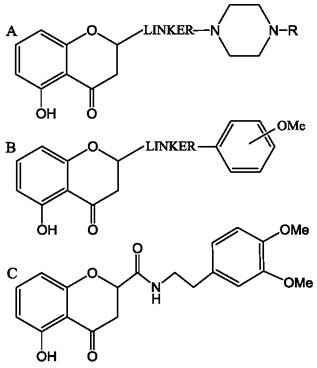
Aurones are also effective inhibitors of ABC transporters. Their efficacy in suppressing the ATPase activity of ABC transporters can exceed that of analogous flavones and chalcones. This is because the benzofuran moiety of aurone molecules more closely resembles the adenine molecule than does the benzopyranone moiety of flavones (Fig. 85). Additionally, it is suggested that the presence of a hydroxyl group at position 4 can mimic the amino group at the corresponding position in adenine.
Fig. 85. Comparison of the benzofuran moiety of an aurone molecule with the adenylyl moiety of an ATP molecule.

2.5.11. Skin Protection against UV Radiation
2.6.5.1. STRUCTURE OF THE Skin Coverings
The skin consists of the epidermis and the dermis, which are separated by a basement membrane (Fig. 86). The upper layer of the skin, the epidermis, is constantly renewed through the division of cells in the basal layer underlying it. Following division, the daughter cells, or keratinocytes, gradually mature, initially forming the stratum spinosum (spinous layer). Moving further toward the skin surface, keratinocytes form the stratum granulosum (granular layer). As they approach the surface, keratinocytes die and transform into corneocytes of the stratum corneum (horny layer). Corneocytes lack cytoplasm and are filled with the protein keratin. The epidermal stratum corneum, formed by corneocytes, serves as the primary skin barrier that prevents the penetration of substances and pathogens. Interspersed between corneocytes are layers of lipid bilayers containing ceramides and sphingosines with long saturated hydrocarbon chains, creating a barrier against substance penetration. Furthermore, the basement membrane—a thin layer containing collagen, Elastin, and fibrillin fibrils—acts as another crucial barrier to the penetration of substances.
Fig. 86. Schematic representation of skin structure. Proportions have been altered for clarity. In actual skin, unlike the diagram shown, the thickness of the epidermis is tens of times smaller than that of the dermis.
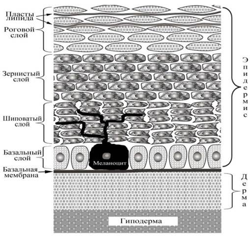
Beneath the basement membrane lies the dermis (propria). The basal layer also contains melanocytes, which use long cellular extensions (dendrites) to deliver melanin-filled melanosomes into the intercellular spaces of the overlying epidermal layers, thereby forming melanin granules that protect the epidermis from the effects of ultraviolet radiation.
The dermis provides the mechanical Properties of the skin, such as strength combined with elasticity and extensibility, which is achieved through the specific Organization of a network composed of Collagen and elastin fibrils. The space between the fibrils is filled with a glycosaminoglycan (mucopolysaccharide) gel containing a large amount of water, allowing the gel to function as a Shock absorber. In addition, the dermis contains Blood and Lymphatic vessels, Sweat Glands, Hair follicles with attached muscle fibers, nerve endings, pressure receptors (Meissner corpuscles), stretch receptors (Ruffini endings), and cold receptors (Krause end bulbs). Cellular elements such as mast cells (tissue basophils), fibroblasts, T-lymphocytes, and macrophages are also present.
Beneath the dermis lies the subcutaneous tissue, or hypodermis, which is the deepest layer of integumentary tissue of mesenchymal origin. The hypodermis consists of loose connective tissue and adipose tissue containing adipocytes. Furthermore, the hypodermis houses fibroblasts, macrophages, blood vessels, nerve endings, and pressure receptors (Pacinian corpuscles).
2.6.5.2. Mechanisms of the Protective Action of Flavonoids against UV Radiation
Prolonged, intense exposure of the skin surface to ultraviolet radiation leads to oxidative stress, DNA damage, and inflammatory processes. UV irradiation can cause various skin diseases, among which one should mention not only premature Aging but also serious conditions such as melanoma and non-melanoma skin cancers. The ultraviolet components of the solar spectrum can be conventionally divided into three ranges: short waves, medium waves, and long waves (Table 16).
Table 16. Characteristics of UV ranges.
UV Range Designation |
Wavelength, nm |
Characteristics |
(A) Long waves |
320-400 |
Accounts for 90-95% of the ultraviolet spectrum. Referred to as "aging light". Penetrates to a depth of up to 1 mm. Can induce cancer. Triggers lipid peroxidation, the generation of free radicals and singlet oxygen, which may lead to macromolecular damage, including DNA. Can suppress The Immune System. |
(B) Medium waves |
290-320 |
Accounts for 5% of solar UV radiation. Causes various skin diseases. Penetrates 160-180 µm through the entire epidermis into the dermis. Can induce oxidative stress and DNA damage. Acts as an initiator and promoter of carcinogenesis. |
(C) Short waves |
200-290 |
Absorbed by the ozone layer and does not reach the Earth's surface. Can cause severe damage to various molecules, including DNA. Penetrates to a depth of 60-80 µm. |
Most plant-derived polyphenolic compounds are capable of absorbing radiation in the UV range and can therefore function as a screen (Fig. 87). Indeed, it has been experimentally demonstrated that applying plant extracts to the skin surface significantly reduces the skin's response to ultraviolet irradiation. However, the protective effect of these substances is not limited solely to shielding tissues from the ultraviolet spectrum. Protection is also achieved through their action on cellular regulatory systems.
Fig. 87. Absorption spectra: 1 - aqueous extract of coltsfoot (Tussilágo fárfara); 2 - rutin.

A significant impact on skin aging processes induced by UV radiation is exerted by the increased expression of metalloproteases, which are capable of degrading proteins in the extracellular matrix. Specifically, collagen degradation occurs and the formation of new collagen fibrils is disrupted, leading to impaired mechanical properties of the skin and wrinkle formation. Certain flavonoids—such as apigenin, luteolin, and amentoflavone—can suppress the expression of metalloproteinases by inducing the influx of calcium cations into the cytoplasm, thereby affecting the calcium-dependent MAPK signaling pathway. Furthermore, some catechins, including EGCG, can stabilize collagen fibrils and prevent their degradation by collagenases. It has been demonstrated that various polyphenolic compounds can directly interact with the hydrophobic sites of collagen. This can facilitate the accelerated formation of collagen fibrils from monomers, as shown in the case of taxifolin's effect on fibril formation dynamics. It is known that at a neutral pH (in a phosphate buffer), collagen molecules form fibrils similar to those present in tissues. It was found that in the presence of taxifolin, The process of fibrillogenesis is significantly accelerated. Under an Electron microscope, the fibrils formed in the presence of taxifolin exhibit a higher degree of order and display the characteristic periodic banding pattern of this object (Fig. 88).
Fig. 88. Effect of taxifolin on the formation of collagen fibrils in a solution of molecules of this protein. A - change in light scattering intensity due to fibril formation. The arrow indicates the moment the protein solution is injected into the cell. (1) - control collagen preparation; (2) - same, in the presence of 0.001% taxifolin; (3) - in the presence of 0.01% taxifolin. The arrow indicates the moment the taxifolin solution is injected. B - electron micrograph of collagen bundles formed in the presence of 0.01% taxifolin. B’ - an individual collagen thread formed in the presence of taxifolin. Transverse banding is visible.
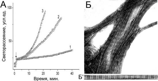
In addition, differential scanning microcalorimetry indicates a concentration-dependent increase in the thermostability of fibrils formed in the presence of taxifolin. These data suggest that taxifolin may enhance skin resistance to damaging factors by accelerating the formation of collagen fibrils and increasing their stability. Differential scanning microcalorimetry also demonstrates that flavonoids or their plant extracts can protect collagen from the effects of ultraviolet radiation (Fig. 89).
Fig. 89. Effect of UV irradiation on collagen thermograms obtained using differential scanning microcalorimetry: (a) - control collagen sample melts at 41°C. Ultraviolet irradiation from an SVD-120A ultra-high-pressure mercury lamp for (b) - 30 sec and (c) - 2 min destabilizes collagen and lowers the melting point; (d) - coltsfoot extract (0.04%) and (e) - a mixture of coltsfoot (0.04%) and aloe (0.015%) extracts partially preserve collagen thermostability. For thermograms (d) and (e), the UV irradiation time is 2 min.

In experiments on keratinocytes and skin areas irradiated with ultraviolet light in the UVB range, it was found that one of the main damaging factors of ultraviolet radiation is the appearance of reactive oxygen species in the cytoplasm. However, as noted, the quantum energy of electromagnetic radiation in the UV-B range is insufficient to initiate the highly intensive oxidation processes and the accumulation of reactive oxygen species (ROS) that are actually observed in the cytoplasm of keratinocytes upon irradiation. It was discovered that the appearance of ROS is associated with the activation of the NF-$\kappa$B factor and the subsequent expression of NADPH oxidase and COX-2, the activity of which causes the accumulation of ROS in keratinocytes. Cytoplasmic cyclooxygenase activity and the accumulation of lipid peroxidation (LPO) products are linked to an increase in the concentration of calcium cations in the cytoplasm upon UV irradiation.
Antioxidants appear incapable of protecting the skin from the consequences of UVB irradiation. For instance, it was experimentally shown that the antioxidant ionol was ineffective in protecting cells subjected to UV irradiation, whereas the cyclooxygenase COX-2 blocker aspirin reduced the LPO concentration in keratinocytes. Because UV-B irradiation induces the activity of COX-2 cyclooxygenase, the concentration of prostaglandins (PG) derived from arachidonic acid increases in skin cells. As a result, inflammatory processes develop, edema is observed, keratinocyte proliferation and epidermal hyperplasia are accelerated, and oxidation products accumulate, leading to oxidative DNA damage. Consequently, chronic irradiation leads to the accumulation of Mutations, resulting in malignant transformation of keratinocytes and the development of carcinogenesis. Conversely, the action of COX-2 inhibitors or agents preventing the expression of this enzyme can significantly hinder the carcinogenic transformation of epidermal cells. Suppressing COX-2 expression while undergoing a course of UV therapy can be effective in treating certain forms of keratosis (Darier's disease). This condition is associated with the overexpression of the sarcoendoplasmic reticulum Ca2+-ATPase, which regulates the concentration of calcium cations in myocytes and certain other cells, including keratinocytes. Ultraviolet radiation decreases the expression of this enzyme, which alleviates the course of the disease, whereas COX-2, conversely, significantly increases Ca2+-ATPase expression, thereby worsening the patients' condition. Suppressing COX-2 expression alongside concurrent UV therapy may also be effective in treating erythema.
The ability of certain flavonoids to suppress COX-2 expression may underlie the mechanisms of these substances' protective action against UV radiation, as demonstrated with baicalein, wogonin, hesperetin, mangiferin, and tangeretin. It is possible that this regulation is mediated through the MAPK pathway, as shown in the case of luteolin. Using the methylated flavonol 5,7-dimethoxyflavone, it was demonstrated that not only COX but also Other components of the regulatory chain, such as PPAR and NF-$\kappa$B, can be subject to regulation, resulting in a decrease in the concentration of pro-inflammatory cytokines (IL-6, IL-8). Furthermore, a reduction in metalloprotease expression and oxidative stress components is observed, and the activity of inflammatory components is suppressed via the NF-$\kappa$B and MAPK regulatory pathways. Ultimately, UV radiation-induced skin aging is prevented.
Flavonoids can also influence other regulatory systems in skin cells. It was recently discovered that silibinin prevents epidermal damage after UVB irradiation by activating the p53 tumor suppressor protein, which leads to the activation of the GADD45$\alpha$ protein (Growth arrest and DNA-damage-inducible protein), helping to protect cells under stress and DNA damage conditions. The flavone chrysin protects epidermal keratinocytes from UVA- and UVB-irradiation damage primarily by restoring the expression of aquaporin 3 (AQP-3), which ensures the normalization of cellular osmotic and salt balance disrupted by irradiation. In a study of the protective effect of eriodictyol on keratinocytes, it was found that this flavanone acts via phosphatase-dependent modulation of the p38 MAPK and Akt signaling pathways. Eriodictyol is capable of directly associating with the respective protein kinases involved in the phosphorylation of these signaling system components. Blackberry anthocyanins protect keratinocytes from UV radiation by significantly enhancing the expression of antioxidant enzymes: catalase, mitochondrial superoxide dismutase, and glutathione peroxidase, thereby preventing the development of oxidative stress. Grape procyanidins also prevent the production of reactive oxygen species in cells, but through a different mechanism—the suppression of p38 (MAPK14) and JNK1/2 (MAPK8) expression. 5,7-Dimethoxyflavone protects the epidermis by inducing melanogenesis and increasing melanin content via the initiation of transcription of corresponding melanocyte genes through a cAMP-dependent signaling pathway. The soy isoflavone daidzein metabolite, 7,3',4'-trihydroxyisoflavone, prevents UV-induced skin cancer by acting on the ATP-binding sites of the Cot and MKK4 protein kinases. Notably, the parent daidzein molecule is incapable of interacting with these proteins and exhibits no anticarcinogenic activity under these conditions.
2.5.12. Bactericidal Properties of Flavonoids
In recent years, there has been an increase in the number of antibiotic-resistant bacterial strains, creating additional difficulties in treating many diseases. There are several ways to solve this problem, including the creation of new drugs or the modification of existing ones, which provides only a brief respite. Promising approaches involve the creation of adjuvant agents aimed at suppressing bacterial resistance mechanisms or agents capable of enhancing the human body's resistance to bacterial infections. For the development of these areas, searching for natural products with the requisite properties is of great importance. Plant materials rich in polyphenolic compounds have been used to treat bacterial diseases for centuries. Some of these possess the necessary ability to reduce the virulence of pathogenic strains or boost the body's defense mechanisms. Thus, substances that inhibit bacterial growth at concentrations below 10 $\mu$g/mL are of great interest to pharmacology. However, certain flavonoids, such as panduratin A, are capable of inhibiting bacterial growth at concentrations below 0.1 $\mu$g/mL, as demonstrated in Staphylococcus aureus (Table 17).
Table 17. The ten most active antibacterial flavonoids.
Flavonoid |
Gram (+) |
Gram (-) |
Flavonoid |
Gram (+) |
Gram (-) |
Panduratin A |
0.06-2.0 |
- |
Sepicanin A |
1.2 |
- |
Isobavachalcone |
0.3-0.6 |
0.3-39 |
Isolupalbigenin |
1.6-3.1 |
- |
Bartericin A |
0.6-2.4 |
0.3-39 |
Flavone |
7.8-31.3 |
1.95-31.3 |
Scandenone |
0.5-0.8 |
2.0-32 |
3'-O-methyl-diplacol |
2.0-4.0 |
> 32 |
Kaempferolrhamnoside |
0.5-16 |
> 16 |
Licochalcone A |
2.0-8.0 |
- |
Note. Minimum inhibitory concentrations of substances in $\mu$g/mL against Gram-negative and Gram-positive bacteria are presented.
Several theories explain The Mechanism of the antibacterial action of polyphenolic compounds. It is hypothesized that catechins can damage the bacterial plasma membrane due to the initiation of hydrogen peroxide production by host cells. For example, the antimicrobial activity of catechins (0.5 mg/mL) against actinomycete and Candida strains was shown to be associated with hydrogen peroxide production. Leveraging this effect, a bactericidal gel containing catechins was developed. However, this mechanism is not universal. Conversely, owing to their antioxidant properties, plant extracts containing polyphenolic compounds can also protect bacteria from the effects of hydrogen peroxide. There are also instances of independent antioxidant and bactericidal actions of plant polyphenols. Thus, an Histology/2.html">EXTRACT FROM THE stem of the tropical plant Schotia latifolia, containing high amounts of proanthocyanidins and Tannins, exhibits potent antioxidant activity, thereby efficiently scavenging agents such as hydrogen peroxide, nitric oxide, and lipid peroxidation products from the medium. Despite this, the extract also demonstrated antibacterial activity against both Gram-negative and Gram-positive bacteria.
There is evidence that the antibacterial activity of flavonoids is associated with damage to the bacterial plasma membrane, resulting in the efflux of potassium from the cytoplasm, as demonstrated in the case of galangin, a bactericidal flavonoid from propolis. Disruptions in the bacterial cell wall structure and cell lysis linked to the disorganization of lipoteichoic acids in the envelope of Staphylococcus aureus were also observed upon treatment with the bactericidal green tea catechin EGCG.
Another reason for the bacteriostatic and even bactericidal action of polyphenols may be the ability of these agents to induce cell aggregation and membrane damage. A similar effect was also observed on phosphatidylcholine Liposomes treated with EGCG. Other catechins with lower bactericidal activity did not cause such an effect on cells or liposomes. Liposome aggregation from various phospholipids was also observed upon treatment with isoflavones. Certain flavonoids, such as quercetin, catechin, and taxifolin, are capable of inducing aggregation and even slow membrane fusion in the presence of iron cations, which can form bridges between two flavonoid molecules "anchored" in adjacent membranes. Calcium cations can facilitate membrane adhesion by forming bridges between lipid phosphate groups. Cell aggregation can lead to growth inhibition and death due to reduced nutrient access and the accumulation of metabolic waste products. Furthermore, in the presence of EGCG, aggregation not only of cells but also of isolated protein molecules extracted from bacteria can occur, resulting in the suppression of their enzymatic activity.
It was discovered that some flavonoids can specifically interact with certain bacterial cell proteins, causing disruptions in their functioning. For instance, epicatechin gallate (ECG) can penetrate the envelope of Staphylococcus aureus and disrupt the functioning of penicillin-binding protein, which increases the susceptibility of penicillin-resistant strains to penicillin Antibiotics. The flavonoid galangin from Alpinia officinarum is capable of overcoming Staphylococcus aureus resistance to antibiotics associated with the activity of $\beta$-lactamase, an enzyme determining bacterial resistance to antibiotics (Penicillins, Cephalosporins, etc.). Quercetin and baicalein exhibit similar, albeit less pronounced, activity.
Flavonoids can disrupt the operation of the genetic apparatus in bacterial cells. For example, catechins, especially EGCG, can inhibit bacterial DNA gyrase activity by interacting with the ATP-binding site of this protein. The high activity of EGCG is attributed to the ability of the benzopyran ring of this catechin to penetrate deeply into the Active Site of gyrase. Soy isoflavones can inactivate topoisomerases I and II in the cytoplasm of Staphylococcus aureus, leading to more than a twofold decrease in cellular DNA and RNA content. The antibacterial activity of quercetin and apigenin is associated with the ability of these flavonoids to inhibit D-Ala-D-Ala ligase through competitive interaction with the DNA-binding site of this enzyme. The flavonoids morin, silymarin, baicalein, silibinin, rimantadine (not to be confused with the well-known antiviral agent remantadine), amantadine, and epicatechin can inhibit the F1F0 ATP synthases of E. coli membranes, thereby disrupting cellular energetics.
Flavonoids are capable of disrupting the functioning of various enzymes involved in the synthesis of bacterial membrane lipids. Thus, a polyphenol extract from the leaves of Acer truncatum, growing in China and Korea, inhibits beta-oxoacyl-acyl-carrier-protein reductase (FabG), which is involved in the NADPH-dependent fatty acid synthesis in bacteria. Flavonoids hinder the interaction of NADPH with this enzyme. As a result, a strong cytotoxic effect is observed not only against Gram-positive and Gram-negative bacteria, but also against certain fungi. Quercetin, apigenin, and sakuranetin (a flavanone present in rice) can suppress the activity of the beta-hydroxyacyl-acyl-carrier-protein dehydratase (HpFabZ) from Helicobacter pylori through hydrophobic interactions with the substrate-binding sites of this protein, which is involved in membrane lipid synthesis. The flavonoids butein, isoliquiritigenin, and fisetin suppress the growth of various bacteria, including Mycobacterium tuberculosis, by inhibiting the Rv0636 dehydratase involved in the functioning of fatty acid synthase (FAS-II) necessary for the production of membrane lipids in the cell. The flavonoids naringenin, eriodictyol, and taxifolin are capable of suppressing the activity of beta-ketoacyl-acyl-carrier-protein synthase (KAS), which is involved in bacterial FATTY ACID Biosynthesis. Consequently, these flavonoids exhibit bactericidal action against Enterococcus faecalis.
Last update: 06/08/2026
Editorial and Educational Adaptation: This material has been compiled based on the primary/original source text. The project team performed an editorial review, corrected technical inaccuracies, structured sections, and adapted the content for an educational format.
What was processed:
- elimination of formatting defects (OCR errors, structural breaks, corrupted characters);
- editorial organization of content;
- standardization of terminology in accordance with academic sources;
- verification of factual statements against the original source text.
All mentions of the author, publication year, and origin of the primary text have been preserved in accordance with the source.