FLAVONOIDS: BIOCHEMISTRY, BIOPHYSICS, MEDICINE - Tarakhovsky Yu. S. - 2013
PART 2. MECHANISMS OF FLAVONOID ACTION
2.4. Effects of Flavonoids on Biological Membranes
The Cytoplasm of any Cell is separated from the external environment by a cell membrane, known as The Plasma Membrane. In Eukaryotic Cells, membranes also separate various intracellular compartments, distinguishing Organelles such as The Nucleus, Mitochondria, Golgi apparatus, Endoplasmic reticulum, vacuoles, METABOLISM/14.html">Chloroplasts, and Lysosomes within The Cell.
Membrane Functions are diverse, but their primary function is barrier formation. Membranes separate two aqueous environments—for example, the extracellular milieu and the cytoplasm, the cytoplasm and karyoplasm (nuclear content), the cytoplasm and the vacuolar lumen, and so forth. Crucially for cell survival, membranes act as selective barriers with permeability tailored to different substances. Virtually all vital substances can cross membranes, but the pathways, mechanisms, and directions of their transport vary, forming the foundation of cellular metabolism. These barrier properties protect the cell from pathogen invasion and enable the targeted transport of metabolic components.
Functioning as semipermeable barriers, membranes are also capable of signal reception and the selective transmission of signals from the cell surface to the cytoplasm, which defines their signaling function. However, many vital processes are directly associated with the membrane itself, occurring either on its surface or within its Hydrophobic core. The membrane is highly structured, with different regions varying not only functionally but also in chemical composition. Its two opposing surfaces also differ significantly in both function and chemical composition, establishing a vector directionality for transmembrane transport.
Along with high structural Organization, membranes are dynamic liquid-crystalline assemblies. Their structural foundation is a lipid bilayer formed by Phospholipids. Both natural phospholipids and their synthetic analogs are known to spontaneously form bilayer structures upon Hydration. The most prominent phospholipid that spontaneously forms bilayers, typically as Liposomes, is phosphatidylcholine. Total phospholipid fractions extracted from natural sources, such as egg yolk Lipids (egg lecithin), soybean lipids (soy lecithin), and Brain lipids from various animals, can also readily form liposomes.
Beyond phospholipids, the structural diversity and functional specialization of cell membranes are largely determined by the presence of Proteins and other molecules, primarily Cholesterol. The concept that Membrane Proteins—whether surface-attached or integrated into the phospholipid bilayer—can diffuse laterally within the bilayer like a fluid, and in some cases aggregate into "islands" within a fluid lipid "sea," is embodied in the fluid-mosaic model of the membrane proposed by Singer and Nicolson in 1972. Despite subsequent modifications and additions, this model remains relevant to this day.
The structural basis of Introduction/36.html">Biological Membranes is a lipid bilayer. Lipids serve as the primary building blocks that dictate many of the Physical Properties of membranes and provide the specific environment necessary for membrane protein function. The term "lipid" originates from the Greek "lipos," meaning fat. They constitute a broad Class of natural Organic compounds extracted from the Tissues of various organisms using organic Solvents (such as chloroform, alcohols, and acetone). According to modern Classification, lipids are divided into eight major classes: glycerolipids, Glycerophospholipids, Fatty acids, Sphingolipids, sterol lipids, prenol lipids, Glycolipids, and Polyketides. The majority of membrane-bound lipids belong to the glycerophospholipid class, often referred to simply as phospholipids.
Lipid molecules feature a polar, Water-interacting (hydrophilic) moiety and a nonpolar, water-insoluble (hydrophobic) moiety consisting of hydrocarbon chains, which are frequently fatty acid residues (Fig. 57). The polar HEAD group may contain one or more negatively charged phosphate groups. Most lipids also bear additional charged hydrophilic groups within their polar region, determining the net molecular charge—typically negative or neutral, although positively charged lipids also occur in nature.
Fig. 57. Most common lipids in eukaryotic membranes. PC - phosphatidylcholine, LPC - lysophosphatidylcholine, PA - phosphatidic acid, LPA - lysophosphatidic acid, PE - phosphatidylethanolamine, PG - phosphatidylglycerol, PS - phosphatidylserine, CL - cardiolipin, Chol - cholesterol, FA - fatty acid,
PI - phosphatidylinositol, PIP3 - phosphatidylinositol trisphosphate, DAG - diacylglycerol, Sph - sphingosine, Sph1P - sphingosine-1-phosphate, SphM - sphingomyelin, Cer - ceramide, GlcCer - glucosylceramide. The glycerol backbone is highlighted in bold.
Flavonoids/flavonoids.files/image060.jpg" width="588"/>
Many of the lipids shown are glycerolipids. Frequently, they also contain a phosphate group and are thus classified as glycerophospholipids. In the phosphatidylcholine (PC) molecule, as in most glycerophospholipids, There is a glycerol backbone esterified with fatty acid residues at positions 1 and 2. Notably, the fatty acid at position 2 often contains one or more cis-conformation double bonds. In sphingolipid molecules (SphM, GlcCer, PI, PIP3), the hydrocarbon chain at position 2 is attached via an amide bond. The polar region of the PC molecule contains two charged groups: a negatively charged phosphate group and a positively charged Choline group. Consequently, the net charge of phosphatidylcholine is zero. This polar head group exists as a zwitterion and exhibits dipole properties. LPC possesses a similar polar group, differing from PC only by the absence of the fatty acid residue at position 2. The SphM molecule also exhibits zwitterionic properties, with a polar region identical to that of PC. Additionally, there is a group of uncharged, neutral molecules, which includes Chol, DAG, Cer, and GlcCer. The negative charge residing on the phosphate group is frequently not counterbalanced by a positive charge, resulting in a net negative molecular charge. Molecules of this type include PA, LPA, PG, PI, and Sph1P. Two negative charges and one positive charge are present in the phosphatidylserine (PS) molecule, whereas cardiolipin (CL) contains two phosphate groups and, accordingly, two negative charges. Among the lipids presented, the PIP3 molecule carries the highest number of negative charges (approximately five).
Table 8. Lipid Composition of various rat Organs.
Lipid |
Tissue |
||
Erythrocytes |
|||
Chol esters |
- |
2 |
- |
TAG |
4 |
7 |
- |
DAG |
1 |
- |
- |
Chol |
4 |
5 |
30 |
CL |
12 |
5 |
- |
PE |
33 |
20 |
21 |
PI |
4 |
4 |
3 |
PS |
- |
- |
3 |
PC |
39 |
55 |
32 |
SphM |
2 |
2 |
8 |
LPC |
- |
- |
1 |
Lower Variability is characteristic of both major abundant lipids (PC, PE) and certain minor lipids, such as PI. Since erythrocytes contain only a plasma membrane, comparing the erythrocyte data presented above with the plasma membrane composition of other cells is of particular interest (Fig. 58).
Fig. 58. Phospholipid composition and transbilayer Asymmetry of phospholipids in the plasma membrane of cells from various human tissues.
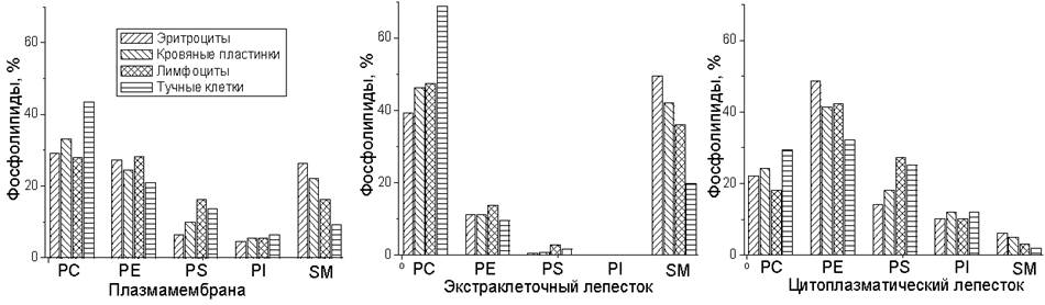
Analysis of membrane lipid composition across various sources reveals substantial variations (Table 8). Significant variability in CL content is related to differences in mitochondrial density among tissues of various origins. Substantial variations in Chol and SphM content are also observed.
The data presented for cells from various human tissues demonstrate that phosphatidylcholine is dominant in the plasma membrane in the majority of cases, although, as seen in the previous table (Table 9), this is not a universal rule for all mammals. Furthermore, PC and SphM predominate in the outer monolayer of Plasma Membranes across all cell types, whereas PE, PS, and PI are markedly enriched in the inner monolayer.
Table 9. Phospholipid composition of rat hepatocyte membranes isolated from various subcellular fractions.
Lipid |
Membranes (mol % of phosphorus) |
||||
Whole tissue |
Nucleus |
Mitochondria |
Microsomes |
Plasma membranes |
|
CL |
5 |
- |
15 |
2 |
- |
PE |
25 |
26 |
34 |
22 |
20 |
PI |
7 |
4 |
7 |
8 |
7 |
PS |
3 |
6 |
1 |
4 |
4 |
PC |
51 |
57 |
41 |
59 |
43 |
SphM |
4 |
6 |
2 |
4 |
23 |
LPC |
1 |
- |
1 |
2 |
2 |
Differences in lipid composition between the outer and inner monolayers of The cell membrane are established during biogenesis. In addition, these asymmetries are maintained by flippases, which catalyze ATP-dependent lipid translocation between the monolayers. Flippases reside not only in the plasma membrane but also in the membranes of the Golgi apparatus, where they ensure the asymmetrical arrangement of lipids and proteins within bilayers destined for delivery to the plasma membrane via Vesicular Transport.
The asymmetric distribution of PS in the plasma membrane plays a critical role in numerous biological processes. For instance, during apoptosis, PS asymmetry is disrupted, and this lipid migrates from the inner monolayer to the outer leaflet. This serves as an "eat-me" signal that activates phagocytes to clear dying cells. Thus, the asymmetric localization of PS in plasma membranes participates in regulating immune responses directed against dead or dying cells, including tumor cells.
The lipid composition of cellular organelles also varies. The main reason for these differences lies in the Specific features of lipid synthesis and intracellular trafficking. Most lipids, including the major structural phospholipids, ceramides, and cholesterol, are synthesized in The endoplasmic reticulum. The synthesized lipids are then transported to other PARTS OF THE cell. For instance, although cholesterol is synthesized in the endoplasmic reticulum (ER), its concentration there is low because it is rapidly delivered to the plasma membrane. Conversely, the ER is rich in lipids that are atypical for the plasma membrane, such as DAG, which serves as a precursor in the synthesis of many lipids. Dolichol, a polyisoprenoid lipid involved in the glycosylation of proteins and lipids, is also present here.
The Golgi apparatus is also involved in lipid synthesis. It specializes in the synthesis of sphingolipids, producing SM, glucosylceramide (GlcCer), and lactosylceramide (LacCer), which are subsequently transported to the plasma membrane.
The flow of membrane material—and with it, lipids—moves not only from the cytoplasm to the cell surface, but also in the reverse direction. The plasma membrane delivers lipids to the cytoplasm via endocytosis. Early endosomes differ little from the plasma membrane, but as they mature, they gradually lose cholesterol and PS while becoming enriched in bis-(monoacylglycero)phosphate (BMP), which is involved in endosomal fusion and the Hydrolysis of sphingolipids. Phosphoinositol phosphates, bearing one or more phosphate groups attached at various positions on the Inositol ring, also play a major role during endosomal maturation by regulating vesicular trafficking processes.
The lipid composition of mitochondria, especially the inner membrane forming the cristae, differs sharply from that of other cellular compartments, yet closely resembles that of Bacteria. First and foremost, it is essential to mention the presence of CL in The inner mitochondrial membrane, which is also characteristic of bacterial cell membranes. The metabolism of CL is likewise associated with the presence of PA and PG in the mitochondrial membrane. SphM and PS are practically absent from the membrane, whereas the PE content is high. The cholesterol content in the inner mitochondrial membrane is generally low, although an exception is found in steroid-synthesizing cells, where cholesterol is also present in the mitochondrial membranes.
2.4.1. Interaction of flavonoids with the phospholipid bilayer
For flavonoids, as for many other BIOLOGICALLY ACTIVE SUBSTANCES, Structure/106.html">Hydrophobicity and, accordingly, The ability to interact with biological membranes are prerequisites for pharmacological activity. However, flavonoid molecules, being polyphenols, possess a significant number of hydroxyl groups that determine the polarity of the molecule and are responsible for weak acidic properties. An inverse correlation between the number of hydroxyl groups and flavonoid hydrophobicity has been demonstrated experimentally. Furthermore, differential scanning microcalorimetry (DSC) studies have revealed substantial differences in the ability of flavonoids to affect lipid melting processes (Fig. 59).
Fig. 59. A - DSC data for multilamellar DMPC liposomes containing flavonoids (flavonoid:lipid = 1:1 mol/mol): (a) - DMPC control, (b) - liposomes with rutin, (c) - with catechin, (d) - myricetin, (e) - taxifolin, (f) - quercetin, (g) - phloretin. B - Chemical formulas of the listed flavonoids.
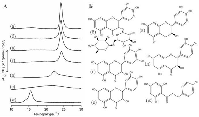
Thus, The Effect of flavonoid Glycosides, such as rutin, on lipid melting was virtually unobservable. This is because glycosides are more water-soluble than their corresponding aglycones and are therefore able to interact only with The surface of The Lipid Bilayer, whereas their influence on the melting of the hydrophobic region is negligible. Catechin and myricetin—flavonoids known as relatively hydrophilic compounds that interact weakly with the phospholipid bilayer—exert a similarly negligible effect on lipid melting.
The Influence of taxifolin and quercetin on lipid melting was significantly greater. The transition width increased, while the transition maximum decreased by several degrees. Studies utilizing fluorescent probes indicate that the effect of quercetin on lipid melting can be characterized as an increase in membrane viscosity. It is hypothesized that quercetin localizes at the interface between the polar and nonpolar Regions of the bilayer. The effect of quercetin on the fluid Properties of the membrane is comparable to that of cholesterol, suggesting the possibility that quercetin molecules penetrate into the hydrophobic core of the bilayer and interact with the lipid hydrocarbon chains.
Consistent with the provided thermograms (Fig. 59), it can be concluded that the effect of taxifolin on lipid melting was smaller compared to that of quercetin. An explanation for these observed differences can be found by comparing their molecular shapes. It is assumed that the planar molecule of quercetin has to overcome smaller steric barriers when penetrating between densely packed lipid hydrocarbon chains compared to the bent molecule of taxifolin.
2.4.2. Localization of flavonoids in the bilayer
Information regarding the localization of flavonoids within the bilayer is quite contradictory. According to some researchers, they are capable of penetrating deep into the bilayer; however, the presence of numerous hydroxyl groups enables polyphenol molecules to form Hydrogen Bonds with lipids, facilitating their interaction with the more polar regions. In reality, much depends on the pH of the medium, which determines the magnitude of the charges on both the flavonoid and the lipid. The lower the pH, the lower the degree of flavonoid deprotonation and the deeper they are able to penetrate into the bilayer.
Catechins bearing gallate groups (ECG, EGCG) are adsorbed by the bilayer to a greater extent than catechins lacking gallates (EC, EGC). Various Methods FOR STUDYING the interaction of catechins with liposomes reveal the following order of affinity of these substances for the lipid bilayer: ECG> EGCG> EC> EGC. This sequence correlates with the lipophilicity of these substances, the magnitude of which is expressed as the partition coefficient (log P) in the octanol:water system.
Many studies on the interaction of flavonoids with membranes have been carried out using catechins. Upon adsorption by the bilayer, all catechins penetrate into the region situated deeper than the phosphate groups and distribute within the plane of the bilayer via lateral diffusion. Molecular modeling shows that membrane penetration results in an increase in the bilayer area by 0.374 nm2 per EGCG molecule. At the same time, an average of 10.8 lipid molecules interact with each EGCG molecule. Lipids adjacent to the EGCG molecule are tightly packed, occupying an area of 0.51 nm2, which is 0.14 nm2 smaller than that of the remaining molecules. All catechin molecules tend to be unevenly distributed within the plane of the bilayer and form aggregates. Catechin aggregation is observed both in molecular models of the lipid bilayer and in experiments with Artificial Membranes. The appearance of flavonoid aggregates in the bilayer can lead to defects in the bilayer structure, compromise the integrity of the hydrophobic barrier, and increase membrane permeability.
Residing within the bilayer, the hydroxyl groups of catechins form hydrogen bonds with oxygen atoms in the lipid molecules. The greater the number of hydroxyl groups, the stronger the interaction. Under these conditions, membrane permeability to fluorescent Dyes, such as calcein, may increase. The formation of hydrogen bonds between the catechol hydroxyl groups of flavonoids and the oxygen atoms of lipid peroxides dictates the Antioxidant Properties of catechins. Overall, it is hypothesized that the formation of these hydrogen bonds may play a significant role in the anticarcinogenic and antibacterial activities of catechins.
NMR studies using nanoscale, isotropically tumbling fragments of a planar phospholipid bilayer, known as bicelles, provide insight into the interaction process between catechins and the bilayer. It has been demonstrated that catechins interact with the phosphatidylcholine bilayer (Fig. 60), with the galloyl group of ECG or EGCG positioned in close proximity to the trimethylammonium group belonging to the phosphatidylcholine molecule. Because this group carries a positive charge, NMR allows the observation of its interaction with the π-electrons of the galloyl group. This interaction between catechins and the cationic charge of the phosphatidylcholine molecule (cation-π interaction) is of great importance in stabilizing the polyphenol molecule within the interfacial region of the phospholipid bilayer. Previously, analogous cation-π interactions between the nitrogen atoms of phosphatidylcholine and phosphatidylethanolamine and the Tryptophan residues of membrane proteins were also investigated using gramicidin molecules as a model.
Fig. 60. Location of various flavonoids within the phospholipid bilayer. For simplicity, a DOPC monolayer is shown. The positions of the phosphate and carbonyl groups are indicated by horizontal dashed lines. The preferred orientation within the monolayer is shown for epicatechin gallate (ECg) from the study, daidzein (Dai) and genistein (Gen) from the study, as well as quercetin (Que) and the protonated form of quercetin sulfate (QueS) from the study. Data were obtained from X-ray scattering, NMR spectroscopy, and molecular modeling.

NMR studies have also shown that catechin molecules are capable of rotating within the bilayer around an axis inclined at an angle of approximately 55° to the plane of the bilayer, with the distance from the nearest lipid phosphate group to the carbonyl carbon of the catechin being 5.3 ± 0.1 Å (Fig. 60). The flavonoids quercetin, genistein, and daidzein are likewise located closer to the polar head groups than to the hydrophobic center of the bilayer. Furthermore, in accordance with recent studies, it is hypothesized that the long axis of the molecule is oriented preferentially parallel to the plane of the bilayer, while the plane of the rings is perpendicular to the bilayer plane. The effect of flavonoids on the bilayer is sometimes compared to that of cholesterol, although it is known that the long axis of a cholesterol molecule is predominantly perpendicular to the plane of the bilayer.
The presence of flavonoids alters the mechanical properties of the bilayer. For instance, at maximum concentrations of genistein (20%) and daidzein (14%)—which represent the solubility limits of these substances in the lipid—the compression modulus of the POPC bilayer decreases by 40%. Simultaneously, the bilayer area increases by 8% and 12%, respectively, thereby enhancing the deformability of the bilayer.
Changes in the physical parameters of the bilayer can be crucial in explaining the ability of genistein and daidzein to prolong the open state of transmembrane protein channels by lowering the energy barrier required for the conformational expansion necessary for channel opening. Recent studies on the effect of flavonoids on the viscoelastic properties of various leukocyte cell membranes, measured using membranotropic fluorescent probes, have demonstrated the ability of quercetin and EGCG to increase bilayer anisotropy and elevate the transmembrane potential. This is hypothesized to be significant for the manifestation of the antioxidant and regulatory properties of these flavonoids.
It is known that in the bodies of humans and animals, flavonoids can undergo substantial chemical modifications. In particular, as discussed in more detail in the section of the book dedicated to the metabolic transformations of flavonoids, these molecules can be methylated, sulfated, and glycosylated. Methylation of one of the hydroxyl groups of quercetin has little effect on the interaction of this flavonoid with the phospholipid bilayer. Considerably greater changes are observed upon sulfation and glycosylation of the molecules. For example, in a neutral medium, the sulfated form of quercetin is less lipophilic than quercetin itself and localizes closer to the aqueous surface. However, in acidic media, protonation of the sulfo group occurs, which promotes the penetration of the molecule into the bilayer. The molecule may also reorient with its long axis spanning across the bilayer if it is sulfated at the 3’ or 4’ position (Fig. 60). The attachment of glucuronic acid significantly shifts the molecule toward the periphery of the bilayer such that the glucuronic moiety resides in the aqueous phase. These modifications can diminish the antioxidant properties of flavonoids against radicals located deep within the bilayer.
2.4.3. Penetration of flavonoids through the phospholipid bilayer
The interaction of flavonoids with biological membranes is a crucial process that dictates their effects on living cells. Flavonoids are capable of modulating the physical properties of the phospholipid bilayer, altering the Thermodynamic parameters of lipid melting, and influencing Lipid Peroxidation processes. Of great importance is also the ability of flavonoids to permeate cell membranes and specifically bind to cytoplasmic receptors. Investigating the interaction of flavonoids with membranes is likewise of current interest due to the potential medical application of liposomes for the storage and delivery of these compounds.
The question arises whether flavonoids are capable of crossing the phospholipid bilayer, or if specialized protein transporters are required for their transmembrane passage, which are known to be necessary in many cases for The entry of various substances into the cytoplasm? To address this question, one can examine the permeation of flavonoids through the multiple bilayers of phosphatidylcholine multilamellar liposomes. It can be hypothesized that the effect of flavonoids on phospholipid membrane melting will differ significantly depending on whether the flavonoid is added to a lipid solution in chloroform—i.e., prior to liposome formation (internal addition)—versus an experiment where the flavonoid is introduced into a suspension of pre-formed multilamellar liposomes (external addition). In the latter case, the flavonoid will freely interact only with the outermost membrane, whereas to interact with the remaining (and larger) portion of the lipid, the flavonoid must cross the hydrophobic barrier of numerous bilayers. However, it was found that a flavonoid added externally to liposomes exerted approximately the same effect on lipid melting (Fig. 61) as when it was introduced into chloroform prior to liposome preparation, i.e., incorporated internally.
Fig. 61. DSC thermogram showing the effect of the flavonoid taxifolin (dihydroquercetin) on the melting of DMPC liposomes. (A) Taxifolin added to the lipid solution in chloroform prior to liposome preparation (“internal”). (B) Taxifolin added to the aqueous suspension of pre-formed liposomes (“external”). Numbers indicate the concentration of the added flavonoid in mol %.
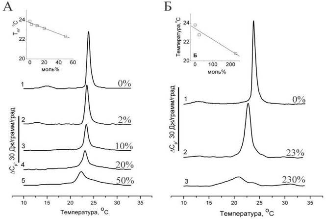
The microcalorimetric data presented above indicate that taxifolin added to the aqueous solution surrounding multilamellar liposomes is capable of influencing the melting process of the entire lipid mass, regardless of whether the lipid is part of the outer liposomal membrane or located in the interior. To explain this observed phenomenon, we can propose two hypotheses: 1) Taxifolin disrupts multilamellar liposomes, thereby significantly increasing the accessibility of lipids to taxifolin action. 2) The liposomes retain their structural integrity, but taxifolin is able to cross the phospholipid bilayer structures, allowing it to affect the melting of numerous membranes located within the multilamellar liposomes.
Freeze-fracture Electron Microscopy (Fig. 62) demonstrates that under our experimental conditions, giant multilamellar liposomes were present in the suspension both initially and in the presence of taxifolin. This indicates that taxifolin at the studied concentrations, when added to pre-formed liposomes, does not exert a disruptive effect on the phospholipid bilayer. At the same time, we found that the Morphology of the membrane fracture surfaces was significantly altered.
Fig. 62. Freeze-fracture Cytology/cytology/93.html">ELECTRON MICROGRAPHS OF DMPC liposomes. All samples were maintained at 37°C for 1 h and then cooled to 18°C prior to cryofixation. (a) Cross-fracture of multilamellar liposomes. (b) The same preparation showing hydrophobic fracture surfaces containing the ripple phase. Arrows indicate the step separating the fracture face of the outermost bilayer from that of the next innermost bilayer. (c) Suspension of liposomes with added taxifolin (23 mol %). (d) and (e) Higher magnification views of the fracture surface areas highlighted by boxes in (b) and (c), respectively.
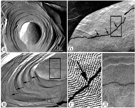
It is well established that the freeze-fracture technique permits The Study of the hydrophobic region of the phospholipid bilayer, as the fracture plane passes through the center of the membrane's hydrophobic core between the two lipid monolayers. Upon cryofixation from 18–20°C, the hydrophobic fracture surface of DMPC membranes exhibits characteristic Periodic structures of the ripple phase. The ripple-phase structure could be observed both on the fracture surfaces of the peripheral bilayer and on the hydrophobic fracture faces of all internal bilayers within the vesicles (Fig. 62).
In the presence of taxifolin added externally to pre-formed liposomes, the periodic ripple-phase structures were absent, and the hydrophobic fracture surfaces of the membranes appeared smooth. Such alterations were equally characteristic of bilayers located at the periphery and those situated in the central core of the multilamellar liposomes. It is known that the appearance of the ripple phase is directly related to the physical state of lipids at specific temperatures. For instance, in DMPC liposomes, the ripple phase forms within the Temperature interval between the pretransition (13–16°C) and the main phase transition (23–25°C) of this lipid.
Various agents that affect lipid melting, such as cholesterol or tocopherol, can modify The structure of the ripple phase and alter the temperature boundaries of its existence. The microscopy data presented here, demonstrating the disappearance of ripple structures in all layers of multilamellar liposomes, indicate that taxifolin can indeed penetrate the multiple phospholipid bilayer structures and influence the phase state and morphology of the membranes. Furthermore, the micrographs confirm that the multilamellar STRUCTURE OF THE liposomes is preserved at all taxifolin concentrations used in the experiment. The transmembrane permeation of taxifolin can be attributed to its high solubility in organic solvents and, consequently, its ability to penetrate deep into the hydrophobic core of the bilayer. This is further supported by alterations in lipid melting profiles recorded by DSC, as well as changes in the morphology of the hydrophobic fracture surfaces of liposomes observed via electron microscopy.
2.4.4. Effect on Lipid Phase Behavior
Despite possessing a considerable number of hydroxyl groups, flavonoids have rather limited solubility in water. Conversely, as mentioned above, there is ample evidence of their ability to interact with biological membranes and penetrate the phospholipid bilayer. Using differential scanning microcalorimetry, it has been demonstrated that flavonoids can also substantially affect the melting process of the phospholipid bilayer and presumably influence the phase behavior and Structural organization of biological membranes. Recently, we found that quercetin complexes with iron(II) exert the most pronounced effect on the bilayer-to-hexagonal HII phase transition characteristic of phosphatidylethanolamine, accompanied by an increase in the transition temperature by several degrees. Moreover, these complexes, much like free flavonoids, lower the melting temperature and broaden the transition width of both phosphatidylcholine and phosphatidylethanolamine (Fig. 63).
Fig. 63. Effect of quercetin and its Fe(II) complexes on the phase transitions of phospholipids: dimyristoylphosphatidylcholine (DMPC) – A, and palmitoyl-oleoyl phosphatidylethanolamine (POPE) – B. Native lipids (a) melt at 24–25°C. Additionally, at ~ 69°C POPE undergoes a transition from a bilayer structure to a hexagonal HII phase. Changes in lipid melting curves are shown upon The addition of quercetin (b), iron (c), sequential addition of quercetin followed after 30 min by iron (d), or a pre-formed quercetin-iron complex (e). Experimental lipid/quercetin/Fe ratio = 100:10:1. Medium: 10 mM Tris-HCl, pH 7.4.
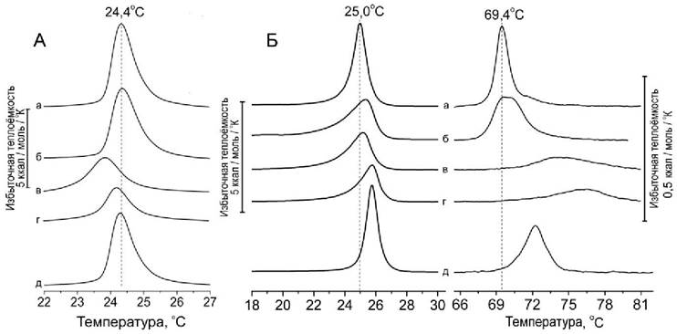
Notably, the pre-formed quercetin-iron(II) complex had a minor effect on lipid phase transitions, indicating weak interaction with the phospholipid bilayer. In contrast, when quercetin was added to the liposomes first, followed by the subsequent addition of iron(II), the resulting complex exerted a much stronger effect than quercetin alone. This is particularly evident in its impact on the POPE phase transition from the bilayer structure to the hexagonal HII phase, which natively occurs at 69°C (Fig. 64, b–d). This was accompanied by an increase in the transition temperature by several degrees, broadening of the transition interval, and a decrease in its peak height.
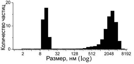
The inability of pre-formed quercetin-iron complexes to interact with the phospholipid bilayer is attributed to the formation of water-insoluble particles. Photon correlation spectroscopy reveals that mixing quercetin with iron(II) produces particles of two distinct size populations in the medium: small particles in the 10–15 nm range and larger particles in the 1–5 µm range (Fig. 64).
Fig. 64. Particle size analysis of quercetin / Fe(II) = 10:1 complexes using photon correlation spectroscopy.
2.4.5. Membrane Aggregation and Fusion
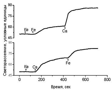
We also observed a phenomenon related to the action of flavonoid-iron complexes on calcium-dependent processes of membrane aggregation and fusion. It is well known that liposome aggregation occurs in the presence of calcium cations. In the presence of flavonoids, liposome aggregation is likewise triggered by divalent iron cations. Sequential additions of iron and calcium show that their aggregation effects are additive, as can be monitored via the light scattering of the liposome suspension (Fig. 65).
Fig. 65. Effect of quercetin (Q), divalent iron (Fe), and calcium cations (Ca) on the light scattering of a phosphatidylcholine liposome suspension. Light scattering was measured at a 90° angle using a spectrofluorimeter. Phosphatidylcholine concentration was 2 · 10-4 M, quercetin and iron 1 · 10-5 M, and calcium 1 · 10-4 M. The initial diameter of the liposomes was 100 nm.
Analysis demonstrates (Fig. 66) that the addition of flavonoids with iron(II) to liposomes results in only a negligible increase in the size of particles with an initial diameter of 100 nm, whereas the addition of flavonoids, iron, and calcium gives rise to a new fraction of significantly larger particles. Freeze-fracture electron microscopy reveals that following the addition of flavonoids, iron, and calcium, interliposomal contacts are formed and giant liposomes appear, providing clear evidence of membrane fusion processes (Fig. 67).
Fig. 66. Liposome size analysis using photon correlation spectroscopy. Flavonoids including quercetin (Q), taxifolin (Tax), catechin (Cat), along with iron (Fe) and calcium (Ca) cations, were added to a suspension of 100 nm liposomes. Phosphatidylcholine concentration was 2 · 10-4 M, flavonoids and iron 1 · 10-5 M, and calcium 1 · 10-4 M.
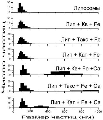
Fig. 67. Freeze-fracture Electron Microscopy of phosphatidylcholine liposomes (100 nm in diameter) before (A) and after (B, C) the addition of quercetin, ferrous iron, and calcium. Interliposomal contacts are indicated by arrows.
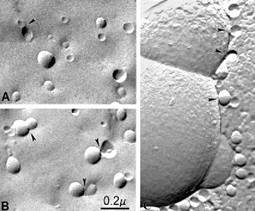
The ability of phosphatidylcholine liposomes to form aggregates, exchange lipids, and fuse has been extensively studied for many years, as these phenomena are directly relevant to liposome-mediated drug and Gene delivery. It has been found that the aggregation of liposomes induced by various agents can be followed by the activation of their fusion process, which depends on liposome size and the physical state of the lipids. However, 100 nm phosphatidylcholine liposomes used in this experiment typically lack the ability to spontaneously fuse, whereas smaller liposomes can aggregate and fuse at temperatures above the lipid melting point, although this process takes many hours or even days.
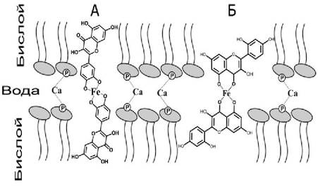
Flavonoid-iron complexes exhibit higher lipophilicity than free flavonoid molecules. This is due to a decrease in the polarity of hydroxyl groups in the flavonoid molecules, with the iron atom becoming the most polar part of the complex. According to our proposed scheme, a 2:1 flavonoid-iron complex can form a bridge between adjacent liposomal membranes. In this process, the flavonoid molecules insert into the hydrophobic lipid region, while the polar part of the complex containing the iron atom acts as a bridge (staple) between neighboring membranes. Calcium cations can also bridge membranes, but through interaction with phosphate groups. Since these mechanisms of membrane interaction are distinct and independent, their effects are additive. It should be noted that to achieve a comparable magnitude of aggregation in these experiments, the calcium concentration was ten times higher than that of iron and flavonoids.
The ability of polyphenolic compounds, such as Tannins, to initiate membrane adhesion is well known. Tannin molecules have been shown to interact with two adjacent membranes and form bridges between them. The efficiency of membrane adhesion in the presence of tannins is due to the fact that these molecules are large enough to penetrate the hydrophobic regions of neighboring membranes and bridge them. In contrast, flavonoid molecules are significantly smaller, and bridging can only be achieved by a flavonoid dimer formed upon metal complexation (Fig. 68).
Fig. 68. The "metal staple" model formed by a complex of quercetin (A), morin (B), and iron (Fe) flavonoids located between the surfaces of two adjacent membranes. For simplicity, only two neighboring lipid monolayers are shown. Calcium bridges between lipid phosphate groups are also depicted.
2.4.6. Structural Heterogeneity of Biological Membranes
2.4.6.1. Lipid Rafts
Cell membranes are complex mosaic structures that support the operation of numerous cellular systems and respond to environmental changes. The size of mosaic elements typically ranges from 2 to 200 nm, often making their direct observation within the cell difficult. Variations in lipid composition are observed across membranes of different origins, as discussed above. Even adjacent, closely positioned regions of the same membrane can differ in composition, indicating the presence of lateral membrane heterogeneity and the formation of lipid domains (clusters) within the bilayer. Such heterogeneity can even be observed in artificial bilayer membranes composed of several lipids with different melting temperatures or those exhibiting preferential mutual interactions. One would expect that in living cell membranes, which feature complex protein and lipid compositions as well as diverse functional specializations, lateral lipid heterogeneity is even more pronounced than in model systems. However, investigating lipid domains in living cells is extremely challenging due to technical limitations.
Among the most intensively studied mosaic elements are membrane rafts, which were first observed in the apical membranes of epithelial cells enriched in sphingomyelin and cholesterol. In recent years, numerous studies on plasma membrane heterogeneity have been conducted in search of Structural domains known as rafts. Lipid rafts are formed by densely packed, ordered lipid domains consisting of cholesterol and saturated sphingolipids, which float in a "sea" of unsaturated and more loosely packed lipids. The hydrocarbon chains of sphingolipids within rafts are somewhat longer than the fatty acid chains of other glycerophospholipids. They possess a higher melting point, are densely packed, and exist in an extended conformation, forming a solid gel ($s_o$ phase), which renders the monolayer thickness in rafts greater than in the rest of the membrane.
Initially, the existence of such structures was discovered due to their increased resistance to non-ionic detergents, such as Triton X-100, although it later became apparent that cholesterol-sphingolipid-specific Protein Complexes present in membranes cannot be directly equated with membrane fragments obtained after detergent Treatment. Recently, some progress has been made in raft Isolation Methods using the milder detergent Brij-96, which, according to some researchers, better preserves the native heterogeneity of the cell membrane bilayer. The presence of millimolar concentrations of Mg2+ and K+, as well as nanomolar concentrations of Ca2+ in the medium, is also of great importance. The definitive definition of rafts as specialized microdomains of cell membranes enriched in cholesterol and sphingolipids was established at the Keystone Symposium. Currently, membrane rafts are defined as "dynamic nanoscale assemblies of membrane proteins and lipids enriched in cholesterol and sphingomyelin that, through concerted protein-protein and protein-lipid interactions, can transition from a metastable resting state to a state of active function."
It has been demonstrated that lipid rafts can be involved in The regulation of various cellular processes, including defense against viral, bacterial, and parasitic infections. Furthermore, rafts play a role in apoptosis, Immune Response initiation, cell signaling, substance transport, and Intercellular Communication. Lipid rafts are implicated in numerous membrane-related pathologies, such as inflammatory processes, immune disorders, liver and cardiovascular diseases, atherosclerosis, and neurodegeneration, as well as the progression of viral and bacterial infections and parasitic invasions, such as malaria.
The immiscibility of lipids with one another is a primary driver of membrane heterogeneity and raft formation. The simplest explanation for this phenomenon relates to the ability of lipids to undergo a temperature-dependent phase transition known as melting. This process can be described as a transition of the phospholipid bilayer from a solid ordered state (So) to a liquid disordered state (Ld). Cholesterol can influence phospholipid melting and induce the formation of an intermediate state known as the liquid ordered state (Lo). It is hypothesized that the planar cholesterol molecule inserts between lipid molecules, disrupts the crystalline packing of hydrocarbon chains, and promotes the Lo state, in which the lateral mobility of lipid molecules approaches that of the liquid state, whereas the conformational mobility of the lipid hydrocarbon chains is restricted and resembles that of the ordered state. This effect depends on the molar concentration of cholesterol and the Chemical Nature of the lipid. In the presence of 25-30 mol % cholesterol, lipid bilayer structures formed from phosphatidylcholine, phosphatidylserine, or phosphatidylethanolamine can adopt the Lo state, especially if the lipid hydrocarbon chains are saturated and sufficiently long (18-22 carbon atoms). However, sphingomyelin interacts with cholesterol significantly stronger than other lipids, exhibiting a propensity to segregate within the plane of the bilayer and form lipid domains (Fig. 69) in the Lo state, which are surrounded by lipids in the Ld state. This capacity for lateral lipid segregation, governed by melting temperatures and interaction selectivity, provides a theoretical and experimental framework for explaining raft formation.
Fig. 69. Structure of rafts. A - Organization of lipids within a raft. On the outer surface of the plasma membrane, the raft is depicted as a complex of sphingomyelin (polar head groups in black) and cholesterol. The COMPOSITION OF THE cytoplasmic monolayer may also differ from that of the surrounding lipids. B - Organization of proteins within a raft. The same membrane region is shown. Transmembrane proteins "a" and "b" form an oligomer and penetrate the raft domain. Peripheral proteins "g" and "d" attach to cholesterol and glycophospholipid molecules (such as glycophosphatidylinositol or glycosphingolipids) located within the raft. On the cytoplasmic side, proteins associate with the raft via the attachment of palmitic acid "c".
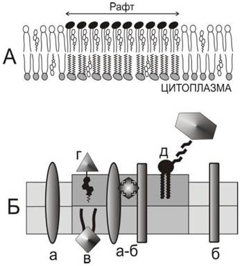
Rafts are bilayer domains enriched with various membrane proteins. A hallmark of these structures is the presence of proteins anchored to glycosphingolipids or glycophosphatidylinositols in the outer monolayer of the plasma membrane, designated as GPI-APs (glycophosphatidylinositol-anchored proteins). Residing within a cholesterol-rich bilayer, these proteins tend to form short-lived dimers with a lifetime of about 200 ms. As their concentration increases, GPI-AP molecules oligomerize into larger structures and segregate into rafts. Additionally, many raft-associated proteins are covalently bound to palmitic acid (palmitoylated). Experimental evidence indicates that palmitoylation may drive the accumulation of certain proteins in rafts, although not all palmitoylated membrane proteins reside within rafts. Furthermore, rafts contain proteins attached (anchored) to the membrane via cholesterol.
Rafts harbor a diverse array of proteins involved in cellular regulation, including receptors for various signaling molecules and channel-forming proteins. An important raft component is also the Hedgehog (Hh) signaling protein, which anchors to the membrane via palmitoylation or cholesterylation. Clusters of this protein in the membrane serve as elements of cell signaling, which is particularly crucial during Embryogenesis. One of its forms, Sonic Hedgehog (SHh), can serve as a therapeutic target in brain tissue injuries, various cancers, and cardiovascular diseases.
Another potential component of rafts is the tumor necrosis factor receptor, TNF receptor, known as FasR (or CD95 - cluster of differentiation 95), which is a component of the DISC (death-inducing signaling complex) apoptotic pathway. Oligomers of this protein have been shown to accumulate in rafts.
2.4.6.2. Caveolae and Caveolar Endocytosis
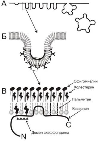
Caveolae are a specialized subset of membrane rafts abundantly distributed in the plasma membrane of endothelial cells, smooth Muscle cells, fibroblasts, and adipocytes. Caveolae participate in cellular uptake of various substances via a process known as caveolin-dependent endocytosis. Appearing as plasma membrane invaginations 60-80 nm in diameter (Fig. 70), caveolae are formed through the polymerization of caveolin proteins attached to membrane rafts. Accordingly, the lipid composition of caveolae exhibits the elevated cholesterol and sphingolipid levels characteristic of rafts. In addition to caveolin, these structures contain cavin (also referred to as PTRF-Cavin or Cav-p60), which is present in mature caveolae and is essential for their function. It is hypothesized that cavin is required for the incorporation of caveolin into caveolae.
Fig. 70. Schematic representation of caveolae and their structural organization. A - In cross-section through the plasma membrane, caveolae appear as individual invaginations or more complex clusters forming rosettes that invaginate into the cytoplasm via caveolin-dependent endocytosis. B - On the cytoplasmic surface of the caveola, caveolin molecules are attached. C - A more detailed view reveals that the extracellular surface of the caveolar membrane is formed by a sphingomyelin-cholesterol complex functioning as a lipid raft. The caveolin molecule, attached to the inner leaflet, possesses a hairpin-like hydrophobic domain that inserts into the bilayer. Additionally, the protein is anchored to the membrane via covalently linked palmitic acid and cholesterol residues, which attach to a specialized region termed the scaffolding domain that possesses a net positive charge.
2.4.7. Regulatory Processes in Rafts and Caveolae
Upon entering the bloodstream, flavonoids interact with endothelial cells, which feature the highest Abundance of caveolae—structures through which the regulation of not only individual cell metabolism, but also various organs and the Organism as a whole, is mediated. Several mechanisms underlie this regulation. For instance, caveolae house the enzyme endothelial nitric oxide synthase (eNOS), which synthesizes nitric oxide from Arginine. The generated NO acts as an important signaling molecule regulating Insulin secretion, smooth muscle tone (and consequently peristalsis of the digestive tract, biliary tract, and Urinary Tract), Blood vessel tone, and blood pressure. Excessive nitric oxide production can trigger inflammatory processes. It has been demonstrated that eNOS can directly interact with caveolin-1, leading to the inhibition of eNOS enzymatic activity. A synthetic peptide corresponding to a region of the caveolin molecule known as the "scaffolding domain" exerts a similar inhibitory effect. Recently, the co-localization of eNOS and caveolin-1 in the plasma membrane and Golgi membranes was visualized using confocal microscopy in endothelial cells, providing direct evidence for an association between these proteins in living cells.
The heat Shock protein Hsp70 (70-kilodalton heat shock protein) also interacts with caveolin. It has been demonstrated that eNOS can likewise bind to Hsp70, which enhances eNOS enzymatic activity and increases nitric oxide levels. Consequently, a balance between the eNOS/caveolin-1 and eNOS/Hsp70 complexes exists within caveolae, governing tissue nitric oxide levels. This balance can be modulated using statin medications, which restore normal nitric oxide levels by increasing the abundance of eNOS/Hsp70 complexes via upregulating eNOS and Hsp70 expression while downregulating caveolin-1 expression. Conversely, in pathologies associated with a marked decrease in caveolin-1 expression, eNOS is activated and nitric oxide levels rise, leading to elevated blood pressure.
Caveolae also harbor the estrogen receptor ERα (estrogen receptor alpha), whose localization coincides with that of caveolin-1. In fact, caveolin facilitates the delivery of ERα to the plasma membrane. Research shows that upon ERα activation by 17β-estradiol, the signal is relayed to caveolin, which subsequently activates the phosphatidylinositol 3-kinase (PI3K) regulatory pathway, resulting in eNOS activation and enhanced NO production.
The eNOS activation observed upon caveolin-1 deficiency can lead to uncoupling of mitochondrial Oxidative Phosphorylation AND increased hydrogen peroxide levels. In this process, caveolin initiates the formation of cytochrome b5 reductase clusters that complex with caveolin molecules, which can be regarded as one of the earliest manifestations of oxidative stress leading to apoptosis. Furthermore, it has been discovered that caveolin overexpression, as observed in Cancer cells, may serve as a defense mechanism against oxidative stress during carcinogenesis.
Caveolae may also house Other Enzymes and signaling proteins that frequently interact with the caveolin-1 scaffolding domain. These include G proteins, adenylate cyclase, phosphatidylinositol 3-kinase, protein Kinases A, C, Src, and H-Rac. Additionally, numerous G protein-coupled receptors (GPCRs) are present in caveolae, notably the endothelin receptor ETB and the angiotensin II receptor.
Caveolae also contain TRAF-2 (TNF receptor-associated factor), a tumor necrosis factor receptor. TRAF-2 is activated by the tumor necrosis factor TNF-α, binds to caveolin-1, and subsequently triggers the activation of the nuclear factor NF-κB, which regulates DNA Transcription and participates in the Pathogenesis of inflammatory processes as well as viral and bacterial infections.
Caveolae are likewise involved in regulatory processes mediated by arachidonic acid metabolites. For instance, caveolae contain phospholipase A2, which releases arachidonic acid from corresponding phospholipids. This process may contribute to The production of endothelium-derived hyperpolarizing factor (EDHF), a poorly understood agent involved in smooth muscle relaxation. Caveolae also harbor cyclooxygenase-2 (COX-2), which converts arachidonic acid into Prostaglandins (PG) that are likewise localized within caveolae. Furthermore, COX-2 is implicated in carcinogenesis and inflammation. COX-2 levels in caveolae can increase substantially in cancer cells. It has also been demonstrated that in Colorectal Cancer cells, a decrease in caveolin content is accompanied by an elevation in COX-2 levels. It is hypothesized that caveolin binds COX-2 and promotes the endocytosis and cytoplasmic degradation of this enzyme.
A study of keratinocytes revealed that phospholipase D2 and aquaporin are present in caveolae. It is hypothesized that the aquaporin-phospholipase D2 complex may be involved in keratinocyte function. A deficiency of aquaporin in keratinocyte membranes can result in impaired Skin integrity and increased permeability to toxic substances. The aquaporin-phospholipase D2 complex may participate in the metabolism of phosphatidylglycerol, a bioactive lipid essential for normal keratinocyte functioning.
Caveolae and lipid rafts participate in the regulation of cytoplasmic Ca2+ levels. It is hypothesized that calcium channel clustering occurs within rafts, allowing for the modulation of their conductance and, consequently, the control of cytoplasmic calcium levels. Caveolae contain a variety of proteins involved in regulating membrane calcium transport, including L-type calcium channels, the Na+/Ca2+ exchanger (NCX1), and the plasma membrane Ca2+ pump. The regulation of these proteins is potentially mediated by caveolin. In Skeletal Muscle, the complex responsible for calcium release from the sarcoplasmic reticulum is associated with caveolin-3. In smooth muscle, calcium influx regulation involves caveolin-1. Within airway smooth Muscles, intracellular calcium participates in Muscle contraction, and hyperreactivity of smooth muscles can lead to asthma. The Development of these pathologies is linked to dysregulated cellular calcium levels mediated by caveolin-1, the membrane abundance of which is regulated by the cytokine TNF-α (whose receptor is also located within caveolae). In cardiomyocytes, intracellular calcium levels are regulated by phospholipase Cβ-Gα, which resides in caveolae alongside the caveolin-3-Gα complex. Experiments demonstrate that calcium waves observed in cardiomyocytes are abolished upon the disruption of this complex, highlighting its role in regulating Calcium Homeostasis in heart muscle cells.
2.4.8. Interaction of flavonoids with rafts and caveolae
In the bloodstream, flavonoids are transported by albumins and Lipoproteins, which interact with the plasma membrane of endothelial cells and can enter lipid rafts as well as caveolae derived from them. Indeed, literature abounds with evidence regarding the involvement of caveolae in the endocytosis and transcytosis of low-density lipoproteins (LDL) and albumins, suggesting the potential for delivering flavonoids directly to these membrane domains. Once localized within lipid rafts or caveolae, flavonoids can influence the function of the membrane-resident regulatory systems mentioned above. Over the past few years, numerous hypotheses have been proposed regarding the exact mechanisms of flavonoids within membranes. Most studies have utilized green tea catechins, whose cellular effects are particularly pronounced—most notably epigallocatechin gallate (EGCG).
The ability of EGCG to interact with membrane rafts and modulate caveolae-mediated cellular signaling has been investigated in numerous laboratories, as these pathways may contribute to the suppression of inflammatory processes in endothelial cells. Specifically, by downregulating the expression of caveolin-1 and cyclooxygenase-2 (COX-2), EGCG inhibits endothelial cell activation triggered by linoleic acid. This effect is mediated by the inhibition of the MAPK signaling pathway kinase ERK1/2 and the kinase Akt. The flavonoid daidzein similarly exhibits the capacity to inhibit caveolin-1 expression and activate caveolar signaling via PI3K and Akt kinases, which are implicated in apoptosis and Various Forms of carcinogenesis.
Rafts contain the receptor for Laminin, a glycoprotein present in the structural matrix surrounding cells in most tissues. In cancer cells, laminin receptor expression is upregulated, which promotes metastasis and invasive activity. It has been discovered that green tea EGCG alters the structure of membrane rafts, thereby preventing the binding of epidermal growth factor (EGF) to its receptor (EGFR) located within these rafts; overexpression of EGFR can drive oncogenesis. The impact of flavonoids on rafts may be of profound significance, as these membrane domains harbor members of the MAP kinase family responsible for regulating cellular apoptosis and carcinogenic progression.
Studies on multiple myeloma cells demonstrated that EGCG can induce cancer cell apoptosis without impairing the viability of healthy cells via interaction with the 67LR laminin receptor, the expression of which is significantly elevated in myeloma cells compared to normal blood mononuclear cells. This interaction triggered lipid raft clustering. Furthermore, EGCG induced the translocation of acidic sphingomyelinase (aSMase) to the plasma membrane and the phosphorylation of protein kinase Cδ (PKCδ) at Ser664. This engaged a specific Intracellular Signaling cascade that ultimately led to myeloma cell apoptosis.
The clustering of raft proteins induced by EGCG or total green tea flavonoid extract has also been observed in human colorectal adenocarcinoma cells. However, in this case, researchers unexpectedly noted an increase in cell viability. This effect was abolished upon membrane cholesterol depletion, underscoring the critical role of rafts in this process. Experimental data indicate that this phenomenon involved the activation of the mitogen-activated protein kinase signaling pathway via MEK and ERK1/2, which govern cell proliferation and differentiation.
The protective effect of EGCG against atherosclerosis is associated with the suppression of endothelial cell inflammation. Heme oxygenase-1 (HO-1), which concentrates in endothelial caveolae, plays a pivotal role in this process. EGCG has been found to accumulate in caveolae, prompting the displacement of caveolin from the inner membrane surface into the cytoplasm. This can enhance the nuclear translocation of the transcription factor Nrf2 (nuclear factor erythroid 2-related factor 2), widely recognized as a primary cellular defense component against oxidative stress. Consequently, the expression of antioxidant defense factors such as heme oxygenase-1 (HO-1) and bilirubin was induced, indicating the activation of cytoprotective mechanisms and the attenuation of inflammatory processes in endothelial cells.
Proteinaceous agents such as hepatocyte growth factor (HGF) and its receptor c-Met, which possesses Tyrosine kinase activity, are Key Components of the regulatory network driving the Invasion and Metastasis of most human cancers. EGCG was found to prevent the tyrosine phosphorylation of the c-Met receptor. Epicatechin gallate (ECG) exhibited similar activity, whereas green tea epigallocatechin (EGC) and epicatechin (EC) did not. It was demonstrated that in prostate cancer cells, c-Met partitions into rafts exclusively upon phosphorylation. Thus, catechins bearing a galloyl moiety prevent c-Met activation, culminating in the disruption of lipid raft Structure and function. The flavonoid luteolin also exhibits c-Met-modulating activity, blocking HGF-dependent c-Met phosphorylation within lipid rafts.
Bile acids are synthesized from cholesterol via cytochrome P450-mediated oxidation. Together with bile, they enter the duodenum and are subsequently reabsorbed in the ileum via the apical sodium-dependent bile acid transporter (ASBT). EGCG, unlike other green tea catechins, inhibits ASBT function, thereby reducing total body cholesterol levels by impairing its intestinal reabsorption. This is accompanied by a decrease in the maximum transport velocity of bile acids (Vmax) and a reduction in ASBT content within the lipid raft fraction, whereas the total ASBT content in the intestinal epithelial cell (Caco-2) membrane remained unchanged. Thus, the hypocholesterolemic effect of green tea EGCG has been shown to stem from its action on lipid rafts, which disrupts ASBT transporter function.
Under conditions of cerebral ischemia, the delivery of therapeutic agents to the affected area is crucial, a process hindered by the blood-brain barrier (BBB) formed by brain capillary endothelium. Green tea polyphenols have been shown to increase BBB permeability in the ischemic lesion zone, thereby significantly improving the condition of damaged tissues. Research demonstrated that this increase in BBB permeability is driven by the downregulation of caveolin-1 expression, resulting in a reduced abundance of this protein in the endothelial plasma membrane. Additionally, elevated expression and increased phosphorylation levels of raft-associated ERK1/2 kinases, which regulate cell proliferation, were observed.
The inhibitory effect of green tea polyphenols on caveolin-1 expression may protect aortic cells from pathological alterations associated with a high-fat diet. This protective effect can be attributed to the action of these flavonoids on membrane rafts, accompanied by the activation of ERK1/2 kinase and the inhibition of the mitogen-activated protein kinase p38 MAPK, pointing to the suppression of autoimmune and inflammatory pathways.
Quercetin can likewise modulate regulatory systems localized within caveolae. Its anti-inflammatory properties are tied not only to its antioxidant activity and inhibition of nitric oxide production, but also to its ability to disrupt lipid rafts and thereby suppress the Regulatory Functions of various kinases, including c-Jun, p38, Akt, Src, JAK-1, Tyk2, and NF-κB. Furthermore, quercetin exerts an inhibitory effect on Serine/Threonine and tyrosine Phosphatases. For instance, quercetin blocks the capacity of toxic polychlorinated biphenyls (PCBs) to stimulate caveolin-dependent signaling pathways that drive inflammation and the progression of atherosclerosis. It is known that caveolae contain the aryl hydrocarbon receptor (AhR), the activation of which triggers caveolin expression, oxidative stress, and the expression of cytochrome P450 1A1 (CYP1A1). Quercetin is capable of blocking these alterations, leading to decreased expression of both caveolin and cytochrome P450. The induction of Cell Adhesion molecules (VCAM-1), E-selectin, and P-selectin is similarly blocked. Thus, the protective action of quercetin is directly linked to its effects on caveolae. However, quercetin’s mechanism cannot be reduced solely to raft disruption. For instance, in colorectal cancer cells, quercetin has been shown to relocate the death receptors DR4 and DR5 into lipid rafts, facilitating their interaction with the tumor necrosis factor-related apoptosis-inducing Ligand (TRAIL) and thereby activating cancer cell apoptosis. The capacity to restore the expression of caveolin-1 and angiotensin AT-1 receptors has also been observed following treatment with the flavonoid phytoestrogen genistein in ovariectomized rats with experimentally induced Hypertension.
Last update: 06/08/2026
Editorial and Educational Adaptation: This material has been compiled based on the primary/original source text. The project team performed an editorial review, corrected technical inaccuracies, structured sections, and adapted the content for an educational format.
What was processed:
- elimination of formatting defects (OCR errors, structural breaks, corrupted characters);
- editorial organization of content;
- standardization of terminology in accordance with academic sources;
- verification of factual statements against the original source text.
All mentions of the author, publication year, and origin of the primary text have been preserved in accordance with the source.