IMMUNOLOGY - Roitt I. - Mir 2000
Chapter 10. Cell-Mediated Immunity
T- AND NK-CELL CYTOTOXICITY
Cytotoxic T Cells and NK cells act in defense reactions aimed at eliminating virus-infected host cells
Cellular cytotoxicity is a vital defense mechanism against intracellular pathogens such as Viruses, certain Bacteria, and Protozoa. Several Cell types can exhibit cytotoxic activity—cytotoxic T cells (Tcs), natural killer (NK) cells, and occasionally myeloid cells—although their target recognition mechanisms differ (Fig. 10.12).
✵ Cytotoxic T cells recognize specific Antigens (e.g., viral Peptides On the surface of infected cells) in association with MHC molecules. The majority of Tc cells bear the CD8+ marker and recognize antigen presented in association with Class I MHC molecules, whereas approximately 10% of MHC-restricted cytotoxic T cells belong to the CD4+ subpopulation and recognize antigen in association with class II MHC molecules.
✵ Natural killer cells recognize cells that lack expression of class I MHC molecules. In addition to negative recognition, NK cells also exhibit positive recognition of their targets via receptors for various ligands. For instance, through Fc receptors (CD16), they are capable of binding Antibodies that have formed immune complexes with antigens on The surface of target cells—a process known as antibody-dependent cellular cytotoxicity (ADCC), or killer (K) cell activity.
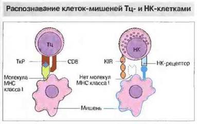
Fig. 10.12. Cytotoxic T cells (Tcs) use their receptors (TCRs) to recognize processed antigen presented in association with MHC molecules on a target cell. Most Tc cells belong to the CD8+ subpopulation and recognize antigen presented in association with class I MHC molecules, whereas a smaller subset belonging to the CD4+ subpopulation can recognize antigen in association with class II MHC molecules. Unlike Tc cells, NK cells bear killer inhibitory receptors (KIRs). Upon recognizing class I MHC molecules on a target cell, these receptors deliver an inhibitory signal that suppresses cytotoxic activity. Positive recognition of targets by NK cells occurs via their own specialized receptors (NK receptors), notably CD2 and CD69, or via antibodies bound through the Fc receptor (CD16).
The primary function of Tc cells is the elimination of virus-infected cells (see Ch. 16). Almost all nucleated cells express class I MHC molecules and, upon viral infection, are capable of presenting pathogen-derived antigens to CD8+ cytotoxic T cells. The mechanisms of such presentation are discussed in detail in Ch. 9. Host cell Proteins, following partial degradation in proteasomes, are transported to The Endoplasmic reticulum, where they form complexes with class I MHC molecules and are subsequently transported to The Cell surface. In this way, every cell displays "samples" of its proteins for inspection and recognition by CD8+ Tc cells. Antigens derived from microbes localized intracellularly are presented in the same manner.
Sometimes, additional interactions are required to stabilize the bond between a Tc cell and its target (Fig. 10.13); these can also facilitate cytolysis. For example, cytolysis of target cells by bound Tc cells can be induced in vitro using antibodies against CD3 or CD2, which are Tc surface markers. Presumably, the binding of physiological ligands to these molecules similarly "switches on" the cytolytic activity of Tc cells.

Fig. 10.13. Some of the ligands involved in the interaction between cytotoxic T cells and their targets.
Certain viruses, particularly Herpesviruses, attempt to evade recognition by Tc cells by downregulating the expression of class I MHC molecules on the surface of infected cells; however, NK cells recognize such virus-infected cells. Consequently, Tc and NK cells can be viewed as two complementary tools of Immunity against viral tissue infections.
NK cell cytotoxic activity is inhibited by class I MHC molecules
NK cells, which originate mainly from large granular lymphocytes (LGLs), account for approximately 5% of peripheral Blood lymphocytes in humans. Most frequently, they have the CD3-CD16+CD56+CD94+ phenotype (see Appendix 2) and a germline (unrearranged) T-cell receptor Gene configuration. Initial studies on the Specificity of NK cytotoxic action established that resistance to it is conferred by certain dominant alleles of the HLA-C locus. It was subsequently shown that NK cells can indeed recognize various MHC allotypes, although any of these molecules, including allotypes of the HLA-A and HLA-B loci, can suppress cytolysis.
A recent and highly promising discovery is the identification of HLA-G molecules, which are expressed exclusively on trophoblast cells and act as potent inhibitors of NK cytotoxicity, conferring resistance to all types of NK cells. Trophoblast cells (the outer layer of the mammalian blastocyst) come into contact with circulating maternal blood once the Placenta is formed; they are allogeneic to the mother because they possess paternal MHC genes. However, the expression of all conventional MHC antigens on these cells, with the exception of HLA-G, is translationally or transcriptionally downregulated. Consequently, HLA-G molecules are essential for protecting the placenta from maternal NK cells.
NK cells recognize class I MHC antigens using two distinct types of molecules (Fig. 10.14). Molecules of the first type belong to C-type Lectins and were initially identified in mice (Ly49) and rats, and subsequently in humans (presumably CD94). Molecules of the second type are members of the immunoglobulin superfamily (IgSF), specifically CD158a and CD158b (which bind to different HLA-C molecules and each possess two IgSF domains), as well as the p70 protein, which contains three IgSF domains. Receptor molecules of this type were first identified in humans, and equivalent structures were later found on rodent cells.

Fig. 10.14. Two Types of receptors mediating the inhibition of NK cell cytotoxicity by class I MHC molecules: immunoglobulin-like molecules (the three-domain p70 protein and the two-domain p50 and p58 proteins) and specific C-type lectins, including Ly49 in mice and CD94 in humans.
The surface of NK and K cells bears several different receptors for target identification
NK cells attack their targets using CD2, CD16, and CD69 molecules as receptors, along with lectin-like receptors similar to those that inhibit cytotoxicity. Through the Fc receptor (CD16), NK cells bind antibody molecules attached to the surface of target cells, thereby mediating ADCC (Fig. 10.15). This is typically interpreted as a manifestation of killer (K) cell activity, but in addition to NK cells, other cell types bearing Fc receptors—notably T cells—can perform this function. Myeloid cells expressing Fc receptors also exhibit K-cell activity, though presumably utilizing different cytolysis mechanisms compared to NK and T cells (see below).
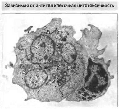
Fig. 10.15. K-cell activity. Electron micrograph of a lymphocyte (right) attacking an antibody-coated target cell (left), ×2500. (Kind courtesy of Dr. R. Penfold.)
Potential targets for K cells include cell surface-expressed viral antigens, MHC molecules, and individual epitopes characteristic of tumor cells. Furthermore, monocytes and (according to some conflicting reports) polymorphonuclear granulocytes can exhibit killer activity against antibody-coated tumor cells. Myeloid cells such as monocytes and eosinophils undoubtedly play a crucial effector role in the destruction of antibody-coated schistosomula (see Ch. 18).
Cytokine-activated killer cells likely belong to NK cells
Immunology is actively developing several approaches to experimental Cancer immunotherapy. One of them is the in vitro Activation of a patient's own lymphocytes using interleukin-2, followed by their reinfusion. Such lymphocytes, isolated from blood or the Spleen, are known as cytokine-induced (or lymphokine-activated) killer (LAK) cells. They exhibit enhanced, MHC-unrestricted cytotoxicity and appear to originate primarily from precursors that are indistinguishable from NK cells. Most likely, LAK cells are a product of activation rather than a distinct cell Lineage. This method of antitumor immunotherapy using LAK cells is currently undergoing clinical trials.
The cytotoxic effect of killer cells is executed either through contact-dependent interaction with targets or via the release of cytokines and granule exocytosis
To destroy a target, Tc, NK, and K cells employ several Mechanisms of action. One is signal transmission upon direct cellular contact via surface structures, and another is indirect signaling mediated by cytokines. In addition, the Cytoplasm of many CD8+ cytotoxic T cells and large granular lymphocytes (NK and K cells) contains protein-rich granules whose release near the cytoplasmic membrane of the target cell causes its damage. Which combination of these three cytotoxic mechanisms is utilized under specific conditions depends on the type of cytotoxic cells, particularly the Tc cell subpopulation.
The granules of cytotoxic T cells contain perforin and granzymes. The granules of NK cells and cytotoxic T cells contain several proteins, including perforin and granzymes (granule-associated Enzymes). As soon as a cytotoxic T cell binds to its target, the granules within the Tc cell migrate toward the contact site on the membrane. Subsequently, Ca2+-dependent exocytosis of the granule contents occurs into the intercellular cleft between the cytotoxic cell and its target.
Perforin is a monomeric protein that induces pore formation in the cytoplasmic membrane. Structurally and functionally, it is closely related to C9 (the ninth component of The Complement System). In addition to perforin, the granules contain Serine esterase, which may be part of the lytic complex. In the presence of Ca2+, perforin monomers bind to the target Cell Membrane and polymerize, forming a transmembrane channel. Despite close contact between its own Plasma Membrane and perforin, the cytotoxic T cell itself is not damaged by it and proceeds to attack subsequent target cells. The proteoglycan chondroitin sulfate A, which is also present in the granules, likely protects the T cell from self-injury. This protein can bind to perforin and thereby induce its inactivation.
Granzymes are a set of serine esterases that are released upon granule exocytosis and subsequently activated. The action of granzymes is not strictly required for cytotoxicity—cells lacking these enzymes are still capable of destroying their targets. Some granzymes can trigger apoptosis programs and DNA fragmentation in target cells by interfering with Intracellular Signaling pathways. The mechanisms of perforin and granzyme involvement in target cell cytolysis are illustrated in Fig. 10.16.
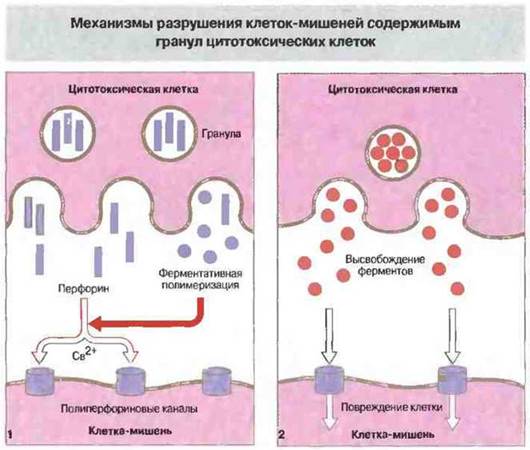
Fig. 10.16. Upon degranulation of cytotoxic lymphoid cells, perforin and various enzymes (granzymes) are released in the immediate vicinity of the target cell's cytoplasmic membrane. This is followed by Ca2+-dependent enzymatic polymerization of perforin, resulting in The formation of polyperforin channels in the target membrane (1). Through these channels, hydrolytic enzymes and other toxic substances secreted by the cytotoxic cell penetrate into the cell interior, causing damage (2).
Target cells can also perceive cytotoxic signals via FasL and TNF receptors. CD4+ cytotoxic T cells have been shown to lack perforin and destroy their targets in a Ca2+-independent manner. Thus, they employ a different cytotoxic mechanism, as proven by experiments on Tc cells from "knockout" mice (generated using gene knockout technology; see below). In such mice, targeted site-specific mutation prevents the expression of the perforin gene. In the absence of perforin, the cytotoxic activity of CD4+ T cells is reduced, but not entirely abolished. The search for an alternative cytotoxic mechanism led to the identification of a group of molecules on the target cell surface whose binding serves as an apoptotic signal. Among these, Fas (CD95) and TNF receptors are of primary interest. This group of molecules also includes CD30 and CD40. The Fas Ligand, or FasL, is expressed on mature CD4+ and CD8+ T cells following their activation. The binding of FasL to Fas receptor molecules induces their aggregation and the recruitment of the MORT-1 protein to their intracellular segments, ultimately leading to target cell apoptosis. Fas is also essential for the survival and elimination of specific lymphocyte subpopulations during their maturation. Structurally, Fas molecules resemble TNF receptors. For instance, both possess intracellular (cytoplasmic) "death domains"—Structure/135.html">Structural motifs found in A number of specialized proteins that regulate cell survival.
The granules of Tc cells may also contain TNFα and lymphotoxin (TNFβ). However, these cytokines alone cannot account for cytotoxicity: an individual Tc cell is capable of inducing target cell death within 3–4 hours, whereas the effects of TNF manifest much later. Nevertheless, the structural similarity between the TNF receptor and Fas points to the potential involvement of TNFα and TNFβ in target destruction (Fig. 10.17).
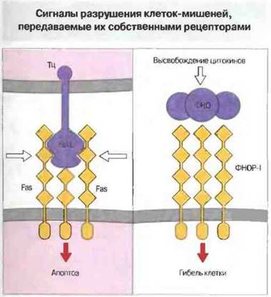
Fig. 10.17. The Fas ligand (FasL) of cytotoxic T cells induces the aggregation of Fas receptor molecules on the target cell surface, leading to the association of intracellular proteins (such as MORT-1), which initiates the cascade of events leading to apoptosis. Fas contains four extracellular domains (as a member of the nerve growth factor receptor superfamily) and a single cytoplasmic "death domain." Alternatively, signaling can be triggered by the binding of TNF to its specific type 1 receptor (TNFR-1), which belongs to the same superfamily as Fas.
Thus, the cytotoxic action of CD8+ T cells relies on granule release and FasL expression, that of CD4+ T cells primarily on FasL expression, and that of NK cells predominantly on granule release. TNF may also contribute to the cytotoxic activity of all these cell types.
The cytotoxic action of myeloid-lineage cells relies on a broad spectrum of target destruction mechanisms
Macrophage-mediated destruction of tumor cells is frequently driven solely by TNFα action. Together with IFNγ produced by T and NK cells, this cytokine elicits a potent synergistic cytolytic effect on susceptible tumors (Fig. 10.18). The mechanisms of cytokine-mediated cytotoxicity are still insufficiently understood. In some cases, cytokines trigger an upregulation of cyclooxygenase and lipoxygenase activity, followed by the intracellular generation of free radicals. In addition, potential cytokine-dependent effects include free radical production via Mitochondrial Electron Transport and the impairment of Protein Synthesis. Myeloid cells are also capable of releasing the same toxic mediators that function during the destruction of microbial cells within phagocytes, notably highly reactive unstable oxygen and nitrogen metabolites (see Chapter 17).

Fig. 10.18. In addition to cytokines, target cell damage can be induced by highly reactive oxygen and nitrogen metabolites, cationic proteins, hydrolytic enzymes, and complement components released by myeloid-lineage cells.
Last update: 13/08/2026
Editorial and Educational Adaptation: This material has been compiled based on the primary/original source text. The project team performed an editorial review, corrected technical inaccuracies, structured sections, and adapted the content for an educational format.
What was processed:
- elimination of formatting defects (OCR errors, structural breaks, corrupted characters);
- editorial organization of content;
- standardization of terminology in accordance with academic sources;
- verification of factual statements against the original source text.
All mentions of the author, publication year, and origin of the primary text have been preserved in accordance with the source.