Chordate Zoology - textbook - Y. V. Tsaryk - 2013
Chapter 5. SUPERCLASS TETRAPODS (LAND VERTEBRATES) TETRAPODA, seu QUADRIPEDA.
ANAMNIOTES (ANAMNIA) and AMNIOTES (AMNIOTA)
5.4. CLASS MAMMALS, or BEASTS, MAMMALIA, seu THERIA
5.4.1. Structural Features of Mammals
Appearance. Mammals are highly diverse in size and appearance. The smallest modern mammal is the Etruscan shrew Suncus etruscus (Soricomorpha), weighing 1.2–1.7 g with a body length of 3.8–4.5 cm. The largest terrestrial mammal is the African elephant, reaching 3.5 m in height and up to 4-5 t in weight, while the largest aquatic mammal is the blue whale, with some individuals reaching a length of 33 m and a weight of 150 t (equivalent to 30-35 elephants). The mammalian body is divided into a HEAD, neck, trunk, paired limbs, and a tail.
Seals, sirenians, and especially cetaceans have a streamlined body, with limbs modified into flippers; a leathery tail fin gives sirenians and whales a fish-like appearance. Semiaquatic mammals (platypus, desman, beaver, coypu, otter) have dense fur that prevents wetting, a shortened neck, well-developed swimming webs between the toes, and a more or less flattened tail. Many equids and cervids share a similar appearance: long legs, a compact torso, and a long, mobile neck, which helps them travel long distances and evade common enemies—large canids. Lagomorphs and rodents are also similar in body shape. Adaptation to rapid movement on hind limbs in open habitats has led to The Development of a similar body shape—weak forelimbs and strong hind limbs, and a long tail acting as a counterweight: in kangaroos (marsupials), elephant shrews
(Soricomorpha) and various rodents—jerboas, gerbils, etc. In burrowing species, the body becomes cylindrical, with shortened limbs and tail (ground squirrels, marmots, voles, etc.). In the transition to a subterranean lifestyle, along with a cylindrical torso, the forelimbs, which are involved in digging, become highly developed (marsupial moles, true moles, etc.).
In arboreal species, adaptations for locomotion include short but strong limbs with sharp claws, and a long bushy tail that AIDS in jumping (squirrels, martens, etc.). The long limbs of primates enable them to move through tree canopies. Opossums, some monkey species, silky anteaters, pangolins, and porcupines use their tails for grasping. In flying phalangers, flying squirrels, and colugos, a lateral Skin fold allows for long gliding leaps. Remarkably, marsupials exhibit life forms similar to many placental mammals.
Thus, representatives of different orders living in similar conditions and sharing a similar lifestyle can have a similar body shape (convergent similarity). Such groups of species are called "life forms" or ecological types.
Integument. The relatively thick skin, as in all vertebrates, consists of two layers, is rich in glands, and has keratinized structures—Hair, claws, etc. (Fig. 5.46). The epidermis is stratified. At its base lies the Malpighian layer, composed of columnar or cuboidal, living, dividing Cells. Closer to the surface, these cells lose their nuclei and ability to divide. They flatten, undergo keratinization, and form a dead outer layer. These cells slough off the surface and are gradually replaced by new ones from the Malpighian layer. Skin coloration is determined by pigments present as melanin granules in the Cells of the stratum germinativum, in intercellular spaces, and in specialized pigment cells (melanoblasts, melanophores). The epidermal layer reaches its greatest thickness in areas of constant friction during locomotion; calluses may form here (footpads, ischial callosities of some monkeys, knee calluses of camels, etc.).
The dermis (corium) is the Deep Layer of mammalian skin. It is rich in Collagen and Elastin fibers that interweave to form a reticular Structure. Adipose tissue—subcutaneous fat—is deposited in the deep layers of the dermis; in some mammals (whales, dolphins), it is highly developed, compensating for the complete reduction of the hair coat.
Such Properties of the dermis as elasticity, strength (due to elastin and collagen fibers), and low thermal conductivity (due to adipose tissue), combined with the mechanical strength of the keratinized epidermal layer and its derivatives, make mammalian skin a reliable means of protecting the body from damage and adverse environmental factors. In addition, the dermis contains the basal parts of glands, hair follicles, sensory nerve structures, well-developed Blood Vessels, and a capillary network that, penetrating into the upper papillary layer of the dermis, provides nourishment to the epidermis and serves a thermoregulatory function. Smooth Muscle fibers can also develop in the dermis.
In addition to hair, Mammary Glands—modified Sweat Glands—are specific to mammals. It is due to the presence of mammary glands that this Class of vertebrates received the name mammals. Mammals possess sweat, sebaceous, and scent glands. Scent glands are derivatives of either sweat or Sebaceous Glands. These glands are usually complex; their basal portion is located in the corium, and their excretory ducts open onto the skin surface or into a hair follicle.
Based on The structure of their excretory ducts, glands are classified into tubular and alveolar. The first type is tubular glands. These include sweat glands, which mostly open onto the skin surface, and occasionally into a hair follicle. The inner end of a sweat gland forms a coil (see Fig. 5.46). The walls of these glands consist of a Simple Epithelium, whose epithelial cells secrete sweat. Sweat consists of 97-99% Water, in which urea, creatine, volatile Fatty acids, and salts (also found in urine) are dissolved. Thus, waste products are excreted with sweat, but The primary function of sweat glands is thermoregulatory (sweat evaporates and cools the body). Sweating is regulated by the thermoregulatory centers of the BRAIN AND SPINAL cord. Sweat glands are well developed in primates and ungulates, and relatively weak in canids, felids, lagomorphs, and rodents. In species with poorly developed sweat glands, thermoregulation occurs differently. For example, when dogs overheat, heat loss is enhanced by rapid shallow breathing (polypnea) and evaporation of water from the saliva of the panting Tongue and the oral mucosa. Sirenians, cetaceans, sloths, pangolins, and moles lack sweat glands.
The second type of glands is alveolar glands, which include sebaceous glands. They have an acinar structure, and their ducts open into hair follicles.
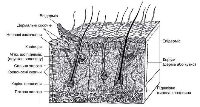
Рис. 5.46. Будова шкіри ссавців
The walls of the glands are formed by Stratified Epithelium. Fatty degeneration occurs in its cells, producing a lipid secretion that lubricates The surface of the skin and hair, helping to maintain elasticity and preventing the penetration of microbes and Fungi. Such glands are absent in cetaceans, sloths, and some shrews.
Scent (musk) glands are Skin glands that secrete a fragrant substance, musk, which primarily serves to attract females, mark territory, and facilitate species recognition. They are modified sweat or sebaceous glands, sometimes of mixed structure, combining alveolar and tubular forms. Scent glands include the anal glands of many caniforms, especially mustelids; the musk glands of musk deer, beavers, desmans, and muskrats; the preorbital glands of many cervids (deer, antelopes, sheep); and the hoof glands of goats, etc. The strong, foul-smelling secretion of the anal glands of American skunks and some polecats serves for self-defense. The combined odors of secretions from musk, sebaceous, and sweat glands help animals distinguish individuals of their own species from others and facilitate the meeting of males and females.
Mammary glands—modified sweat glands—are developed in the females of all mammals. In monotremes, the mammary glands retain a tubular structure and are arranged in groups known as glandular fields: on the abdomen in platypuses, and in the brood pouch in echidnas. They lack nipples, and the gland ducts open into hair follicles; the young lap up droplets of milk from the fur. In all other mammals, the mammary glands have a more complex, acinar structure, and the mammary ducts open onto nipples. In some species, nipples are arranged in two rows from the forelimbs to the groin (shrews, carnivores, rodents), while others have only one pair of nipples (primates, sirenians, elephants, bats) or only inguinal nipples. In most ungulates, the mammary Glands of the right and left sides merge into an udder located in the groin, which has two or four teats. The number of nipples in different mammalian species varies from 2 to 12 pairs and roughly corresponds to the number of offspring to be born.
The hair coat (pelage) is a characteristic feature of mammals. In most animals, it is developed on all PARTS OF THE body (absent on the Lips, and in some species, on the soles) and performs a thermal insulation function, serves as a tactile receptor, protects the skin from damage and certain parasites, improves the aerodynamic and hydrodynamic properties of the body, and provides species-specific coloration. Only in a few mammals (cetaceans, elephants, rhinoceroses, hippopotamuses) is the hair coat virtually absent; they are temporarily covered with hair primordia during embryonic development in the female's body.
Mammalian hair develops from an epidermal primordium that gradually invaginates into the corium (Fig. 5.47). The outer layers of the epidermal primordium form the hair follicle and sebaceous glands, while the inner layers form the hair shaft itself. The hair grows due to the proliferation of basal cells in the bulb—the Base of the hair. As they are gradually pushed upward, the Hair cells undergo keratinization; the entire hair, except for the basal region, is a dead structure. A fully formed hair consists of a shaft projecting above the skin surface and a ROOT embedded in the skin. Within the hair shaft, the medulla has a porous structure and consists of flat, keratinized cells with air spaces. A particularly large amount of air is characteristic of the hair of animals inhabiting regions with cold winters; this enhances the insulating properties of their coat. The loose medulla is surrounded by a dense cortex, formed by keratinized cells elongated along the longitudinal axis of the hair. This layer provides strength and elasticity to the hair and contains pigments. Externally, the cortex is covered by a cuticle of flat, transparent keratinized cells overlapping like shingles. The root of the hair expands at the bottom into a bulb composed of living cells. A dermal papilla with blood vessels invaginates into the bulb from below, supplying nutrients to its cells. The hair root is enclosed in a hair follicle, which is an invagination of the epidermal layer. The layer of the follicle closest to the hair is called the root sheath, and the outer layer is the Connective Tissue sheath. Smooth Muscles are attached to its lower part, and their contraction alters the angle of the hair. Sensory nerve endings are also located in the hair follicle.
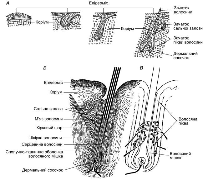
Рис. 5.47. Розвиток (А), будова (Б) та іннервація (В) волосини
In mammals, the coat is heterogeneous. The largest, usually solitary, stiff hairs that project prominently above the fur are called vibrissae. They serve as tactile Organs, located on areas of the body that the animal most frequently brings into contact with surrounding objects (the tip of the snout, abdomen, limbs) and having numerous nerve endings in the hair follicle. The upper layer of the fur is formed by guard hairs, while the undercoat consists of thread-like, often spirally coiled, fine underfur hairs, which play a crucial role in thermal insulation. In a few mammals, the fur consists of a single hair type: the summer coat of deer and wild boars is composed solely of guard hairs, while the fur of burrowing mammals (moles, mole-rats) consists entirely of underfur. Bristles (pigs) and spines (echidnas, hedgehogs, porcupines) are modified guard hairs.
Generally, hair is arranged in an orderly fashion on the body. It is typically inclined in a specific direction, which streamlines the body against air and water currents. The primary direction of the hair tract is from head to tail. The pattern of the coat changes in areas where skin folds and stretches frequently occur. The lay of the fur is closely linked to the animal's lifestyle and mode of locomotion. For example, sloths constantly hang from trees upside down, and their hair is directed from the abdomen toward the back, which can be attributed to both gravity and the shedding of rain. In subterranean dwellers (moles, mole-rats), which often move both forward and backward, the short hair stands vertically.
A similar coat is found in inhabitants of dense grassy thickets and leaf litter (shrews).
Fur wears out and needs replacement, which occurs during molting. In the tropics, animals molt continuously as the hair wears down (in monkeys, this can occur throughout the year). A similar phenomenon in temperate latitudes is observed in burrow dwellers, whose fur quickly rubs off in certain areas, leading to partial molting in these patches. In most temperate-latitude species, the coat is typically shed twice a year—in autumn and spring; molting is accompanied by A change in fur structure and often its color. When the summer coat is replaced by the winter one, the density of the hair increases, dramatically improving the thermal insulation properties of the winter coat compared to the summer one.
The terminal Phalanges of the digits in most mammals are protected by horny claws, which are epidermal derivatives (Fig. 5.48). In arboreal forms, they are sharp and strongly curved, while in burrowing forms, they are elongated and flatter. In all felids (except the cheetah), the claws are retractile; the claw, along with the terminal phalanx, is pulled toward the dorsal surface of the penultimate phalanx by special tendons, preventing it from dulling during locomotion. In many primates, claws have evolved into Nails, covering the tips of the digits only from above; a soft pad is developed underneath, enhancing the tactile properties of the fingers. The specialization of claws led to The formation of hooves—thick horny structures that almost completely encase the terminal phalanx. Hooves are particularly well-developed in fast-running species (horses, antelopes, goats, etc.).
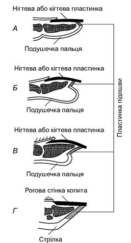
Fig. 5.48. Longitudinal sections through the claw of a monkey (A) and the nail of a human (B), the claw of a dog (C) and the hoof of a horse (D)
As a result of the significant proliferation of cornified epithelium, horns are formed in rhinoceroses and hollow-horned ruminants (Bovidae)—hollow horny sheaths situated on bony cores that fuse with the frontal bones. Antlers of deer are bony structures derived from the dermis; animals shed them annually. In many mammals, horny scales similar to those of reptiles develop on the tail and limbs (marsupials, shrews, rodents). In pangolins, large, rhombic horny scales cover the entire body, overlapping like shingles. In armadillos (xenarthrans), the carapace is formed by bony plates (derived from the corium) covered on top by horny plates, which are epidermal derivatives.
Thus, the integument of mammals performs many Functions. Secretions of skin glands maintain its elasticity, protect against wetting and infection; the scent of secretions plays an important role in intraspecific relations. The stratum corneum of the epidermis protects the skin from mechanical damage and reduces water loss. The hair coat and subcutaneous adipose tissue reduce heat loss, helping to maintain a constant body Temperature. In addition, fat reserves in the subcutaneous tissue serve as an energy reserve. The activity of sweat glands determines the skin's involvement in Water-Salt METABOLISM and thermoregulation. Hair and skin pigments provide species-specific coloration of animals.
In aquatic mammals, the skin and hair coat enhance the hydrodynamic properties of their body. Cetaceans have very thick, hairless skin with a smooth and elastic epidermal layer and a substantial corium, whose papillae penetrate particularly deep into the epidermis. The spaces between the intricately interwoven elastin and collagen fibers of the corium are filled with fat. This skin design ensures high elasticity: by yielding under pressure, the skin dampens turbulent eddies that disrupt the smooth (laminar) flow of water around the animal's body. This is further aided by reflex waves of contraction in the subcutaneous Muscle tissue that run along a dolphin's body as it accelerates. The dense fur of aquatic mammals (desmans, beavers, otters, minks, etc.) has a well-developed underfur of crimped hair. The guard and guide hairs are arrow-shaped and project above the underfur; in water, their upper portion deflects in the direction opposite to movement, resting on the elastic layer of underfur. Therefore, the hair coat of these animals forms an elastic system analogous to the elastic skin of cetaceans.
The Skeleton of mammals is characterized by a diversity of structure, corresponding to the significant variety of their modes of locomotion.
THE Vertebral Column consists of cervical, thoracic, lumbar, sacral, and caudal regions (Fig. 5.49). Its characteristic feature is the platycoelous (flat-surfaced) shape of the vertebrae, between which cartilaginous intervertebral discs are located. The neural arches are well-developed. There are seven vertebrae in the cervical region, and the length of the neck depends on their length; only the manatee and the sloth Choloepus hoffmani have six, while sloths of the genus Bradypus have 8-10. Cervical vertebrae are very long in the giraffe and very short in cetaceans, which lack a neck constriction. Ribs are attached to the vertebrae of the thoracic region, forming the rib cage. The Sternum that encloses them is flat; only in bats and burrowing mammals with highly developed forelimbs (e.g., moles) is there a small ridge (keel) for the attachment of pectoral muscles. The thoracic region contains 9-24 (usually 12-15) vertebrae, with the last 2-5 thoracic vertebrae having false ribs that do not reach the sternum. The lumbar region has from two to nine vertebrae with well-developed transverse processes. The sacral region is formed by 4-10 fused vertebrae, of which only the first two are true sacral vertebrae, while the rest are caudal. The number of free caudal vertebrae ranges from 3 (in the gibbon) to 49 in the long-tailed pangolin. The degree of mobility of individual vertebrae varies. In small running and crawling animals, it is high throughout the length of the spine, allowing their bodies to bend in various directions and even curl into a ball. Less mobile vertebrae in the thoracic and lumbar regions are found in large, fast-moving animals. In mammals that move on their hind limbs (kangaroos, jerboas, elephant shrews), the largest vertebrae are at the base of the tail and the sacral region of the spine, gradually decreasing in size anteriorly. In ungulates, conversely, the vertebrae, especially their spinous processes, are larger in the anterior part of the thoracic region, where powerful Muscles of the neck and partially of the forelimbs are attached.
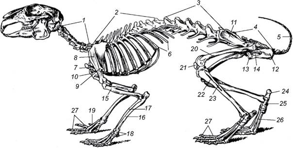
Fig. 5.49. Skeleton of a rabbit:
1 - cervical vertebrae; 2 - thoracic vertebrae; 3 - lumbar vertebrae; 4 - sacrum; 5 - caudal vertebrae; 6 - ribs; 7 - manubrium of sternum; 8 - scapula;
9 - acromion process of scapula; 10 - coracoid process of scapula; 11-13 - ilium (11), ischium (12), and pubis (13) of the innominate bone; 14 - obturator foramen; 15 - humerus; 16 - ulna; 17 - radius; 18 - carpus; 19 - metacarpus;
20 - Femur; 21 - Patella; 22 - Tibia; 23 - Fibula; 24 - calcaneus; 25 - talus; 26 - metatarsus; 27 — phalanges
The mammalian Skull is of the synapsid type. It features a zygomatic arch formed by the maxillary, jugal (zygomatic), and squamosal bones. The mammalian skull differs from that of reptiles by a significantly larger cranial capacity, a reduced number of bones (due to their reduction and fusion), and its articulation with the vertebral column via two condyles (Fig. 5.50). The lower jaw is formed by only a single paired bone—the dentary, which articulates directly with the zygomatic process of the squamosal bone. The articular bone of the reptilian lower jaw is reduced in size and transformed into one of the mammalian Middle ear ossicles—the malleus. The second part of the mammalian middle ear apparatus is formed by the quadrate bone, which transformed into the incus; the third auditory ossicle—the stapes—evolved from the upper portion of the hyoid arch—the hyomandibula—already in amphibians and is retained in all terrestrial vertebrates.
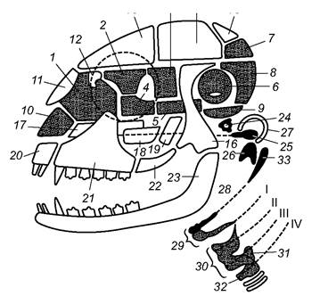
Fig. 5.50. Diagram of the mammalian skull (dark - replacing bones, light - membrane bones):
1 - ethmoid; 2 - orbitosphenoid; 3 - alisphenoid; 4 - presphenoid; 5 - basisphenoid;
6 - petrosal; 7 - supraoccipital; 8 - exoccipital; 9 - basioccipital; 10 - remnant of the cartilaginous skull (nasal septum); 11 - nasal; 12 - lacrimal; 13 - frontal; 14 - parietal; 15 - interparietal; 16 - squamosal; 17 - Vomer; 18 - palatine;
19 - pterygoid; 20 - premaxilla; 21 - Maxilla; 22 - jugal; 23 - dentary; 24 - stapes; 25 - incus; 26 - malleus;
27 - tympanic bone; 28, 29 - remnants of the hyoid and branchial arches forming the Hyoid bone and laryngeal cartilages; 30 - thyroid Cartilage; 31 - arytenoid cartilage; 32 - styloid process - remnant of the hyoid that fuses with the Zygomatic bone, I-IV - visceral arches
In the braincase, four occipital bones fuse into a single Occipital bone (occipitale); it surrounds the foramen magnum and forms two occipital condyles for articulation with the vertebral column. The otic bones fuse into the paired (right and left) petrosal bone (petrosum) (Fig. 5.50, 6). The cranial base is formed by the unpaired basisphenoid (basisphenoideum) and presphenoid (praesphenoideum) bones, and anterior to them, in the olfactory region, the unpaired Ethmoid bone (ethmoideum) develops.
The interorbital septum and the anteroventral part of the braincase are formed by paired bones: the orbitosphenoid (orbitosphenoideum) and the alisphenoid (alisphenoideum).
The skull roof is formed by paired dermal bones: the nasal, lacrimal, frontal, and parietal bones, as well as the unpaired interparietal bone. A significant part of the lateral wall of the braincase is formed by the paired dermal squamosal bones, each of which gives rise to a robust zygomatic process that connects to the zygomatic bone, which, in turn, fuses with the maxilla. The zygomatic arch formed in this way borders the Orbit from below. The cranial base is reinforced by paired dermal bones—the palatine and pterygoid bones—and a small unpaired vomer. In many species, the premaxillae fuse with the maxillae. The middle ear region is covered by the tympanic bone (tympanicum), which is unique to mammals and apparently derived from the angular bone of the ancestral lower jaw.
All mammals are characterized by the formation of a hard palate that separates the nasal passage from the Oral Cavity; it is formed by the palatine bones and the palatine processes of the premaxillae and maxillae. Thanks to the hard palate and its posterior extension—the soft palate (a connective tissue membrane)—the choanae are shifted toward the Larynx, allowing the animal to breathe while the oral cavity is filled with food. Unlike in birds, the sutures between mammalian bones persist throughout life. The Teeth are heterodont and set in alveoli.
The Pectoral Girdle is simplified and connected to the Axial Skeleton only by muscles and ligaments. The large scapula is well-developed, featuring a spine on its lateral surface (increasing the area for muscle attachment). The coracoid is reduced and fuses with the scapula as the coracoid process (remaining a separate bone only in monotremes). The clavicle is present only in those mammals whose forelimbs retain The ability to move in multiple planes (moles, bats, primates, bears, cats, etc.), while in others (canids, ungulates) it is absent, as their forelimbs move only in a plane parallel to the body axis. The Pelvic Girdle consists of two innominate bones formed by the fusion of the ilium, pubis, and ischium. The pelvis is closed: the pubic and ischial BONES OF THE left and right sides fuse with each other along the midline, forming a symphysis.
The Skeleton of the paired limbs retains the typical pentadactyl structure characteristic of terrestrial vertebrates. Unlike in reptiles and similarly to amphibians, the mobile joint of the mammalian forelimb is located between the forearm bones and the proximal row of Carpal Bones, while in the hindlimb, it lies between the lower leg and the proximal row of Tarsal Bones (the ankle joint). Due to adaptation to various modes of locomotion, the ancestral limb structure has undergone significant modifications across different mammalian orders: the relative length of limb segments, the shape and thickness of bones have changed, and the number of digits has been reduced, among other adaptations (Fig. 5.51).
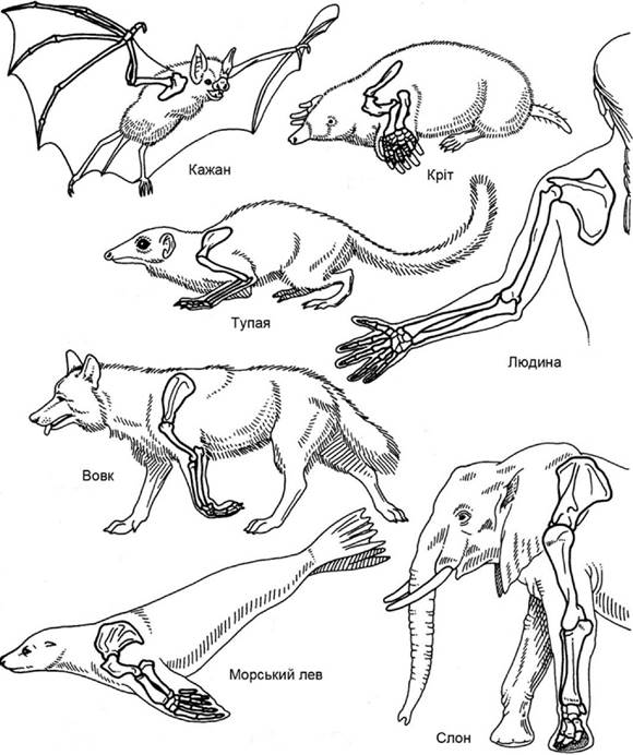
Fig. 5.51. Limb skeletons of various mammals
In bats, the unusually elongated phalanges of digits 2–5 support a stretched membrane, forming a wing. A mole's forelimbs are true digging tools; the metacarpus and metatarsus of monkeys are adapted for grasping, while the hindlimbs of kangaroos and jerboas are adapted for jumping; the single-toed limbs of horses are adapted for fast running on compacted ground, and the flippers of cetaceans and sirenians, with shortened segments and an increased number of phalanges, resemble the fin of a lobe-finned fish. Limb elongation, which increases the speed of animal movement, is usually achieved by lengthening the metatarsal and Metacarpal bones (Fig. 5.52). The transition from plantigrade to digitigrade, and in ungulates, to unguligrade locomotion, accelerates movement.
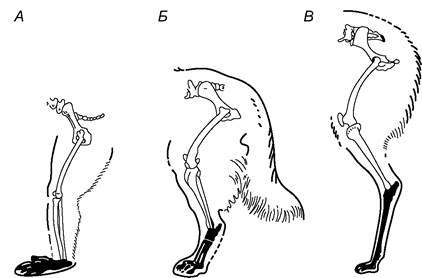
Fig. 5.52. Limb elongation due to the elevation and lengthening of the FOOT bones (shaded):
A—monkey (baboon), plantigrade; B—dog, digitigrade; C—llama, unguligrade
Mammals possess an advanced bone histological structure. The long bones of the limbs, which bear the greatest load, have a well-defined dense layered (zonal) structure in their main shaft (diaphysis). Their walls are built of osteons—concentric tubes consisting of bone lamellae bound together by dense
bundles of collagen fibrils. Bone cells (osteocytes) are located between them, and a nerve and a blood vessel pass through the internal cavity of each osteon, the Haversian canal, supplying the bone cells with nutrients and oxygen (Fig. 5.53). The spaces between osteons are filled with interstitial lamellae. A bone with this structure is characterized by low weight and high strength. Its surface is covered by the periosteum—a dense Fibrous connective tissue membrane containing Vessels and nerves that subsequently penetrate into the Haversian canals. The internal cavity of a long bone is filled with Bone Marrow, a hematopoietic organ. The upper and lower ends of a long bone—the epiphyses—are composed of spongy bone, which is formed by a complex system of trabeculae and plates aligned along the lines of maximum dynamic stress, compression, and tension (Fig. 5.54). Bone strength depends on the degree of its mineralization—the proportion of calcium salts impregnating the Bone tissue. It is highest in bones that withstand maximum mechanical load. The degree of mineralization varies among different animals: in cetaceans, due to the relative weightlessness of the body in water, the skeleton contains significantly fewer mineral salts than the skeleton of terrestrial mammals.
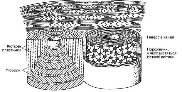
Fig. 5.53. Diagram of compact Bone Structure. Two osteons: left—bone lamellae and fibrils; right—bone cells
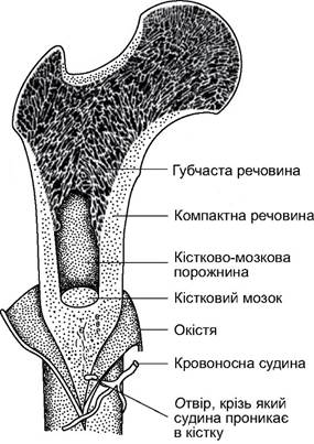
Fig. 5.54. Diagram of a long bone structure
Mammalian muscles are highly differentiated and vary significantly across different orders and families depending on their modes of locomotion. The masticatory musculature, associated with food capture and mechanical Processing, has reached a high level of development and differentiation. The complex subcutaneous musculature is involved not only in thermoregulation (altering hair position, curling the body into a ball) but also in animal communication: it controls the movement of tactile hairs (vibrissae) and is responsible for facial expressions, which play an important role in information transfer, especially in canids and primates. The dome-shaped Diaphragm—a muscular partition—separates the thoracic and abdominal cavities; its evolutionary appearance allowed for a dramatic intensification of pulmonary ventilation. Many mammalian muscles contain Myoglobin (muscle Hemoglobin), which provides an oxygen reserve. Its concentration is highest in the hard-working cardiac and skeletal muscles. The highest myoglobin content is found in aquatic mammals, enabling them to remain underwater for extended periods. In terrestrial mammals (ungulates, carnivores, mouse-like rodents), myoglobin accounts for up to 15–25% of all hemoglobin (blood + body tissues), whereas in cetaceans, the share of myoglobin corresponds to approximately half of all hemoglobin. In the sperm whale, the myoglobin content in muscles is 8–9 times higher than in terrestrial mammals.
Mammalian movements are diverse. As noted, the speed of locomotion depends on limb length and stride frequency. The positioning of the limbs beneath the body, with the elbow joint facing backward and the knee joint facing forward, increased stride length and muscle efficiency, as it drastically reduced the energy expended on supporting the body above the ground. The transition from plantigrade posture to supporting weight on the tips of the digits not only contributed to limb elongation but also increased propulsive force. Maintaining The flexibility of the spine is also crucial for accelerating movement.
Among many modes of locomotion, the most common are: the gallop, during which the hind legs are thrown far ahead of the forelegs in jumps (ungulates, lagomorphs, many mouse-like rodents, etc.); the walk and the trot—an accelerated diagonal movement of the limbs, or the pacing, where limbs on the same side move in pairs. Bipedal locomotion occurs among primates; in jerboas and kangaroos, it takes the form of ricochetal saltation on the hind limbs. Arboreal forms climb and leap. When swimming, aquatic mammals (desman, otter, muskrat, etc.) use their hind limbs and partially their tails. In cetaceans, the body flexes vigorously in the water, and propulsion is generated by the tail, with the pectoral flippers acting as stabilizers. True seals swim similarly, but generate thrust with their hind flippers. Eared seals move relatively easily on land by supporting themselves on both fore and hind flippers, whereas true seals are incapable of this due to the Structural Features of their pelvic girdle skeleton (Fig. 5.55). Finally, bats have masterfully conquered flight.
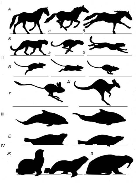
Fig. 5.55. Locomotion features of mammals:
I—typical modes of movement: a—walk, b—Two phases of a gallop. A—horse; B—cheetah; II—ricochetal running of plains dwellers: C—southern gerbil;
D—jerboa; E—eastern grey kangaroo; III—swimming Phases of the bottlenose dolphin; IV—terrestrial locomotion of pinnipeds: F—harbor seal; G—fur seal; H—walrus
The speed of locomotion varies greatly. Shrews and voles reach speeds of 4–7, elephants and rabbits up to 40, and fur seals up to 27 km/h while swimming. Some antelopes can run at speeds of up to 80 km/h; a lion can reach the same speed, though only over short distances. The cheetah is considered the fastest mammal: when pursuing prey, it reaches speeds of 105–115 km/h (accelerating to 75 km/h in 2 seconds). Bats fly at speeds of 25–30 km/h, while the fastest flyers—bulldog bats—can reach up to 60 km/h.
Digestive System. The diet of mammals is extremely diverse. The vegetative parts of woody, shrubby, and herbaceous plants form The basis of the diet for ungulates, proboscideans, lagomorphs, and rodents (voles, marmots, ground squirrels, and beavers); rodents (especially jerboas, zokors, mole-rats, etc.) readily consume the underground parts of plants. Sirenians feed on aquatic plants. Seeds and fruits serve as food for mice, squirrels, chipmunks, and dormice; seeds and fruits are also consumed in large quantities by bears, deer, wild boars, and martens. Sable populations are determined not only by the availability of animal prey (small rodents) but also by the yield of pine nuts and other fruits. Fruit bats (Megachiroptera) feed on fruits. Certain specialized groups of bats and some marsupials feed on flower nectar, while South American vampire bats suck the blood of large mammals. Invertebrates constitute the diet for most shrews and bats (except fruit bats), as well as for mice, jerboas, ground squirrels, squirrels, and small to even large carnivorans (bears). Marine plankton is the primary food source for the largest modern mammals—baleen whales. Among carnivorans, only a few live exclusively on large live prey (mostly felids), while the majority diversify their diet with plant foods. Carnivorans often attack prey larger than themselves and successfully capture it by exploiting individual advantages—strength, suddenness of attack, and agility (mustelids, leopards, etc.)—or by hunting in packs (wolves, lions).
Evolutionarily, dietary specialization has emerged, which has not only influenced the structure (Fig. 5.56) and Functions of the body of various mammals but has also determined their behavioral patterns, territory use, the formation and nature of social groups (herds of ungulates, colonies of ground squirrels and marmots), and overall mobility. As in birds, the food requirement of mammals is linked to their warm-bloodedness. Since the body temperature of mammals is lower than that of birds, they require a relatively smaller amount of food. The smallest animals (for example, the Eurasian least shrew, weighing 1.5–2.5 g) consume 2 to 4 times their body weight in food daily, alternating feeding periods with Sleep; they cannot survive without food for more than 5–8 hours. Larger mammals consume a relatively smaller amount of food, feed at specific times of the day, and have more or less prolonged periods of nocturnal or diurnal rest. The daily activity rhythms of mammals are, to a certain extent, determined by their dietary specialization.
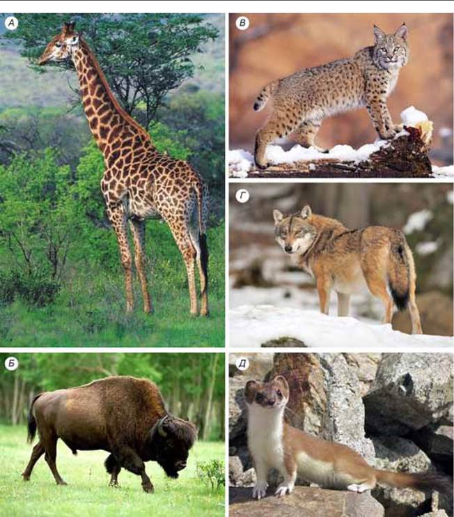
Fig. 5.56. Differences in the appearance of mammals associated with their feeding habits.
Artiodactyls: A — giraffe (feeds on tree leaves); B — bison (feeds on herbaceous vegetation).
Carnivores: C — lynx (stalks and waits for prey); D — wolf (tracks and pursues prey); E — least weasel (catches small rodents in their burrows)
The acquisition, gathering, and mechanical processing of food are carried out using the oral apparatus—lips, tongue, jaws, and teeth—as well as the limbs, and in proboscideans, the trunk. The shape of The Skull and the proportions of its main parts—the jaws, nasal region, orbits, and cranial cavity—are determined by the Development of the Sensory Organs, brain, and jaw apparatus, and are closely linked to the method of food acquisition and diet composition. For example, there are differences in the STRUCTURE OF THE lips in two rhinoceros species: in the white rhinoceros, a wide muzzle with broad lips facilitates feeding on herbaceous vegetation, whereas in the black rhinoceros, which primarily consumes tree and shrub branches, the muzzle is somewhat pointed, and a finger-like prehensile appendage is developed on the upper lip (Fig. 5.57, A, B). A narrow muzzle and a long tongue with a brush-like tip in the nectar-feeding bat (see Fig. 5.57, C), or the elongated snout and long, sticky tongue of anteaters, pangolins, and aardvarks, are Examples of similar adaptations.
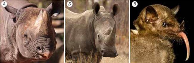
Fig. 5.57. Lip shape in black (A) and white (B) rhinoceroses. Narrow muzzle and long, brush-tipped tongue of the nectar-feeding bat Glossophaga (C)
The digestive tract of mammals is characterized by greater length, more pronounced differentiation of its regions, and a greater variety of digestive glands than those of reptiles and birds. This ensures highly efficient utilization of diverse food types. This efficiency is also enhanced by the maximum development among vertebrates of so-called symbiotic Digestion: the utilization of Bacteria, fungi, and Protozoa in the digestive tract to digest plant-based, fiber-rich foods that cannot be broken down and hydrolyzed by the host's own Enzymes. These symbionts also serve as an additional source of Nutrition for the host (see below).
The oral cavity begins with the vestibule—the space between the fleshy lips (which are unique to mammals), Cheeks, and jaws. In hamsters, chipmunks, and monkeys, cheek pouches are formed here, where animals gather food to carry to their caches. The lips usually contain tactile cells. Fleshy lips are absent in monotremes and cetaceans.
The jaws of modern mammals bear teeth of various shapes and functions. Teeth are divided into incisors (incisivi), canines (canini), premolars (praemolares), and molars (molares). Incisors, canines, and premolars have two generations (deciduous teeth are replaced by permanent ones), while molars have only one. In seals and toothed whales, the teeth are undifferentiated. Mammalian heterodonty is an important adaptation that enhances feeding and digestion.
Different mammalian orders differ in the Composition and Structure of their teeth. The ancestral condition was likely a continuous row of relatively weakly differentiated teeth. This has been partially preserved in shrews, bats, and to some extent in primates and carnivorans. However, it has undergone adaptive changes in each group. In small shrews, which have an elongated, narrow snout, the tooth rows with forward-pointing incisors form a kind of forceps, convenient for grasping and holding small prey (insects, worms, etc.), while their sharp-cusped molars crush the chitinous exoskeleton. The dentition of carnivorans has undergone greater differentiation:
sharp incisors, large canines, and molars with shearing edges. Canines, as a means of capturing and subduing prey, reached their greatest development in the extinct saber-toothed cat.
In monkeys, feeding mainly on soft fruits has led to weaker Differentiation of the dentition: the canines are small, and the molars have cusped or flat grinding surfaces. In rodents, the incisors have sharp cutting edges; the number of incisors is reduced (two in each of the upper and lower jaws), canines have disappeared, and the molars have acquired a cusped or flat grinding surface with enamel ridges; such a surface is capable of grinding the toughest parts of plants. In rodents, the incisors, and in some species also the molars, grow continuously throughout life. Elephants have only a pair of incisors in the upper jaw (tusks) and one large true molar in each half of the upper and lower jaws; the total number of teeth is only six. Other mammals have more. To determine the number and identify species, dental formulas are used, which reflect the number of teeth in one half of the upper and lower jaws. Tooth types are designated by the initial letters of their Latin names. The dental formula is written as follows:
wild boar I 3/3 C 1/1 PM 4/4 M 3/3=44 teeth;
hare I 2/1 C 0/0 PM 3/2 M 3/1=28 teeth.
In baleen whales, teeth develop in the Embryonic Stage but later disappear. Simultaneously, epidermal ridges form on the sides of the oral cavity along the outer margins of the upper jaws; these ridges subsequently grow, become keratinized, and divide into a series of triangular plates whose bases are embedded in the gum tissue. The number of plates varies from 160 to 500 in different species. These plates, known as baleen, bear fringes of interwoven keratinous fibers and serve to filter planktonic organisms: by closing its jaws and raising its tongue, the whale forces water out of the oral cavity.
Keratinized palatal ridges in rodents and ungulates participate in grinding plant food. At the bottom of the oral cavity lies a fleshy tongue, which is involved in chewing and swallowing food, and in ungulates, also in food gathering. In felids, keratinized papillae on the tongue allow them to scrape meat off the bones of their prey. The long, vermiform, and sticky tongue of anteaters is an instrument for extracting ants and termites from their nests (reaching 60 cm in length and anchoring to the sternum). The mucous secretions of three pairs of large and several small Salivary Glands opening into the oral cavity form saliva, which contains enzymes that break down starch during chewing. These enzymes are more abundant in the saliva of herbivores than in carnivores. Taste buds are located in the mucosa of the Oral Cavity and tongue. Chewed and sufficiently salivated food passes from the oral cavity through the Pharynx and Esophagus into The Stomach. The esophagus of most mammals is a thin-walled tube with smooth muscles. Only in ruminant ungulates does it possess striated muscles, which allow food (cud) to be voluntarily regurgitated from the stomach back into the oral cavity for additional chewing. The stomach is separated from the esophagus and intestine and is well-developed in all mammals. Its size, shape, and structure vary across different orders (Fig. 5.58). In monotremes, the stomach is a simple sac-like structure and lacks digestive glands. The single-chambered stomach of carnivorans, primates, and shrews is retort-shaped, and its wall epithelium contains numerous digestive glands. Ruminant diets are dominated by coarse plant forage, so their stomach consists of the rumen, reticulum, omasum, and abomasum (Fig. 5.59): the first three compartments (rumen, reticulum, and omasum) form the so-called forestomach and are lined with stratified epithelium; the forestomach lacks digestive glands, and only bacterial Fermentation occurs there with the participation of symbionts that can exist only in a neutral or weakly alkaline environment. The breakdown of plant food by symbionts occurs in the rumen, where only lightly chewed food accumulates; fermentation intensifies after the cud is re-chewed and moistened with saliva, which has a weakly alkaline reaction. Fermentation and mechanical grinding of food continue in the reticulum and omasum. Processing with gastric juice occurs only in the abomasum, which has an acidic environment.
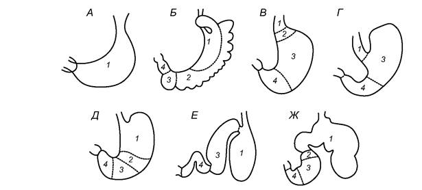
Fig. 5.58. Diagram of the stomach structure in mammals:
A — echidna; B — kangaroo; C — human; D — hare; E — hyrax; F — dolphin; G — bull:
1 — stratified epithelium of the esophagus; 2-4 — simple epithelium with cardiac (2), fundic (3), and pyloric (4) glands
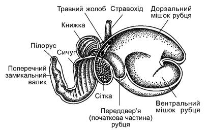
Fig. 5.59. Stomach of a ruminant mammal (sheep):
1 - esophagus; 2 - reticular groove; 3 - dorsal sac of the rumen; 4 - ventral sac of the rumen; 5 - atrium (initial part) of the rumen; 6 - reticulum; 7 - omasum; 8 - abomasum; 9 - pylorus; 10 - torus pyloricus
In sloths, the stomach serves primarily as a food storage site; it accounts for 20-30% of body weight and is always filled with food, which slowly enters the intestines and moves through them at a very low speed (sometimes taking up to a week). The complex stomach of cetaceans, whose teeth cannot grind food, provides mechanical processing during peristaltic movements of its walls. In anteaters, food grinding occurs in the pyloric region of the stomach, which has a tough cuticular lining with highly developed muscles. The gastric juice of cetaceans and anteaters is remarkably active and breaks down (hydrolyzes) even highly resistant substances such as Chitin (which other animals typically cannot digest).
The gastric glands of the stomach are not uniform. In the region closest to the esophagus, there are branched cardiac glands; the middle region is dominated by the highly important for digestion fundic glands, which are weakly branched, and the pyloric region adjacent to the intestine contains pyloric glands. The gastric juice secreted by these glands contains Hydrochloric acid (up to 0.4-0.5%) and enzymes—Pepsin (which breaks down Proteins), lipase (which breaks down fats), and several others.
The mammalian intestine (Fig. 5.60) is divided into the Small Intestine, Large Intestine, and rectum; the initial part of the small intestine is called the duodenum; at the junction of the small and large intestines, a large cecum branches off, which is reduced in size or completely absent in a few carnivores. As in the stomach, the intestinal walls consist of three layers: an inner mucosa with digestive glands, a muscularis with longitudinally and circularly arranged smooth muscle fibers, and an outer serosa. The glands of the small intestinal walls secrete various digestive enzymes; this is also where the DIGESTION AND ABSORPTION of major nutrients take place. Liquefied food containing hard-to-digest particles enters the large intestine. There, and especially in the cecum, fermentation processes occur involving bacteria, fungi, and protozoa, similar to the processes in the complex stomach of ruminants. Reabsorption of water and the formation of feces take place in the rectum.
In herbivorous animals, the problem arises of obtaining Essential Amino Acids, which are found only in animal proteins. Sciurids (squirrels, ground squirrels, etc.) solve this problem by actively preying on small animals. Hamsters and voles, whose stomach lacks digestive glands and is lined with a cuticle—seemingly to protect the walls from damage by coarse fodder—use the stomach as a fermentation vat where protozoa capable of breaking down Cellulose multiply. Passing with the food mass into the intestine, they are digested and provide the host with the necessary proteins. Similar processes have reached an even larger scale in the complex stomachs of ungulates, which could even be called "protistophages," as they utilize Ciliates and other protozoa that multiply in the rumen, reticulum, and omasum. Lagomorphs have only the cecum and large intestine as fermentation vats; however, the symbionts living there cannot be digested because these regions lack the necessary enzymes (pepsin) and acidic environment. They exhibit The phenomenon of coprophagy (eating feces), as this is the only way they can
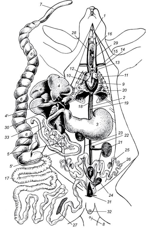
Fig. 5.60. General layout of the Internal Organs of a female rabbit:
1 - Cytology/practical/98.html">Submandibular salivary gland; 2 - esophagus; 3 - stomach; 4 - Liver (reflected upward); 4' - Gallbladder; 5 - Pancreas;
5' - pancreatic duct; 6 - cecum; 7- its Appendix; 8 - anus; 9 - left ventricle of The Heart;
10 - right ventricle of the heart; 11 - left atrium; 12 - right atrium; 13 - aortic arch; 14 - left Subclavian Artery; 15 - left subclavian vein; 16 - left jugular vein; 17 - colon; 18 - posterior vena cava; 19 - aorta; 20 - left lung; 21 - left Kidney; 22 - left Adrenal gland; 23 - right Ureter; 24 - Urinary Bladder; 25 - left Ovary; 26 - left oviduct; 27 - Spleen;
28 - Thyroid Gland; 29 - Trachea; 30 - Bile duct; 31 - Vagina; 32 - urogenital opening; 33 - duodenum
digest the intestinal symbiotic organisms. In this case, they consume feces, which are highly rich in animal proteins.
The length of the intestine and The ratio of its regions in different mammals correspond to their diet. In bats, the intestine is 1.5–4.0 times longer than the body; in shrews, 2.5–4.5 times; in wolves, 6.5 times; in muroid rodents (gerbils, guinea pigs), 5–12 times; in horses, 12 times; and in sheep, 29 times longer than the body. A diet rich in fiber is always accompanied not only by an overall lengthening of the intestine but also by an increase in the relative size of the large intestine and especially the cecum.
The digestive glands—The Liver and pancreas—participate not only in digestion by producing active enzymes, but also in general metabolic and excretory processes, as well as in their hormonal regulation. The ducts of the liver (bile duct) and pancreas empty into the anterior part of the small intestine—the duodenum. The relative size of the liver decreases as body size increases. For example, the liver of a shrew weighing 18–20 g accounts for 5–6% of its body weight, whereas a whale's liver is only 1%. The pancreas increases in size and secretory activity with an increase in the proportion of plant-based food.
A number of specific adaptations ensure the survival of mammals during seasonal changes in food availability. Some of these adaptations are shared with other animals. In particular, the accumulation of reserve substances (fat in the subcutaneous tissue and body cavity, Glycogen in the liver) during favorable seasons (fattening) and their utilization during unfavorable periods. In some mammals, the mass of fat reserves can reach up to 40% of their body weight. Many species exhibit a distinct seasonal shift in diet. For example, moose, hares, and many other herbivores, which feed on green plant parts in summer, consume dry grass, shoots, and the bark of trees and shrubs in winter. Some mammals are characterized by food hoarding, similar to that of social insects.
During unfavorable periods, many mammals enter a state of torpor (hibernation), which is an alternative to remaining active by hoarding food. Torpor is associated with either a shortage or unavailability of food; it typically occurs during summer droughts; it is not observed in tropical rainforests with abundant food supplies or in the tundra, where the duration of the unfavorable period is extremely long. It has been recorded that under particularly harsh conditions—such as severe frosts and blizzards—martens, squirrels, and some other species do not leave their shelters for several days, sleeping through the weather in their nests.
The body prepares for the period of hibernation. Many PHYSIOLOGICAL AND BIOCHEMICAL processes undergo reorganization under the influence and control of the neurohumoral system, which reflects endogenous rhythms and responds to seasonal changes in living conditions—primarily the decline in food availability. This mechanism ensures the preliminary preparation of the body and allows it to adapt to the specific conditions of the year. Changes in behavior and metabolism ensure the accumulation of significant fat reserves in the subcutaneous tissue and body cavity, ascorbic acid (Vitamin C) in tissues, and glycogen and vitamin E in the liver prior to hibernation. Animals seek out or construct shelters in which they will hibernate: bats in caves, dormice in tree hollows, bears in dens, and various rodents in burrows.
There are several types of hibernation, namely:
1) winter sleep is characteristic of bears, badgers, raccoons, and raccoon dogs. During sleep, the animals' respiration and Circulation rates decrease, body temperature drops by only 2-7°C, and the overall metabolic rate decreases by 50-70% (Slonim, 1961). Disturbed animals wake up easily and quickly; sleep is also interrupted during prolonged thaws. Female bears give birth to cubs during winter sleep. The duration of winter sleep is determined by weather conditions and food availability. For example, brown bears in the temperate zone stay in their dens from November to April, whereas in the Caucasus they do so only from December to February (and in some regions, they do not sleep at all during warm, snowless winters);
2) true hibernation interrupted by temperature increases. Animals enter a state of torpor accompanied by a significant reduction in respiration and circulation rates and a drop in body temperature, losing the ability to respond to stimuli (sounds, Touch, etc.). They wake up slowly only during prolonged thaws. In particular, during these periods, bats may hunt insects in caves, and hamsters and chipmunks utilize their stored food reserves;
3) true continuous hibernation is accompanied by a deeper torpor, in which respiration and circulation rates decrease 1020 times, metabolism drops 20-40 times, and body temperature falls to 5-1°C (Kalabukhov, 1956; Slonim, 1961). Even the sharpest external stimuli (sounds, pain, etc.) and brief temperature increases do not cause arousal. During the hibernation period, body weight decreases by 30-40%, with fat reserves being used first. This type of hibernation is characteristic of hedgehogs, some bats, and many rodents (marmots, ground squirrels, jerboas, dormice). As vegetation dries up, ground squirrels enter torpor as early as summer; this estivation transitions into winter hibernation, and the animal becomes active only the following spring, having spent 6-8 months in a state of torpor.
Respiratory system. In mammalian gas exchange, the Lungs play the leading role, while the mucosal surface of the airways and, to a lesser extent, the skin also participate; only 1% of the oxygen required by the animal enters through the skin. The Nasal cavity is divided into the vestibule, respiratory, and olfactory regions. The function of the vestibule is to trap relatively large particles (dust) entering with the air. The vestibule is more developed in inhabitants of steppe and desert regions, where the air is particularly dusty. Fine dust particles are trapped in the respiratory region of the nasal cavity, which is lined with a mucous membrane of ciliated epithelium containing numerous blood capillaries; here, not only is the air further cleared of mechanical impurities, but it is also disinfected by the bactericidally active substances of the mucus. Simultaneously, as it passes between the folds of the nasal conchae, the air is humidified and warmed by the blood flowing through the capillaries, which releases some of its oxygen. The olfactory region, expanded by projections of the lateral walls, forms a labyrinth of cavities lined with olfactory epithelium. Passing through the choanae into the pharynx, the air enters the larynx. At the base of the larynx is a ring-shaped cricoid cartilage, characteristic of all terrestrial vertebrates. The anterior and lateral walls of the larynx are formed by the thyroid cartilage, which is unique to mammals. The arytenoid cartilages are located on the sides of the dorsal part of the larynx. Adjacent to the anterior margin of the thyroid cartilage is the thin epiglottis (Fig. 5.61), which covers the entrance to the larynx during the passage of food through the pharynx. The vocal cords are paired folds of the mucous membrane located between the thyroid and arytenoid cartilages. The voices of most mammals are restricted mainly to the low-frequency range, which, however, does not prevent them from transmitting complex and rich information.
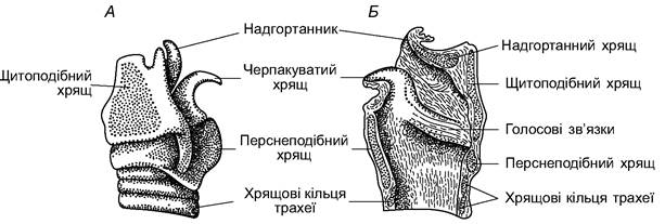
Fig. 5.61. Larynx of a cow: A - lateral view; B - sagittal section
Adjacent to the larynx is the trachea, which is supported by cartilaginous rings. The trachea divides into two Bronchi, which differ from it only in their smaller diameter; within the lungs, the bronchi branch into even smaller tubes, forming the bronchial tree (Fig. 5.62). The smallest tubules—bronchioles—open into expansions lined with respiratory epithelium: alveoli measuring 25-400 μm, in the walls of which numerous capillaries branch. This lung structure increases their respiratory surface area, making it 50-100 times larger than the body surface area. The relative surface area through which gas exchange occurs in the lungs is larger in animals with high activity and mobility.

Fig. 5.62. Bronchial branching in the lungs of a brown bear
A relative increase in lung size is observed in high-altitude and aquatic mammals. Whales possess sphincter muscles that close the entrance to the alveoli, allowing them to retain air even at significant depths.
The respiratory mechanism in mammals is dual. In so-called costal (rib) breathing, the volume of the chest cavity is altered by the intercostal muscles; in diaphragmatic breathing, the same volume is changed by the lowering and raising of the muscular thoracoabdominal partition—the diaphragm. Both mechanisms function in various combinations in all mammals; the former predominates in canids, while the latter is dominant in ungulates. The respiratory rate in small animals with a higher metabolic rate is greater than in large ones; it is 8-16 breaths per minute in horses, 100-150 in rats, and 200 in mice. Respiration rate increases during movement. Respiration also participates in thermoregulation. Rapid, shallow breathing in canids (polypnea) increases evaporation from the surface of the Upper Respiratory Tract, promoting heat loss. Conversely, accelerated deep breathing, by enhancing Gas Exchange in the lungs, increases metabolic heat production at low temperatures.
Circulatory system. In mammals, as in birds, the SYSTEMIC AND PULMONARY circulations are completely separated. A single left aortic arch arises from the left ventricle of the four-chambered heart. In most species, a short brachiocephalic (innominate) artery branches off from it, dividing into the right subclavian and the carotid (right and left) Arteries; the left subclavian artery branches off independently. The dorsal aorta, which is a continuation of the left arch, gives off vessels to the muscles and internal organs (Fig. 5.63).
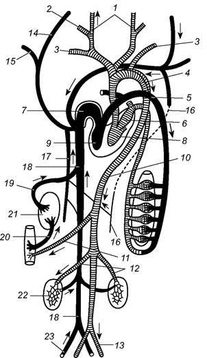
Fig. 5.63. Diagram of the mammalian circulatory system:
1, 2 - external and internal carotid arteries; 3 - subclavian artery; 4 - left aortic arch; 5 - pulmonary artery; 6 - left atrium;
7 - right atrium; 8 - left ventricle; 9 - right ventricle; 10 - dorsal aorta; 11 - visceral artery; 12 - renal artery;
13 - iliac artery; 14 - jugular vein; 15 - subclavian vein; 16 - left azygos vein; 17 - right azygos vein; 18 - posterior vena cava; 19 - hepatic vein; 20 - HEPATIC PORTAL VEIN; 21 - liver; 22 - kidney; 23 - iliac vein
Only in a few mammals are both anterior venae cavae equally developed; in most species, the right anterior vena cava receives the brachiocephalic (innominate) vein, formed by the fusion of the jugular and left subclavian Veins. Asymmetrical remnants of the posterior cardinal veins of lower vertebrates—the so-called azygos (vertebral) veins—are characteristic only of mammals. In most species, the left azygos vein is connected to the right azygos vein, which empties into the right anterior vena cava. Notably, there is no renal portal system, which is associated with the specific nature of excretory processes.
Lymphatic vessels open into the venous vessels near the heart. They begin as lymphatic capillaries that collect interstitial fluid (Lymph). The Lymphatic system of mammals lacks lymphatic hearts (pulsating vessel segments) but possesses Lymph Nodes (glands), whose function is to cleanse the lymph of pathogens with the help of phagocytic cells—lymphocytes. In its chemical composition, lymph is similar to Blood Plasma, though poorer in proteins. In the lymphatic vessels associated with the digestive tract, the lymph is enriched with Lipids, whose molecules cannot penetrate the tight walls of blood capillaries but easily pass through the more permeable walls of lymphatic vessels. The formed elements of lymph consist of various types of lymphocytes (white Blood Cells).
Hematopoietic organs are specialized. Bone marrow produces erythrocytes, granulocytes, and platelets; the spleen and lymph nodes produce lymphocytes; the reticuloendothelial system produces monocytes.
Substances such as agglutinins, lysins, precipitins, and antitoxins neutralize or destroy harmful substances that enter the blood. The small erythrocytes of mammals lack nuclei, which increases their oxygen-carrying efficiency, as they consume 9-13 times less oxygen for their own respiration than avian erythrocytes and 17-19 times less than amphibian erythrocytes. The relative heart size is larger in more active and smaller animals. In large species, the heart mass accounts for 0.2-0.7% of body mass, in small ones up to 1.0-1.5%, and in bats, it reaches 1.3%.
The heart rate per minute is 600 in mice, 140 in dogs, and 24 in bulls and elephants. In aquatic animals, the heart rate decreases upon diving (in seals, from 180 at the surface to 60-30 underwater), allowing for a more economical use of oxygen stores in the lungs and air cavities. At the same time, this facilitates the equalization of blood pressure, which changes significantly over a short period during the rapid descent of cetaceans (100-140 m/min). In all aquatic mammals, The amount of myoglobin in the muscles is sharply increased. The oxygen stores in their bodies are distributed as follows (%):
Distribution of oxygen stores in humans and whales
Lungs |
Blood |
Muscles |
Other organs |
|
Human |
34 |
41 |
13 |
12 |
Whale |
9 |
41 |
41 |
9 |
Excretory system and water-salt balance. In mammals, urea is excreted as the primary product of Nitrogen metabolism; in this respect, mammals are closer to amphibians. Water-salt balance is maintained primarily through the Kidneys and is regulated by Hormones of the posterior Pituitary Gland, which affect diuresis (urination) and the reabsorption of water from the primary urine (filtrate) in the renal tubules. Additionally, both the skin with its sweat glands and the intestinal tract participate in the water-salt metabolism of mammals.
The kidneys of mammals, like those of other amniotes, are metanephric; they have their own excretory ducts—Ureters, which empty into the urinary bladder. In males, the bladder duct opens into the copulatory organ, while in females, it opens into the vaginal vestibule. In monotremes (egg-laying mammals), the excretory ducts empty into the cloaca. The kidneys are bean-shaped and lie on either side of the vertebral column. A Longitudinal section of the kidney reveals that it consists of two layers. The outer (cortical) layer contains the filtration apparatus—the glomeruli. A glomerulus is a cluster of capillaries surrounded by Bowman's capsule. An excretory tubule originates from the capsule and consists of four sections: the proximal convoluted tubule, the Loop of Henle, the Distal convoluted tubule, and the collecting duct. Together, these structures form a nephron (Fig. 5.64). The collecting ducts are shared by a group of nephrons, forming the renal pyramids, or lobes, which are clearly visible on section. The openings of the collecting ducts empty into the renal pelvis, from which the ureters originate.
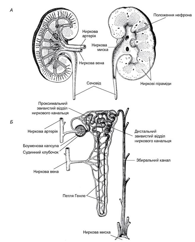
Fig. 5.64. Mammalian kidney: A - cross-section of a human kidney; B - structure of a nephron
The filtrate in Bowman's capsule is protein-free blood plasma, which still contains a significant amount of substances useful to the body (sugars, Vitamins, amino acids, salts, water). These are reclaimed in two ways: by diffusion through the walls of various sections of the nephron tubules, which is governed by basic physical laws (concentration gradient between the tubules and the surrounding kidney tissue), and by active reabsorption involving enzymes bound to the membranes of the tubule wall cells. Active reabsorption of sugars, vitamins, amino acids, chlorides, and partially sodium ions occurs in the proximal convoluted tubule. Water and sodium ions are reclaimed in the loop of Henle, which is highly developed in mammals. Its limbs, through which the countercurrent flows of primary urine pass, are surrounded by tissues where the Osmotic Pressure of the interstitial fluid is highest near the hairpin turn of the loop (renal medulla) and lowest in the cortex. In the medulla, water is drawn out, diffusing into the renal interstitium and then into the blood vessels, thereby concentrating the urine. The transport of sodium ions occurs in the same section via enzymatic systems (active reabsorption). The reabsorption of water and other substances is extremely high. For example, a human filters approximately 180 liters of primary urine through the renal glomeruli daily, but excretes only 1-2 liters of final urine over the same period. The rectum also plays a role in water balance, as its walls absorb water from fecal matter (a feature particularly characteristic of semi-desert and desert animals).
Inhabitants of humid biotopes with abundant water resources have a high rate of water turnover. In contrast, dwellers of semi-desert biotopes obtain most of their water by consuming the juicy parts of succulent plants. Their cutaneous and pulmonary water loss is minimal; for example, at a temperature of 20°C, the relatively moisture-loving tamarisk gerbil loses 170 cm3, whereas the great gerbil, a resident of arid biotopes, loses only 50 cm3 per 1 kg of body mass per hour. True desert mammals are capable of feeding on almost dry food and practically never drink throughout their entire lives, satisfying their needs with metabolic water produced within the body. During forage-rich and wet seasons, camels store fat, which they deplete during periods of drought and food scarcity. The metabolism of this fat yields a certain amount of water. During rest and sleep, camels lower their body temperature, which also reduces water expenditure.
Reproductive System and breeding. Mammals are distinguished by a high degree of parental care at all stages of offspring development. They are characterized by intrauterine development, during which the embryo grows at the expense of the mother's body, receiving nutrients through a specialized organ—the Placenta. Only monotremes lay large, yolk-rich eggs.
In all mammals, the bond with the mother does not end after birth, as the offspring feed on milk. However, even after weaning, the bonds between parents and offspring typically persist for some time. This provides an opportunity for teaching the young—transferring the individual experience accumulated by the parents to the offspring. This intergenerational connection, which allows for the accumulation of population- and species-specific experience, is called signal heredity or signal transmission of information. This phenomenon is common among birds and is particularly characteristic of mammals. It reaches a qualitatively different level in humans, with their intellect and second signaling system, the foundation of which lies in socio-productive relations.
The reproductive organs of mammals are more complex in structure than those of other amniotes (Fig. 5.65). The Testes are paired, located in the posterior part of the Abdominal cavity (monotremes, some shrews, xenarthrans, proboscideans, cetaceans, sirenians, rhinoceroses) or have descended into the Scrotum—a cutaneous pouch connected to the body cavity via the Inguinal Canal (marsupials, caniforms, ungulates, primates). Adjacent to each Testis is an elongated body—the Epididymis—which is a cluster of efferent ducts of the testis (a remnant of the anterior section of the mesonephric kidney). Arising from the epididymis is the Wolffian duct—the vas deferens—which empties into the urogenital canal at the base of the Penis. Before entering the canal, the vasa deferentia form lobulated expansions—the Seminal Vesicles (vesicula seminalis), whose secretion contributes to the formation of semen. In mouse-like rodents, this secretion is discharged after ejaculation into the female reproductive tract and, upon hardening, plugs it after copulation.
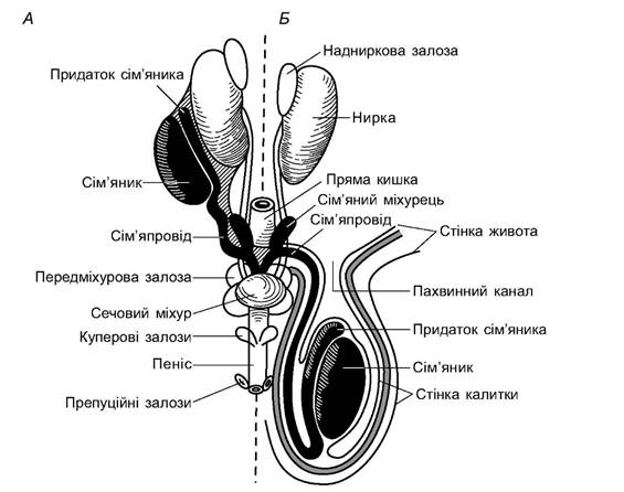
Fig. 5.65. Male reproductive organs of mammals: A - testes located in the abdominal cavity; B - testes in the scrotum
Located at the base of the penis is the Prostate Gland (prostata), whose secretion forms the liquid medium of semen. The penis contains corpora cavernosa that surround the urogenital canal; the engorgement of their cavities with blood provides the rigidity of the copulatory organ. In some mammals (carnivorans, seals, rodents, etc.), its rigidity is enhanced by a special bone (os penis). Cowper's and preputial glands are also associated with the penis; the former contribute to the liquid portion of semen (ejaculate), while the latter secrete an odorous substance that aids in mate recognition and sexual arousal.
The paired Ovaries of females always lie within the body cavity and are attached to the dorsal wall of the abdominal cavity by mesenteries. The size of the ovaries is noticeably smaller than in other vertebrates. The paired oviducts, homologous to the Müllerian ducts, open into the body cavity near the ovaries via infundibula lined with ciliated epithelium. The upper, thin, and convoluted section of the oviduct is called the Fallopian tube, while the lower, expanded section with muscular walls is the Uterus. In monotremes, the uteri open into the urogenital sinus of the cloaca; in all other mammals, the cloaca has disappeared. In marsupials, the lower part of the oviducts is modified into paired vaginas, which sometimes fuse at their ends and open into the vaginal vestibule (urogenital sinus). In placental mammals, the lower sections of the oviducts fuse into a single, unpaired vagina (vagina). If each uterus opens into the vagina by a separate orifice, it is called a duplex uterus (many rodents, elephants, etc.); in a bipartite uterus, the right and left uteri are fused at their distal ends and open into the vagina through a single common orifice (rodents, some caniforms, pigs), and in a bicornuate uterus, they are fused for about half of their length (shrews, carnivorans, ungulates, cetaceans). In some bats and primates, the right and left uteri are completely fused into a single simplex uterus, into which the right and left Fallopian tubes open (Fig. 5.66).

Fig. 5.66. Diagram of the evolution of oviducts in mammals:
I - monotreme; II - lower marsupial (opossum); III - higher marsupial (kangaroo);
IV - VI - placental mammal with duplex (IV), bicornuate (V), simplex (VI) uteri:
1 - Fallopian tube; 2 - uterus; 3 - vagina; 4 - urogenital sinus; 5 - urinary bladder; 6 - cloaca; 7 - rectum
The vagina leads into a short, expanded urogenital sinus (vaginal vestibule), into which the duct of the urinary bladder also opens. In the wall of the vaginal vestibule, There is a small projection containing erectile tissue—the Clitoris, which is homologous to the male copulatory organ. The vaginal vestibule opens externally via a slit-like urogenital opening, bounded by folds of skin—the labia. Cutaneous glands located here secrete an odorous substance.
Egg cells—oogonia—develop in the ovaries from the germinal epithelium; they grow and differentiate into oocytes; each oocyte is surrounded by follicular cells, forming a primary follicle. By the breeding season, the oocyte begins to grow, accumulating yolk, while its envelope expands, and a fluid-filled space forms between it and the oocyte. The follicle develops into a Graafian follicle, which bulges On the surface of the ovary. Then, the follicle ruptures, and the egg Cell is released into the body cavity (ovulation occurs), from where it passes into the Fallopian tube, where Fertilization takes place. The scar formed on the surface of the ovary is filled with blood clots and becomes infiltrated with lutein cells, which contain a yellow pigment and perform endocrine functions. Thus, the corpus luteum is formed in the ovary, secreting the hormone progesterone. If fertilization occurs and Pregnancy ensues, the corpus luteum continues to develop, and progesterone stimulates the implantation of the embryo into the uterine wall, while simultaneously inhibiting the formation of new Graafian follicles and preventing ovulation until the end of pregnancy.
During development, the embryonic membranes of marsupials merely lie against the uterine wall, causing partial histolysis of its superficial layers. In some species, a rudiment of the fetal part of the placenta is formed by the fusion of the outer wall of the allantois with the serosa, though without the formation of villi that penetrate deep into the uterine wall. The embryo is nourished by the egg yolk and secretions from the cells of the uterine wall. In placental mammals, a true placenta develops: the fused Regions of the serosa and the outer wall of the allantois form the chorion (the fetal part of the placenta), whose outgrowths (villi) are embedded in the uterine wall; at this site, the uterus becomes spongy and forms the maternal part of the placenta. At this stage, the histolysis of uterine tissues intensifies, and the embryonic blood vessels establish closer contact with the maternal circulatory system. This facilitates the nourishment of the embryo with substances from the mother's blood (hemotrophic nutrition). The Formation of the placenta is referred to as embryonic implantation. The placenta can have various structures: it is called diffuse when the villi are distributed evenly over the entire chorion, cotyledonary when they are clustered in separate groups, or discoid when the villi are concentrated in one area, forming a disc, etc. The degree of connection between the fetal and maternal placentas varies; based on this feature, deciduate and non-deciduate placentas are distinguished. In the former case, the connection is so intimate that during parturition, a portion of the uterine wall detaches and is expelled as the afterbirth.
In some species, mammal young are born helpless, while in others, they are capable of active movement. Newborn marsupials are exceptionally small. They are underdeveloped and are carried to term in special pouches. Consequently, the gestation period in marsupials is short: 12 days for the opossum and 38–40 days for the giant kangaroo. Generally, the larger the animal, the longer the gestation period. It also depends on the ecology of the species. Shrew-like mammals give birth to underdeveloped young after 13–19 days of pregnancy; gestation lasts 54–73 days; in animals that give birth in burrows with warm nests, gestation is short (house mouse – 18 days, common vole – 16–23 days, muskrat – 25–26 days); it is longer in larger marmots (30–40 days). In animals that lack well-constructed shelters, gestation is longer: about 130 days in the coypu, 120 in the leopard, and 200 days in the brown bear. The longest gestation periods occur in animals whose young follow the mother immediately after birth: in ungulates, from 150 (pigs) to 290 (cattle) days; in the white rhinoceros, 540 days; in elephants, over 600 days; it is also long in pinnipeds (walrus – 200 days) and cetaceans (270–365 days).
Sometimes, pregnancy is extended by a latent period during which the embryo temporarily ceases development. This embryonic diapause allows birth to be delayed until a favorable season. It has been identified in so-called diestrous species, i.e., those with two periods of sexual activity. For example, in the badger, the first estrus occurs in July–August, and the second in October. Eggs fertilized in July remain dormant until late autumn, whereas those fertilized in October develop without diapause; the young are born in spring. In the ermine, females fertilized during the spring estrus give birth after two months, while those fertilized in August–September do so only after 8–9 months. In the roe deer, eggs fertilized During the first estrus (July–August) develop slowly until December, whereas those fertilized during the autumn estrus (November–December) experience no developmental delay. In the marten and sable, the true estrus occurs in mid-to-late summer, while the second (false) estrus occurs in early spring. It is associated with the implantation of diapaused eggs (birth occurs in spring).
The rate of postnatal (post-birth) development in mammals during the initial stages is linked to the COMPOSITION OF THE milk with which the mother feeds her young. Mammalian milk contains all the substances necessary for development: proteins, fats, CARBOHYDRATES, vitamins, and salts. The higher the protein and fat content in the milk, the faster the rate of development.
Mammalian reproduction is rhythmic, meaning it is ordered in time. Its potential is determined by the duration of the reproductive cycles, which begin after sexual maturity. The timing of sexual maturation varies widely. Small animals are the most prolific: voles reach sexual maturity at 1.5–2 months of age, mice at 2–3 months, muskrats at 5 months, and hares at about one year. Larger animals begin to breed later: wolves, martens, foxes, and sables in their second year of life; tigers, bears, and many species of seals and cetaceans in their 3rd or 4th year; deer in their 2nd to 4th year; and rhinoceroses and elephants at 10–15 years. Sexual maturity in great apes occurs at 10–12 years of age. Breeding frequency and periodicity are linked to mortality rates and lifespan. High fecundity is an important adaptation for small mammals, which are physiologically vulnerable and have numerous dangerous predators. It compensates for their high mortality rate. Many mouse-like rodents are potentially capable of breeding year-round, even under the harsh conditions of the Arctic. For example, winter breeding occurs in the Norway lemming. It has also been recorded multiple times in the Siberian lemming. Winter breeding has been observed in mice, voles, gerbils, and hamsters in forest, steppe, and desert zones. Rats and house mice living in human dwellings are also capable of breeding throughout the year. The wide amplitude of fluctuations in fecundity and mortality helps explain the highly volatile population sizes of the aforementioned animals. Other species typically breed during a specific period of the year. The duration of the reproductive cycle depends on the length of gestation. Births always coincide with a period favorable for rearing young, predominantly in the spring. During the breeding season, ovulation and estrus in females recur cyclically until fertilization occurs. These estrous cycles typically consist of four phases: proestrus (prooestrus), estrus (oestrus), metestrus (metaoestrus), and diestrus (disoestrus). The estrous cycle in small mouse-like rodents lasts about 6 days, in dogs 9–14 days, and in ungulates (goats, sheep, cows) 17–24 days.
Reproductive readiness and the timing of mating to a specific period are regulated by a complex mechanism that responds to environmental cues. In temperate and high latitudes, this cue is The change in photoperiod (day length). For species with spring estrus, an increase in day length serves as the signal (many animals); for artiodactyls that mate in autumn, it is the shortening of the day; and for wolves, it is the short winter day. Changes in the light regime affect the neurohypophysis, which stimulates the anterior pituitary gland to release gonadotropic hormones that, in turn, affect the Gonads. At low latitudes, other periodic natural phenomena preceding the onset of a favorable breeding season can act as signaling factors (precipitation in deserts and savannas, the ripening of primary food plants or their fruits in tropical forests, etc.). Even in tropical rainforests with their constantly warm and humid climate, mammalian reproduction is typically seasonal, although it occurs at different times of the year for different species.
Due to high levels and diverse forms of parental care, the overall fecundity of mammals is relatively low. In small mouse-like rodents with high mortality rates, litters can reach up to 12 young (typically 5-6), and breeding continues year-round (up to six litters per year). Hares and squirrels produce 2-3 litters per year, each usually consisting of three to eight young (up to 12). Wolves, foxes, cats, sables, martens, and ermines breed once a year, with litters of 3-6 young. Bovids, deer, seals, and dolphins give birth to 1-2 young once a year. Elephants, baleen whales, tigers, and some other large felids breed once every 2-3 years, typically giving birth to 1-2 offspring. Fecundity varies depending not only on environmental conditions (food availability, weather) but also on population density: as density increases, the proportion of barren (non-breeding) females rises, and litter sizes decrease.
The lifespan of elephants is 70-80 years, canids – 10-15, large felids and whales – 30-40, sciurids – 8-10, and small mouse-like rodents – 1-3 years.
During the breeding season, caniforms and mouse-like rodents form pairs or complex family groups (parcels in rodents, prides in lions). These groups, consisting of several males, females, and young, are better equipped to raise offspring and often persist after the breeding season. In herding artiodactyls, seals, and cetaceans, temporary "harems" are formed during the mating season, consisting of several females led by a dominant male. After mating, females with their young sometimes form separate groups. Outside the breeding season, male and female bats roost separately.
The Endocrine System of mammals is fundamentally similar to the hormonal system of other vertebrates. The differences lie not so much in the composition and quantity of hormones, but in the enhancement of their activity and specialization. This is especially true for Hormones Involved in the reproductive cycle, pregnancy, and Lactation (estrogens and prolactins in females, androgens in males). The close integration of endocrine gland activity with each other and the Central Nervous System ensures the coordinated progression of all vital bodily processes (including the rate and nature of metabolism, and their adaptation to changing environmental conditions). As in birds, the development of secondary sexual characteristics in mammals is determined not only by genetic sex differences but also by gonadal hormones.
Nervous system. While sharing common features with other vertebrates, the mammalian brain exhibits fundamental characteristics that distinguish it as a special cortical type.
In the mammalian brain, the Forebrain reaches the greatest size and complexity. Most of the brain matter is concentrated in the Cerebral Cortex, while the corpora striata are relatively small. The forebrain cortex is formed by the expansion of Nervous Tissue from the walls of the Lateral ventricles. The brain vault formed in this way is called the secondary vault, or neopallium; its primordia appear
in amphibians and are more prominent in reptiles and birds. The neopallium consists of Nerve Cells and unmyelinated fibers (the Gray matter of the brain). Both hemispheres are interconnected by a commissure of white (myelinated) fibers. This commissure is called the corpus callosum. Neuron cell bodies in the cerebral cortex are arranged in layers, forming unique screen-like structures (Fig. 5.67). This brain Organization allows for the spatial mapping of the external world based on sensory input. Screen-like structures are characteristic of the most vital mammalian brain centers, whereas in other vertebrates they occur rarely, primarily in visual centers. The neocortex of the cerebral hemispheres serves as the center of Higher Nervous Activity, coordinating the function of other brain regions (Fig. 5.68). The frontal lobes are responsible for animal communication, including acoustic communication; in humans, they are associated with speech, i.e., the second signaling system.
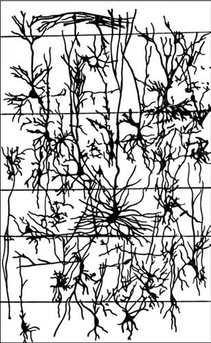
Fig. 5.67. Layered arrangement of nerve cells in the mammalian cerebral cortex
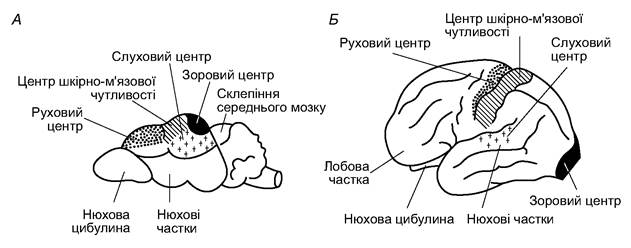
Fig. 5.68. Functional centers of the cerebral cortex in a shrew (A) and a human (B)
The cortex of almost all mammals forms a greater or lesser number of sulci, which increase its surface area. In the simplest cases, there is only a single Sylvian fissure separating the frontal lobe from the temporal lobe; later, the Rolandic fissure appears, separating the frontal and occipital lobes, etc. In primates and toothed whales, the number of sulci is particularly large. The mammalian neopallium, to a greater extent than the Midbrain cortical complex of birds, mediates higher nervous activity by accumulating traces of individual excitations and their combinations, thereby enriching the so-called working memory. This enables the Organism to choose the optimal decision in a new situation based on this memory. Most often, these decisions represent new combinations of already known elements. The Emergence of higher associative centers—the neocortex—in mammals did not lead to the elimination of centers governing instinctive acts, but merely subjected them to higher control.
Other parts of the forebrain are relatively smaller, yet they also perform important functions. The olfactory lobes are located in the anteroventral region (olfactory bulbs and anterior basal nuclei). The corpora striata regulate instinctive reactions under the control of the cerebral cortex.
The midbrain is small; its roof is divided by transverse grooves and is called the corpora quadrigemina. The anterior colliculi form a weakly defined visual cortex, while the posterior colliculi are controlled by the forebrain and serve as auditory centers (Fig. 5.69). The Cerebellum is large and consists of a median vermis and adjacent hemispheres with lateral appendages. As in other mammals, the cerebellum is associated with maintaining muscle tone, posture, balance, and movement of body parts. The close connections between the cerebellum and the cerebral cortex indicate the existence of higher control over it.
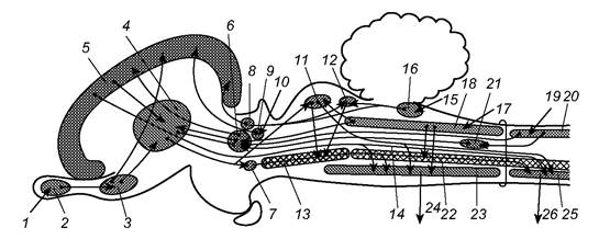
Fig. 5.69. Diagram of connections between brain divisions in mammals:
1 - Olfactory nerve; 2 - olfactory bulb; 3, 4 - Basal Ganglia; 5 - neopallium; 6-9 - dorsal (6) and ventral (7) parts, medial
(8) and lateral (9) geniculate bodies of the thalamus; 10 - Optic nerve; 11, 12 - anterior (11) (visual) and posterior (12) (auditory) colliculi of the midbrain tectum; 13 - midbrain reticular formation; 14 - motor pyramidal tract; 15 - auditory nerve; 16 - auditory center of the Medulla Oblongata; 17 - sensory NERVES OF THE skin and muscles of the head; 18 - sensory nuclei of the Brainstem; 19 - sensory nerves of the skin and muscles of the body; 20 - sensory (dorsal) horns of the Spinal Cord; 21 - sensory nuclei of the medulla oblongata; 22 - reticular formation of the medulla oblongata and spinal cord; 23 - motor nuclei of the brainstem; 24 - motor nerves to the muscles of the head; 25 - motor (ventral) horns of the spinal cord;
26 - motor nerves (to body muscles)
The medulla oblongata gives rise to most of the Cranial Nerves (V–XII). It houses the centers for respiration, Cardiac Activity, digestion, and other vital functions. From the sides of the Fourth ventricle cavity, bundles of nerve fibers separate, projecting to the cerebellum and forming its inferior cerebellar peduncles. Nerve tracts connect the medulla oblongata with the spinal cord.
As in other vertebrates, the relative size of the brain increases with decreasing body size and increasing thermoregulatory demands. Thus, in large shrews, brain mass is about 0.6% of body mass, while in small ones it reaches up to 1.2%; in large cetaceans, it is about 0.3%, and in small ones, up to 1.7%. The brain mass of primates is 0.6–1.9% of body mass, and in humans, it is about 3%. In all mammals, the mass of the forebrain exceeds that of other brain divisions: in various groups, it accounts for 52–72% of the total brain mass; in primates, this figure rises to 76–80%, and in humans, up to 86%.
The ratio of brain mass to spinal cord mass is highest in humans (45:1), high in primates and cetaceans (10–15:1), and lower in canids, shrews (3–5:1), and cervids (2.5:1). In reptiles, it is always less than 1. The spinal cord, via conducting pathways (White matter), is connected to the motor center of the cerebral cortex, which exerts higher control over motor acts and the management of complex movements.
Mammals have 12 pairs of cranial nerves; the XI pair—the Accessory nerve (nervus accessorius)—develops. In addition to innervating the main Sense Organs (Olfaction, Vision, Hearing) and The Muscular System, cranial nerves participate in the formation of the Autonomic nervous system, which controls so-called vegetative processes that are not subject to voluntary control. The parasympathetic nervous system is formed by cranial nerves of the medulla oblongata and Spinal Nerves of the sacral region. The sympathetic nervous system consists of the ganglia of spinal nerves from the cervical, thoracic, and lumbar regions of the spine (Fig. 5.70). Major Organ Systems receive endings from both systems.
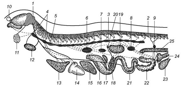
Fig. 5.70. Diagram of the autonomic nervous system of mammals:
1, 2 - parasympathetic centers in the midbrain and medulla oblongata (1) and the sacral spinal cord (2); 3 - Sympathetic trunk; 4 - cervical ganglia; 5 - stellate ganglion; 6 - celiac ganglion; 7, 8 - superior (7) and inferior (8) mesenteric ganglia; 9 - pelvic ganglion; 10 - eye;
11 - salivary glands; 12 - thyroid gland; 13 - lung; 14 - heart; 15 - liver; 16 - stomach; 17 - spleen; 18 - pancreas; 19 - kidney; 20 - adrenal gland; 21, 22 - small (21) and large (22) intestines; 23 - urinary bladder; 24 - uterus; 25 - rectum. Solid lines - parasympathetic innervation, dashed lines - sympathetic innervation
The dorsal columns of white matter consist of fibers ascending to the brain, carrying impulses from sense organs and enteroreceptors (afferent information). In the cranial tracts, fibers carrying impulses from the brain to muscles and other effector organs (efferent information) predominate. The control of higher brain centers over spinal cord function reaches its highest level in mammals.
Dual innervation is explained by oppositely directed effects. If impulses from one system exert an excitatory effect on an organ's function, impulses from the other typically inhibit it. This antagonistic action, by refining regulation, significantly enhances the ability to withstand depressing or highly exciting external influences (stress), thereby increasing the organism's chances of survival across a wide range of conditions.
Sensory organs. Sensory organs are differentially developed in various mammalian orders. Vision is of primary importance for inhabitants of open spaces, whereas olfaction and hearing are paramount for nocturnal and crepuscular animals (inhabitants of forest and shrub biotopes, burrowers, and aquatic species).
The mammalian SENSE OF SMELL is more effective than that of other terrestrial vertebrates. The high resolution of chemoreceptors allows them to distinguish specific substances (odors) or their combinations characteristic of a species, a group of individuals, or a single individual. Olfactory acuity varies across different orders and specific mammalian species. Marsupials, shrews, rodents, xenarthrans, and most carnivorans and artiodactyls are so-called macrosmatic animals, characterized by a highly developed sense of smell; they use it for spatial orientation, foraging, and interspecific and intraspecific communication. Most primates and a number of other mammals have a less sensitive sense of smell (microsmatic animals).
The olfactory organs are located in the posterosuperior part of the nasal cavity, which features a complex system of turbinates covered by a mucous membrane of olfactory epithelium containing receptor cells with characteristic cilia. The axons of these cells bundle together to form fibers that enter the olfactory bulbs, which are connected to brain centers via a chain of Neurons. The complexity of the turbinate structure corresponds to the acuity of smell (Fig. 5.71).
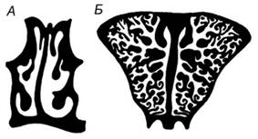
Fig. 5.71. Cross-section of the posterior nasal cavity in mammals:
A - microsmatic (human); B - macrosmatic (roe deer)
In cetaceans, the presence of Olfaction and Taste was once denied, leading them to be labeled as anosmatic. Recent studies, however, have demonstrated that dolphins possess scent glands that open near the anus; these animals can determine the direction of a passing pod by tracking secretion trails, and they perceive the scent of blood as a danger signal. In the oral cavity of baleen whales, paired depressions at the tip of the upper jaw are homologous to the Jacobson's organ of other vertebrates. At the base of the tongue in toothed whales, elongated pits resemble the taste papillae of other mammals. Apparently, these help whales recognize chemical cues and navigate by distinguishing currents with different chemical compositions. Although the cetacean brain is characterized by reduced olfactory lobes, it still retains structures in the cerebral cortex associated with the analysis of chemical signals.
Hearing plays a vital role in the lives of mammals. This is matched by the complex structure of the vocal apparatus, which is capable of producing A wide variety of sounds, often forming complex, temporally organized acoustic sequences. In terms of frequency range, mammals surpass birds, making extensive use of both ultrasound (over 20 kHz) and low frequencies. Hearing and acoustic signaling serve crucial life functions, including foraging, predator detection, species recognition, individual identification within a group (herd or pack), parent-offspring communication, and much more. Auditory characteristics vary across different orders. For instance, bats primarily use ultrasonic frequencies in the range of 40-80 kHz for echolocation, yet they also emit low-frequency sounds down to 12 Hz (infrasound, which is inaudible to the human ear). The acoustic range of baleen whales is even broader, spanning from a few hertz to two hundred kilohertz. Baleen whales emit low-frequency sounds (1-2 kHz) of considerable intensity and duration. The ability to echolocate is also found in shrews and some burrowing rodents. A single species may use different frequency bands for different purposes: echolocation and prey detection at high and ultrasonic frequencies, and intraspecific communication at relatively low frequencies.
The mammalian Organ of Hearing consists of three parts: the outer, middle, and Inner ear. The outer ear (pinna) and the external auditory meatus act as a kind of acoustic antenna and filter, capable of capturing and amplifying biologically significant sounds while dampening extraneous noise. A similar role is played by the middle ear (Fig. 5.72), where three auditory ossicles—the malleus, incus, and stapes—form a lever system that transmits vibrations from the larger tympanic membrane to the smaller membrane of the oval window of the inner ear, thereby amplifying these vibrations.
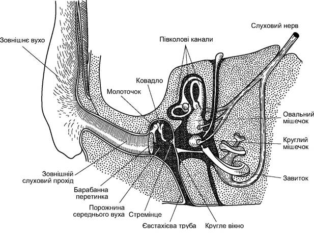
Fig. 5.72. Diagram of the mammalian auditory organ
The auditory bulla, which houses the middle ear cavity, forms a series of resonating chambers that amplify biologically significant sounds. In some species, particularly burrowers, these chambers are sometimes filled with a spongy bone mass that dampens extraneous noise. The middle ear is connected to the back of the oral cavity by the Eustachian tube, which ensures the equalization of air pressure on both sides of the tympanic membrane.
The inner ear is located within the petrous part of the Temporal bone and consists of vestibular and auditory divisions. The vestibular division comprises three semicircular canals and the utricle; it serves as the Organ of Balance and spatial orientation. The auditory division is formed by the saccule and the associated cochlea, which houses The Organ of Corti; the functions of this organ include the primary analysis, predominantly frequency-based, and encoding of acoustic signals, which, after processing, are transmitted to the auditory center of the brain. The cochlea is a spirally coiled membranous tube lying within the bony
casing, filled with endolymph. Along the entire length of the cochlea runs the basilar membrane, upon which transverse fibrils (auditory strings) are stretched. These are contacted by the sensory cells of the organ of Corti, which detect the vibrations of the auditory strings tuned to different frequencies. The impulses received by the sensory cells are transmitted to neurons whose axons form the auditory nerve. This mechanism provides a fine Analysis of the frequency spectrum and Temporal Organization of the acoustic signal captured by the outer ear and transmitted, amplified, through the middle ear to the inner ear.
Vibrations of the vocal cords in the larynx of mammals produce sounds. Ultrasonic signals of bats are generated by the oral or nasal apparatus. In cetaceans, sound production involves the larynx as a whole, the edges of the arytenoid cartilages, the air sacs of the nasal passage, and the blowhole. In addition to vocalizations, some mammals use mechanical sounds: teeth clicking and grinding (canids, some rodents, cervids, primates), antler clashing, foot-drumming on the ground (many burrowing animals, ungulates), the rattling of quills (porcupines), and so on.
The visual system is the third major sensory organ of mammals. For some animals that are predominantly diurnal and inhabit open habitats, most environmental information is received through the visual channel. The Importance of vision decreases for inhabitants of forests, dense undergrowth, and grasslands. In burrowing species, the eyes sometimes cease to function, becoming covered with skin (some moles, mole-rats), or only detect changes in light intensity (mole voles, Prometheus voles). In cetaceans, the eyes are used only for short-range orientation. Mammalian eyes are positioned either laterally, providing a nearly circular field of view where binocular vision is limited to a small sector, or frontally. In the latter case, the overall field of view is reduced, but the binocular field increases. The first type prevails in ungulates and mouse-like rodents, which must constantly watch for predators; the second is characteristic of arboreal primates, which need to accurately judge distances when leaping from branch to branch, as well as some caniforms, especially felids, which must precisely gauge the distance to their prey when ambushing. Relative eye size increases in animals with sharper vision and in those with nocturnal activity.
The mammalian eye has an outer coat (sclera) of fibrous tissue. Anteriorly, the sclera transitions into the transparent cornea. Beneath the sclera lies the choroid, containing blood vessels that supply necessary nutrients. Between the sclera and the choroid in some animals, there is a layer of crystalline cells forming a tapetum, which reflects light rays and causes the eye to "glow" with reflected light (canids, cervids). Anteriorly, the choroid thickens to become the iris and the ciliary body (muscles), which facilitate ocular accommodation by altering the shape of the lens. The iris acts as a diaphragm, regulating the illumination of the retina by changing the size of the pupil. The lens, lenticular in shape, is relatively small in diurnal mammals and increases dramatically in nocturnal animals. Adjoining the inner side of the choroid is the retina, consisting of outer pigmented and inner photoreceptive layers. Cones lack oil droplets. Interspecific differences lie in variations of the rod-to-cone ratio, fluctuations in the total number of receptor cells, and their ratio per single optic nerve fiber. In burrowing animals, the number of receptor cells and nerve fibers is minimal: in the mole-rat, the entire retina contains 800 thousand receptors and 1,900 fibers in the optic nerve (a ratio of 420:1). In nocturnal species and undergrowth dwellers, it is higher: in the hedgehog, there are 6.7 million receptors to 8,400 fibers (760:1); in the yellow-necked mouse, 19.6 million and 28,800 (680:1). This number is even higher in inhabitants of open landscapes: for example, in the European hare, 192.6 million receptors and 167,400 fibers (115:1); in the rhesus macaque (primates), 124.4 million receptors to 1.2 million fibers (105:1); and in the bat (vespertilionids), only 8.9 million receptors to 6,900 fibers (1130:1). The average number of receptor cells per single optic nerve fiber is lowest in primates; this allows for the detection of finer details in objects.
Many mammals are capable of distinguishing colors, though apparently to a lesser extent than birds. This is associated with the generally less diverse coloration of mammals. At the same time, mammals recognize the shapes of objects or their parts, as well as movements, posture, and facial expressions. This is achieved not through a more complex retinal structure, but by the visual analyzer in the brain, which is more complex in mammals than in other vertebrates. A leading role is played by the visual center of the cerebral cortex, whereas the importance of the
midbrain visual center (superior colliculi of the corpora quadrigemina) is reduced. The shift of primary visual processing to the cerebral cortex opened up possibilities not only for visual spatial orientation, but also for the elaboration and enrichment of visual communication between individuals. A language of form, posture, gestures, and facial expressions emerged and became widely used in mammals. This plays a role in regulating social relations within populations and forming groups with coordinated behavior (see below). Under The Influence of natural Selection, the coloration and body shape of animals acquired a camouflage function (cryptic coloration) or served as a display during threat behavior.
On areas of the body that frequently come into contact with surrounding objects, mammals have especially long and stiff hairs called vibrissae. Their roots are associated with nerve endings. Vibrissae usually reach their greatest development on the muzzle.
Last update: 19/08/2026
Editorial and Educational Adaptation: This material has been compiled based on the primary/original source text. The project team performed an editorial review, corrected technical inaccuracies, structured sections, and adapted the content for an educational format.
What was processed:
- elimination of formatting defects (OCR errors, structural breaks, corrupted characters);
- editorial organization of content;
- standardization of terminology in accordance with academic sources;
- verification of factual statements against the original source text.
All mentions of the author, publication year, and origin of the primary text have been preserved in accordance with the source.