Review of Medical Physiology - William F. Ganong 2002
Functions of the Nervous System
Olfaction and Taste
Taste - Receptor Organs and Pathways
Taste buds
The Organs of taste perception are taste buds—oval corpuscles measuring 50–70 µm. Each taste bud consists of four Cell types (Fig. 10-4): basal Cells; type I and type II support cells; and type III cells, which serve as taste receptor cells forming synaptic connections with sensory nerve fibers. Cells of types I through III feature microvilli that project into the taste pore, an opening in the lingual epithelium. The neck regions of all three cell types are joined to each other and to the adjacent Tongue epithelium by tight junctions, leaving only the crown of apical microvilli exposed to contact with oral fluids. Each taste bud is innervated by about 50 nerve fibers, and each nerve fiber receives impulses from an average of five taste buds. Basal cells originate from the epithelial microenvironment of the taste buds. They differentiate into new receptor cells, which have a half-life of approximately ten days. When a sensory nerve is severed, the taste buds it innervates gradually degenerate and disappear. Upon nerve regeneration, the cells of its microenvironment self-organize into new taste buds, likely As a result of a specific chemical induction by the regenerating fiber.
Class="center">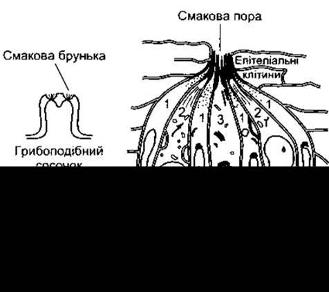
Fig. 10-4. Diagram of The Structure of a taste bud, showing cells of types I through III. The left panel illustrates the localization of taste buds within fungiform and circumvallate papillae of the tongue (modified and reproduced with permission from Shepherd GM: Neurobiology, 2nd ed. Oxford Univ Press, 1988).
In humans, taste buds are located in the mucous membrane of the epiglottis, palate, Pharynx, and predominantly in the walls of the fungiform and circumvallate papillae of the tongue. Fungiform papillae are rounded in shape and most numerous near the tip of the tongue; circumvallate papillae are relatively large and arranged in a V-shaped pattern along the border between the posterior and middle thirds of the dorsal tongue. Each fungiform papilla contains up to five taste buds, which are typically situated near the apex of the papilla (see Fig. 10-4). The larger circumvallate papillae contain up to 100 taste buds, mostly localized on the lateral surface of the papilla. The numerous, smaller filiform papillae contain no taste buds. The total number of taste buds is approximately 10,000.
Gustatory pathways
Sensory nerve fibers from the taste buds in the anterior two-thirds of the tongue run via the chorda tympani, fibers from the posterior third of the tongue reach the Brainstem via the Glossopharyngeal nerve (Fig. 10-5), and fibers from extra-lingual taste buds travel within the Vagus nerve. The taste fibers of these three nerves are myelinated, yet conduct impulses at a relatively low speed; on each side, they converge in the Nucleus of the solitary tract in the Medulla Oblongata (Fig. 10-6). Here, they synapse with second-order Neurons whose axons cross the midline, join the medial lemniscus, and terminate—along with fibers for tactile, pain, and Temperature Sensation—in specific Relay nuclei of the thalamus. From there, impulses project to the gustatory cortical areas located near the Base of the postcentral gyrus. Taste does not have a distinct, dedicated cortical projection field; instead, it is represented in the postcentral gyrus region alongside cutaneous sensation of the face.
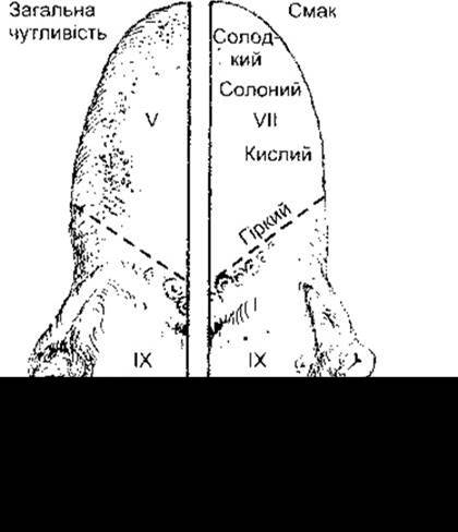
Fig. 10-5. Sensory Innervation of the tongue. Cranial Nerves are indicated by numerals: V - trigeminal; VII - facial; IX - glossopharyngeal.
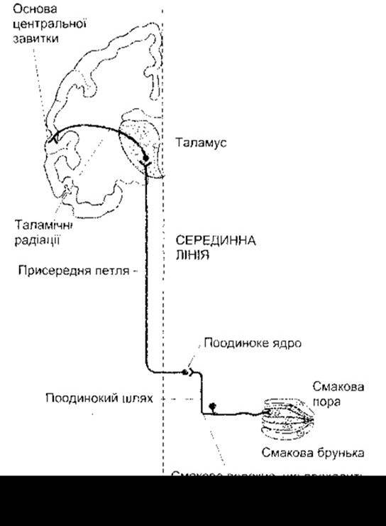
Fig. 10-6. Schematic diagram of the gustatory pathways.
Primary taste modalities
Humans recognize four primary tastes: sweet, sour, bitter, and salty. Although the receptive areas for different tastes on the tongue overlap considerably, bitter taste is perceived mainly by the posterior region, sourness by the lateral surfaces, sweetness by the tip, and saltiness by the anterior part of the tongue dorsum (see Fig. 10-5). Sour and bitter substances—and to a lesser extent, sweet and salty ones—are also perceived by the palate. All four taste modalities can likewise be detected by the pharynx and epiglottis. Histologically, taste receptor cells in regions sensitive to different tastes do not differ from one another; however, recordings of potentials from single taste cells have revealed physiological differences among animal taste cells. These studies have demonstrated that some cells respond better to bitter stimuli, others to salty, sweet, or sour ones. Certain cells respond to more than one taste, and a few respond to all four.
METABOLISM/2.html">THE CONCEPT OF an additional taste modality, known as umami, has recently been formulated. It is believed to convey the sensation of glutamates, particularly monosodium glutamate, which are widely used in Asian cuisine. The receptor for this taste is considered to be one of the metabotropic glutamate receptors, specifically mGluR4.
Substances eliciting primary taste sensations
Acids produce the sour taste. Sour taste receptors are more likely stimulated by H+ cations than by their corresponding anions. For any given acid, the intensity of the sour taste is proportional to the concentration of H+ ions; however, at equal H+ concentrations, organic acids taste sourer than mineral acids. This is presumably due to a greater cell permeability for organic acids compared to mineral acids.
The salty taste is produced by Na+ ions. Certain Organic compounds also taste salty, notably the dipeptides lysyltaurine and ornithyltaurine, with lysyltaurine tasting saltier than NaCl at equivalent molar concentrations.
Most sweet substances are organic compounds. The most familiar are sucrose, maltose, lactose, and glucose; however, Polysaccharides, glycerol, certain alcohols and ketones, as well as a significant number of substances structurally unrelated to the above—such as chloroform, beryllium salts, various amides of aspartic acid—also taste sweet. Artificial sweeteners such as saccharin and aspartame are used in weight-loss diets because they provide a satisfactory sweetening effect at very low concentrations. Today, over 100 million tons of artificial sweeteners are produced worldwide annually.
Two Proteins isolated from African berries—thaumatin and monellin—have been shown to be 100,000 times sweeter than sucrose at equivalent molar concentrations. Although structurally distinct, Antibodies raised against one of these proteins show cross-reactivity with the other. This indicates a similarity in their molecular spatial conformation, which presumably underlies the binding to sweet receptors.
Quinine sulfate is commonly used as a standard for testing bitter taste. The taste of this substance is perceived at a concentration of 8 µmol/L, although strychnine hydrochloride has an even lower threshold of stimulation (Table 10-2). A bitter taste is also exhibited by other organic compounds, particularly morphine, nicotine, caffeine, and urea. Inorganic Compounds of magnesium, ammonium, and calcium elicit a bitter sensation via their cations. Consequently, there is no single molecular structure that can be linked to the bitter taste.
Taste detection and intensity discrimination thresholds
Human ability to discriminate taste intensity, much like olfactory intensity discrimination, is rather limited. Distinguishing a difference in taste intensity requires A change in the concentration of the tastant of at least 30%. The taste threshold varies for different substances (see Table 10-2).
Mechanism of receptor excitation
Taste receptor cells are chemoreceptors that respond to substances dissolved in the saliva bathing the Oral Cavity. These substances act on the microvilli located within the taste pore, triggering receptor cell potentials that lead to the generation of action potentials in sensory neurons.
Table 10-2. Taste thresholds of certain substances
Substance |
Taste |
Threshold concentration, µmol/L |
Sour |
100 |
|
Sodium chloride |
Salty |
2 000 |
Strychnine hydrochloride |
Bitter |
1.6 |
Glucose |
Sweet |
80 000 |
Sucrose |
Sweet |
10 000 |
Saccharin |
Sweet |
23 |
The excitation mechanism of receptor cells varies depending on the taste stimulus. Research on taste receptors is advancing rapidly, and many candidate receptor molecules have already been cloned. A protein designated as mammalian degenerin-1 is a likely receptor for sour substances. An alternative mechanism involves the depolarization of sour taste receptor cells by acids through the activation of H+-gated cation channels; Na+ depolarizes salt-sensing receptor cells via Na+ channels related to epithelial Na+ channels. These channels are blocked by amiloride (see Chapter 1), which is why the direct application of this diuretic to the human tongue blocks the sensation of saltiness. Sweet substances activate adenylyl cyclase via a heterotrimeric G protein; the resulting increase in intracellular cAMP concentration decreases Plasma Membrane permeability to K+ by phosphorylating K+ channels on the basolateral surface of The Cell membrane, thereby causing cellular depolarization. Bitter substances decrease cAMP concentration in their respective receptor cells, likely via a heterotrimeric G protein, and increase the concentrations of IP3 and DAG (see Chapter 1).
Recently, a novel G protein from bitter taste receptor cells, termed α-gustducin, was cloned. It has The ability to decrease intracellular cAMP levels by activating phosphodiesterase. Furthermore, α-gustducin has also been detected in sweet taste receptor cells, where excitation is associated with an increase in intracellular cAMP levels. The precise MECHANISM OF ACTION of α-gustducin remains unclear.
A protein with the property of binding taste molecules has also been cloned. It is produced by von Ebner's glands, which secrete their fluid into the clefts surrounding circumvallate papillae (see Fig. 10-4). The function of this protein is presumably similar to that of odorant-binding proteins, involving the accumulation and transport of tastants to receptor cells.
Flavor
The vast combinations of flavors so appreciated by gourmets are formed from four primary components. In some cases, specific tastes require thermal stimulation (for example, in "hot" spices). Additionally, smell plays a crucial role in the overall sensory experience of food, along with texture and temperature, which contribute to its overall "flavor."
Variability and Aftereffects
There are significant interspecies and individual differences in the distribution of taste buds specialized for perceiving different tastes. In humans, There is a fascinating variability in the perception of phenylthiocarbamide: in dilute solutions, this substance tastes bitter to 70% of the Caucasian population and is tasteless to the remaining 30%. The inability to taste phenylthiocarbamide is inherited as an autosomal recessive trait. Testing for this trait is of considerable importance in human genetic studies.
Taste sensations are characterized by aftereffects and contrast phenomena, which resemble visual afterimages and contrasts. Some of these phenomena may have a chemical basis,
while others are of central origin. A taste-modifying protein called miraculin has been isolated from a plant source. Its action on the tongue converts the taste of sour substances into sweet.
Humans and animals develop a strong aversion to novel foods if consuming them has been followed by illness. The survival value of such aversion becomes evident in the Prevention of poisoning.
Anomalies
Taste disorders include ageusia (absence of taste sensations), hypogeusia (reduced taste sensitivity), and dysgeusia (distortion of taste sensations). Hypogeusia can be caused by numerous medical conditions. Additionally, sulfhydryl-containing drugs such as captopril and penicillamine cause temporary loss of taste. The cause of this effect associated with sulfhydryl compounds remains unknown.
Last update: 10/08/2026
Editorial and Educational Adaptation: This material has been compiled based on the primary/original source text. The project team performed an editorial review, corrected technical inaccuracies, structured sections, and adapted the content for an educational format.
What was processed:
- elimination of formatting defects (OCR errors, structural breaks, corrupted characters);
- editorial organization of content;
- standardization of terminology in accordance with academic sources;
- verification of factual statements against the original source text.
All mentions of the author, publication year, and origin of the primary text have been preserved in accordance with the source.