Antibiotics (Properties, Application, Interactions) - M.P. Cherenko 1999
Developmental Defects and Malformations
Structure/97.html">Definitions, Classification, Etiology, Pathogenesis
Developmental defects, or anomalies, refer to congenital deviations beyond the normal anatomical limits in the structure (shape, size, number) of human Tissues and Organs, which are often accompanied by functional impairments or even threaten the viability of the Organism. Severe forms of Congenital Malformations affecting multiple organs and systems are referred to as monstrosities or major terata.
The Study of the origins and pathology of developmental defects constitutes a distinct medical discipline known as teratology (from the Greek teratos meaning monster, and logos meaning study or science).
Developmental defects are a prevalent form of pathology; according to WHO data, their specific weight in the general population ranges from 2.7% to 16.3% across various countries and has shown an upward trend in recent decades.
There is a vast array of developmental defects—both visible and invisible—of a morphological (MACROSCOPIC AND MICROSCOPIC) and biochemical (congenital Metabolic Disorders) nature. Examples of the latter include agammaglobulinemia, galactosuria, hemophilia A, albinism, alkaptonuria, and many others. The vast majority of biochemical defects stem from disruptions in the body's enzyme systems. Biochemical disorders in humans are typically studied by internists.
Macroscopic morphological defects fall within the purview of surgeons, whereas histomorphological (microscopic) defects generally come to their attention only when they serve as a source of pathological processes, primarily tumors. Such microscopic defects include structurally and topographically diverse pathological Cell clusters in tissues and organs collectively known as heterotopias. Among the most well-known are the so-called hamartias (from the Greek hamartano meaning to err, and choriso meaning to separate), which give rise to hamartomas and choristomas, such as capillary hemangioma of the Skin and pulmonary chondroma, respectively.
There is currently no universally accepted comprehensive Classification of Congenital Malformations due to their diverse nature, localization, origin, and the varying severity of Anatomical and physiological disorders they cause. Depending on the classification criterion used, several groups of defects are distinguished. Based on their anatomical localization, they can be divided into:
1) external (e.g., cleft lip, anal atresia, etc.);
2) internal (e.g., ventricular or atrial septal defect of The Heart, pyloric stenosis—marked narrowing of the gastric pylorus, etc.);
3) combined or mixed (e.g., cleft lip and palate combined with a diaphragmatic hernia, etc.).
Depending on the number of defects in an individual, they are classified as follows:
1) single defects—organ- or system-specific (e.g., hypospadias, chondrodysplasia, etc.);
2) multiple defects—affecting multiple organs or systems (e.g., cleft palate combined with encephalocele; defects involving both the digestive and cardiovascular systems, or the nervous and endocrine systems, etc.);
3) defects of a single organism versus defects involving two organisms (conjoined twins or double monsters).
From a clinical perspective, defects are also frequently classified According to the anatomical and physiological division of the body into systems, regions, and organs (e.g., Central Nervous system defects, gastrointestinal tract defects, Urinary System defects, facial anomalies, skin defects, etc.).
Depending on their sequence of appearance, defects are further distinguished as primary or secondary. For instance, a primary developmental defect in the CEREBROSPINAL FLUID PATHWAYS OF THE Brain (such as stenosis or aplasia) leads to secondary pathology—Hydrocephalus accompanied by cranial deformation.
THE ORIGIN OF developmental defects is rooted in various disruptions during The Development of tissues and organs, specifically:
1) agenesis and aplasia—complete absence of an organ (either with a total lack of its primordium or with only a vascular pedicle present);
2) hypoplasia—underdevelopment of the mass of an organ, specific parts of it, or the entire body (hypotrophy);
3) congenital hypertrophy of an organ (such as gigantism or elephantiasis) resulting from an increase in its volume or cellular mass (hyperplasia);
4) heterotopia—presence of a cell cluster, tissue fragment, or organ component in an atypical tissue or organ site (hamartia and choristia);
5) ectopia and dystopia—abnormal anatomical localization of an organ (e.g., lingual or intrathoracic ectopia of The Thyroid Gland);
6) stenosis—narrowing of the lumen or channel of a vessel, the pulmonary artery, Esophagus, intestine, etc.;
7) atresia—congenital closure or absence of normal bodily openings or tubular organs (e.g., rectal or anal atresia, esophageal atresia);
8) incomplete Separation of organs or organisms (e.g., Syndactyly—fusion of fingers or toes; conjoined twins fused by various body parts, such as the chest in thoracopagus or sternopagus, the HEAD in craniopagus, or the sacrum in pygopagus, etc.);
9) persistence — the preservation after birth of ducts that normally function only during the Embryonic period (patent ductus arteriosus between the aorta and the pulmonary artery, persistence of the patent omphalomesenteric or urachal duct);
10) duplication of an organ or a part thereof (duplication of individual loops or segments of the intestine, fingers — polydactyly, Uterus, Peritoneum, etc.);
11) inversion of THE POSITION OF an organ or organs due to a disturbance in their rotation process — heart, intestine (dextrocardia; situs viscerus inversus — of the abdominal organs);
12) arrest of the fusion of paired segments of an organ or the sides (edges) of embryonic tubes, channels, cavities, etc. (maxillary processes, Abdominal cavity, Urethra); premature overgrowth or closure of the body cavity, organ orifices, etc.;
13) atavism — the appearance in humans of tissue structures in places where they are present in animals (e.g., hairiness of the entire face or individual areas of the body; tail rudiments — tail vertebrae; ichthyosis — scaly skin; polymastia and polythelia — an increased number of Mammary Glands, nipples; true Hermaphroditism, etc.).
Depending on their etiology, congenital malformations are conventionally divided into three groups: hereditary, exogenous, and multifactorial (G.I. Lazyuk, 1991).
Hereditary malformations are associated with structural defects of genes and Chromosomes (Mutations). Exogenous malformations are caused by the action of teratogenic factors on the embryo and fetus. Multifactorial malformations are caused by the combined action of genetic and exogenous factors, each of which is independently incapable of causing the malformation.
All numerous etiological factors of malformations can be divided into two groups — endogenous and exogenous.
The group of endogenous factors includes: mutations of hereditary structures, endocrine diseases, overripeness of Germ Cells, and advanced parental age.
The group of exogenous factors comprises: physical, chemical, and biological factors.
Mutations of hereditary structures — genes and chromosomes — can be either stable or unstable, and occur both spontaneously and under METABOLISM/18.html">The Influence of various exogenous factors on hereditary structures (so-called induced mutagenesis). Although Gene Mutations are much less common than chromosomal (microscopic Chromosomal Disorders) and genomic disorders (abnormal chromosome numbers), the majority of congenital malformations are the result of gene mutations, particularly polygenic ones. Many authors consider mutations to be the primary cause of malformations. In their view, exogenous factors also cause developmental malformations by affecting hereditary factors, i.e., through mutagenic effects.
Extensive studies on the origin of developmental malformations conducted in recent years provide grounds to assert that the vast majority of malformations have a polygenic-multifactorial origin.
Mutations of hereditary formations (genes, chromosomes) can occur spontaneously during the physiological activity of the organism, both under the action of various endogenous metabolites and As a result of the harmful effects of numerous exogenous factors.
Endocrine diseases, hormone secretion disorders (hyperproduction of Steroids by the fetal Adrenal Glands, Diabetes Mellitus in the mother, etc.) also lead to the development of malformations (virilizing syndrome, diabetic embryopathies, etc.). Overripeness of germ cells (their prolonged existence before fusion into a gamete) and advanced parental age are also frequently the causes of congenital malformations.
Among the numerous malformations caused by mutations of hereditary structures, particularly chromosomes and The Genome, are such endocrine disorders as Klinefelter syndrome (47 XXY phenotype) and Shereshevsky-Turner syndrome (45 phenotype), Down syndrome (trisomy of chromosome 21), and certain heart defects, among others.
Among the external causes contributing to the development of congenital malformations, mention should first be made of the progressive deterioration of ecology and the unsatisfactory state of the environment (pollution of air, Water, and soil by human industrial waste, etc.). At the same time, negative environmental factors can act both directly on the human organism, particularly the fetus, and indirectly by transforming its internal environment. This circumstance makes it practically impossible to clearly demarcate external and internal teratogenic agents. The direct causes of most malformations have not yet been elucidated, and by no means all adverse environmental and internal factors have been studied sufficiently. However, The impact of many of them on the development of malformations is well known.
The greatest role in the genesis of malformations is played by physical factors, especially X-ray and radioactive radiation, certain chemicals, medical drugs, infectious diseases, a deficiency of Amino Acids, Proteins, Vitamins, and other nutrients in the diet or the pregnant woman's body, hormonal imbalances in her body, Hypoxia, and mechanical impacts on the fetus, among others.
The negative effects of X-ray and radioactive radiation on living organisms, especially their hereditary mechanism (genome), have been well known since the discovery of these rays. However, their teratogenic effect became particularly evident after studying the consequences of the atomic bombing of the Japanese cities of Hiroshima and Nagasaki.
Subsequent experimental work in this direction and observations of people in other areas where the population was exposed to radioactive radiation confirm the Conclusions drawn by Japanese scientists. It should be noted that there is no threshold level of teratogenicity for radioactive rays, meaning that their effect, even in the smallest dose, is potentially teratogenic.
Among Chemical factors, ethyl alcohol is considered particularly teratogenic. Maternal alcoholism is the cause of developmental malformations in 30% of children, among whom heart defects are observed in 30–49% of cases (N.P. Bilokon, V.P. Podzolkov, 1991). Chronic alcohol intoxication causes damage to the biological structure of germ cells, frequently leading to hypogonadism, cryptorchidism, congenital hernias, heart and nervous system defects, etc.
Many other chemical compounds (salicylates, antimetabolites, cytostatics, insecticides, tin oxide, arsenic and chromium compounds, etc.), many of which are also medicinal drugs (narcotics, tranquilizers, hormonal preparations, etc.), are likewise capable of causing fetal developmental disorders. A striking example of such pharmaceutical action is The Effect of thalidomide, which was synthesized in Germany and distributed in the 1950s–1960s in Western Europe as a sleeping pill. Women who used this drug frequently gave birth to children with limb malformations.
Numerous infectious, especially viral, diseases also cause the development of many malformations, notably defects of the heart, Skull and brain, and Gonads. These include such infectious diseases as Influenza, measles, rubella, Toxoplasmosis, mumps, hepatitis, and rheumatic carditis, among others.
Non-infectious maternal diseases accompanied by hypoxemia (Heart Failure, anemia), by causing Fetal hypoxia, contribute to the development of malformations in the fetus.
Partial forms of starvation, particularly a deficiency of Amino Acids and Proteins, and vitamins (Folic acid, etc.), can be the cause of malformations, especially of The Nervous System.
Many malformations arise as a result of mechanical causes acting on the fetus — pressure from amniotic membranes and folds, oligohydramnios, mechanical origin, etc. Among disorders of this origin are limb and nervous system defects, Torticollis, etc.
Congenital malformations are the result of impaired Embryonic and Fetal development at various stages, including gametogenesis, Fertilization, embryonic morphogenesis (The Emergence of specialized cells from poorly differentiated ones and Organogenesis), and the postnatal period.
The primary cellular mechanisms of teratogenesis involve disruptions in cell proliferation, migration, and differentiation. The consequences of these impaired processes include such common defects as organ agenesis and hypoplasia, failure of embryonic structures to fuse properly leading to dysraphism (clefts), heterotopia, functional immaturity of tissues and organs, and the persistence of embryonic structures (unclosed ducts), among others.
It should be noted that although developmental malformations can occur throughout the entire prenatal period (gametopathies, blastopathies, embryopathies, and fetopathies), they most frequently arise during so-called critical periods, when the embryo is highly vulnerable to environmental teratogens. These comprise the first 6 weeks of Embryogenesis (malformations occurring near the end of the 2nd week of this period are incompatible with life, whereas those arising during the 3rd to 6th weeks are largely compatible with life).
These periods coincide with the most intensive phase of organ and system formation. The earlier a malformation occurs, the more severe it tends to be. Since a damaging factor can induce an organ defect only until its formation is complete—and the timelines for the development of different organs vary—an important concept for teratological theory and practice is The Doctrine of terminal teratogenetic periods, formulated by E. Schwalbe in 1906, which defines the chronological timeframe within which an organ malformation can develop.
For instance, the termination period for conjoined twins is the first 2 weeks post-fertilization, whereas for atrial septal defects, it extends up to the 44th day of gestation. Cryptorchidism and persistent duct structures are defects characteristic of a later period (fetopathies).
EXTERIOR MALFORMATIONS
Central Nervous System (CNS) Malformations. Varying widely in their anatomical and physiological manifestations, Malformations of the CNS—the BRAIN AND SPINAL cord—most commonly result from delayed closure of the embryonic neural tube, which normally occurs by the end of the 4th week of intrauterine development, or, less frequently, from premature closure of the tube.
Malformations predominantly develop at either end of the neural tube: the anterior (rostral) or posterior (caudal) neuropore. Premature closure of the anterior end of the neuropore leads to an arrest in brain development, resulting in Acrania and anencephaly (absence of skull and brain formation).
Severe malformations of early embryogenesis also include cyclopia—the presence of a single, centrally located Orbit on a deformed skull containing fused and underdeveloped eyeballs, accompanied by a proboscis-like Nose situated above the eye—and synotia combined with agnathia, characterized by the fusion of the auricles at their lower ends in the region of the absent Mandible.
A delayed closure of the anterior neural tube is accompanied by a failure of the cranial bones to close, allowing a brain hernia (encephalocele) to protrude through the bony defect. Such a hernia may consist solely of the Meninges (pia mater) forming a sac filled with cerebrospinal fluid (CSF), in which case it is termed a meningocele or hydromeningocele, or it may include brain tissue itself, designated as an encephalocele. Brain hernias are most commonly posterior or occipital (Fig. 116, b), and less frequently anterior or frontal, protruding above the nasal bridge (Fig. 116, a) or near the inner canthus of the eye.
Malformations of The Skull and brain can also be caused by the premature fusion of cranial bones, known as craniosynostosis (the closure of one or more cranial sutures). This leads to skull deformation (such as microcephaly and oxycephaly) and impaired brain function, primarily driven by hydrocephaly (elevated CSF pressure within the brain and cranial cavity).
Microcephaly is characterized by abnormally small head and brain dimensions, cerebral atrophy, and ventricular dilation by CSF.
The features of oxycephaly include a disproportional skull shape, predominantly tower-shaped (high, narrow, and elongated in the sagittal plane), intellectual disability, blindness, and dilation of the cerebral ventricles.
Congenital hydrocephalus occupies a significant place among skull and brain malformations. It refers to a condition characterized by an intracranial accumulation of an increased volume of cerebrospinal fluid at or immediately after birth. Manifestations of congenital hydrocephalus include an enlarged head size, widely open fontanelles and cranial sutures, and markedly dilated intraventricular spaces containing large amounts of CSF (hydromacrocephaly). The causes of hydrocephalus primarily stem from obstructive phenomena within the CSF pathways, as well as hypersecretion of CSF or an imbalance between its production and venous absorption.
Blockade of the CSF pathways may be caused by the underdevelopment (atresia) of the foramina between the ventricles or by ependymitis of infectious or toxic origin.
Among infections, toxoplasmosis was previously assigned a major role in the development of these malformations, though this is now considered an overstatement.
The Treatment of hydrocephalus involves early surgical shunting of the cranial CSF system into the peritoneal cavity. With early surgical intervention and meticulous management of the shunt, favorable therapeutic outcomes can be achieved.
A delayed closure of the caudal end of the embryonic neural tube can result in rachischisis—a clefting of the spinal Column along its entire length due to the failure of dorsal vertebral arches to form, leaving the unclosed neural tube lying within an open groove—or myelocele. This defect resembles the former, with the distinction that in a myelocele, a portion of the dorsal arches is formed (primarily in the thoracic region), and the neural tube is partially formed or covered along its extent.
Class="center">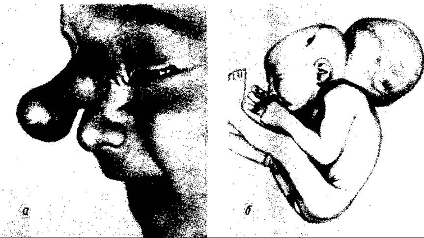
Fig. 116. Brain hernia:
a — anterior nasal (after M.B. Sytkovsky et al.); b — occipital
In addition to these two severe, life-incompatible malformations, more frequently encountered conditions include meningocele—the protrusion of the leptomeninges through a localized defect in the spinal column and dura mater, forming a fluid-filled blister beneath the skin—and meningomyelocele, which involves the herniation of both the meninges and the Spinal Cord itself through a vertebral defect beneath the skin (similar to an encephalocele).
Meningoceles and meningomyeloceles are predominantly localized to the lumbar region of THE Vertebral Column.
A frequent variant of spinal cord and vertebral column malformations is so-called occult spinal dysraphism, or Spina bifida occulta. This condition consists of a defect in the vertebral arches within the lumbosacral region, which is covered by Connective Tissue without any herniation of the meninges or spinal cord.
This malformation is typically detected incidentally during the examination of a patient for another condition, or more frequently, due to localized cutaneous markers overlying the spinal defect, such as hypertrichosis, capillary hemangiomas, lipomas, dermal sinuses, or Scoliosis.
This group of malformations (meningomyelocele) also includes A number of other defects, such as central distension and unilateral cleft lip. A cleft palate may occur either in combination with a cleft of the upper lip and alveolar process or as an isolated defect. It can be either complete or incomplete.
A complete cleft involves both the Hard and Soft palate (complete cleft palate), whereas an incomplete cleft may be restricted to the soft palate alone, the soft palate and part of the hard palate (more commonly), or solely the hard palate (less commonly).
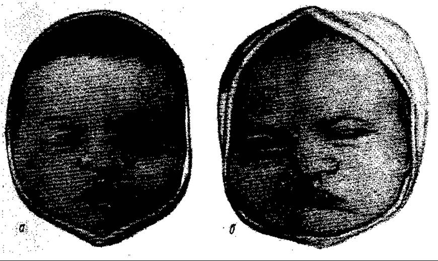
Fig. 117. Unilateral cleft lip: a — partial incomplete cleft; b — complete cleft of the lip, alveolar process, and palate
Clefts of the upper lip, maxillary alveolar process, and palate are caused by embryonic growth arrest and a failure of the maxillary processes to fuse, resulting from yet insufficiently understood extrinsic and intrinsic Factors affecting the embryo (during the 8th–12th weeks of development), including genetic factors. The Role of heredity is supported by a family history of these anomalies in affected patients. It is believed that hereditary malformations are transmitted in an autosomal recessive manner.
A cleft lip, especially a complete unilateral cleft (Fig. 117) and, even more so, a bilateral cleft (Fig. 118), leads to alterations in the nasal wing, tip, and septum.
The combination of a cleft lip and palate is also accompanied by significant anatomical abnormalities, including an increased volume of the Pharynx and Nasal cavity, underdevelopment of the alveolar process and palatine processes of the Maxilla, and Hypertrophy of the nasal conchae.
These congenital anomalies cause severe feeding and respiratory impairments, as well as the rapid Development of Respiratory tract infections (potentially leading to Pneumonia), ear infections, nasal cavity disorders, and intestinal issues.
Cleft lip and palate are treated surgically. Orthopedic treatment of hard palate clefts using obturators, which was once common, is currently used only in rare adult cases (typically due to severe comorbid conditions).
Patients with upper lip clefts undergo surgery in the first weeks of life (less frequently in the first months), sometimes even before discharge from the maternity hospital.
Patients with combined cleft lip and palate are operated on either in two stages—repairing only the lip cleft at an early age and closing the palatine defect at 3–6 years—or sometimes via a single-stage Procedure, though not earlier than one year of age.
Very early surgery for palatine cleft closure (uranoplasty), aside from being a complex and demanding procedure, carries the risk of maxillary underdevelopment and deformity.
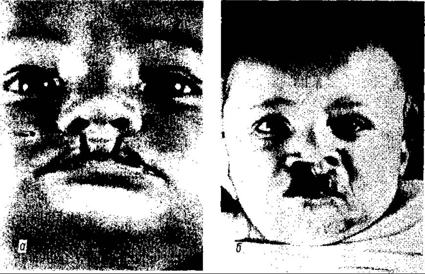
Fig. 118. Bilateral cleft lip: a — lip only; b — lip and jaw (F. Burian)
Postoperative mortality does not exceed 1–2 %.
Plastic repair of both lip and hard palate defects is performed using local soft tissues surrounding the defect (autoplasty) through tissue mobilization (of the lip, as well as the mucosa and periosteum of the hard palate), transposition, and suturing according to various techniques (O. I. Yevdokimova, A. A. Limberg, Yu. I. Bernardsky, et al.).
Surgical intervention is complemented by physical therapy and speech therapy Methods.
NECK MALFORMATIONS
Thyroglossal duct cyst and midline cervical fistula. A thyroglossal duct cyst develops from an unclosed thyroglossal duct (ductus thyreoglossus), which during embryonic development connects the primitive foregut lumen to the thyroid gland.
Already during the 1st trimester of embryonic development, The connection between the thyroid gland and the gut breaks down, and the duct obliterates, leaving a small depression at the Base of the Tongue known as the foramen coecum.
If The process of regression and obliteration of the thyroglossal duct is disrupted, the duct remains patent, typically in its distal (aboral) section, and rarely along its entire length. When it remains patent throughout its entire length, it opens into the Oral Cavity (on the tongue). Since the inner lining of the duct consists of Stratified Epithelium capable of mucus secretion (derived from the gut), mucus gradually accumulates, turning the slit-like lumen into a spherical, fibrous-walled cyst. This secretion buildup is particularly accelerated when the duct epithelium is irritated by microflora, which easily enters via lymphatic and Blood Vessels or directly from the oropharynx (if the duct opens into the oral cavity), or through lymphatic and blood vessels during INFECTIOUS DISEASES OF the patient's oropharynx and Upper Respiratory Tract.
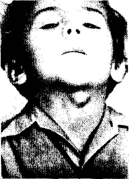
Fig. 119. Thyroglossal duct cyst
Infection of the duct accelerates its transformation into a cyst and frequently leads to cyst suppuration. Acute suppuration of the cyst with the spread of inflammation to the soft Tissues of the neck—which predominantly occurs when the cyst or duct opens into the oral cavity—results in the cyst rupturing onto the skin of the neck and turning into a fistula. A midline cervical fistula is an infectious complication of a thyroglossal cyst, representing a secondary manifestation rather than a primary one.
A thyroglossal duct cyst appears as a spherical mass ranging from 1.5–2 to 3–4 cm in diameter, located along the midline of the neck (due to the fact that the thyroglossal duct originates from the main, medial primordium of the thyroid gland) in the segment between the Hyoid bone and the upper border of the thyroid gland, predominantly at the level of the upper portion of the thyroid Cartilage (Fig. 119). The cyst content consists of clear white mucous fluid or liquid pus in cases of infection.
Although the cyst originates from a congenital substrate, clinically it usually manifests at the age of 5–12 (predominantly 7) years. It can appear earlier or later, even in older age (following oropharyngeal or upper respiratory tract infections due to the activation of secretion in residual unclosed clefts, i.e., sinuses of the thyroglossal duct). The cyst is spherical, firm-elastic in consistency (due to fluid content), and has a smooth surface. It is painless upon Palpation unless inflamed (suppurated). Like a goiter, it moves upward during swallowing along with the Larynx. This specific feature distinguishes it from other (extrathyroid) tumor-like masses of the neck.
Diagnosis is facilitated by fine-needle aspiration (yielding mucous or purulent fluid) and scanning with 131I or 99Tc. The latter accumulates not in the cyst, but in goiter nodules developing in the pyramidal lobe of the thyroid gland (the pyramidal lobe originates from the distal part of the ductus thyreoglosus).
Median fistulas are diagnosed based on both medical history and physical examination: history of a cyst and prior inflammation; typical (median) Location (only occasionally does the fistula opening slightly deviate from the midline of the neck); purulent discharge from the fistula, sometimes accompanied by skin inflammation; and fistulography (using lipiodol or verographin).
The connection between the fistula and the oral cavity is established by injecting methylene blue or brilliant green into the fistula (while placing a gauze swab in the oral cavity); if the fistula is complete (connected to the oral cavity), the colored antiseptic enters the Mouth, staining the gauze swab.
Among median cysts, about 6% are dermoid cysts that do not originate from the ductus thyreoglosus and are clinically indistinguishable from median cysts, but contain different contents (sebaceous detritus).
Treatment for median cysts and fistulas is surgical. The cyst or fistula is excised along with the central part of the hyoid bone, through which the upper end of the thyroglossal duct (or fistula) passes. The duct is ligated near the ROOT of the tongue, and the ligature is cut below it.
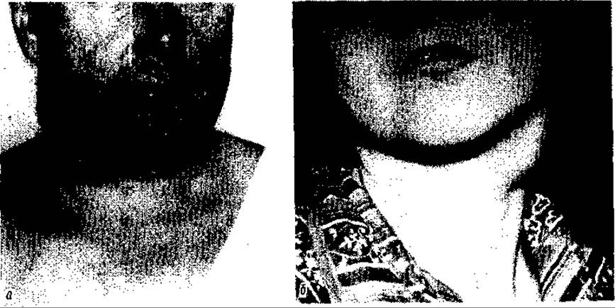
Fig. 120. Lateral cervical cysts (a, b)
From a cosmetic standpoint, the intervention should be performed via a curved transverse approach (open upward) located below the cyst or around the fistula. Median dermoid cysts are excised without resection of the hyoid bone.
Lateral cysts (Fig. 120) and fistulas of the neck most likely originate from the elements of the so-called branchial or branchiogenic pouches (pockets) of the primitive pharynx. These pouches, which appear to be a remnant of the phylogenetic Development of the animal kingdom in humans, serve as the source for The formation of many neck organs—elements of the auditory apparatus, larynx, Tonsils, Thymus gland, Parathyroid glands, etc. The inner pouches originate from the endoderm. The outer pouches—grooves lying between the inner ones and directed inward—develop from the ectoderm. These pouches are separated by the so-called closing membranes. In normal embryogenesis, the pouches disappear within the first 2 months of development.
It is believed that premature closure of the inner pouch from the inside results in the formation of lateral cysts with mucous contents. Conversely, such closure of the outer pouch leads to the formation of a dermoid cyst.
In some cases, so-called perforation openings appear in the closing membrane between the pouches (which do not form during normal development), and these constitute the anatomical substrate of lateral cervical fistulas.
Other hypotheses regarding the origin of lateral cysts also exist. R.I. Venglovsky (1909) suggested that they develop from remnants of the thymopharyngeal duct (ductus thymopharyngeus), which arises from the II–III branchial arches.
Some researchers view the origin of these cysts as a malformation in the Formation of the cervical Lymph Nodes (E.S. King, 1972).
Lateral cysts are predominantly unilateral and located in the upper third of the lateral aspect of the neck, anterior to the sternocleidomastoid Muscle. However, cysts may also be found beneath the lower border of this muscle. They usually develop during adolescence and youth, and not infrequently in adulthood. Similar to median cysts, the growth of a lateral cyst is stimulated by infection. This is evidenced not only by their occurrence in adolescents and adults, but also by the presence of purulent contents within them. Their source is presumably derived mainly from the second, and less frequently the third, branchial pouches. The cyst appears as an oval-shaped tumor that locally deforms the neck, protruding 2–4 cm above the skin level. The tumor does not move during swallowing. It has a smooth surface, and its consistency ranges from soft to firm-elastic with a fluid thrill (ballottement) sign. Palpation is painless in the absence of acute inflammation, and the overlying skin is unchanged. The cervical lymph nodes are not enlarged unless the cyst is complicated by Phlegmon. The latter is quite common and typically follows a severe course (high BODY Temperature AND intoxication). The cyst lies deep; anteriorly, it is covered by the pretracheal Muscles and the edge of the sternocleidomastoid muscle, while its posterior surface rests against the neurovascular bundle of the neck, directly on the Internal jugular vein.
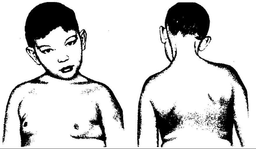
Fig. 121. Congenital muscular torticollis
Treatment of the cyst is surgical: its removal via an oblique-transverse incision 8–10 cm long directly over the cyst, transecting the Cervical fascia along the anterior border of the sternocleidomastoid muscle, retracting the latter laterally and the pretracheal muscles medially.
Fistulas of the lateral surface of the neck. Unlike cysts, cervical fistulas are detected immediately after birth. They are more frequently single, although multiple and bilateral fistulas (up to a maximum of 6—3 on each side) are also observed.
The highest-located cyst is found at the ear lobule or anterior to the tragus; it connects the fistula to the auditory tube and originates from the first branchial pouch (the perforation opening of its closing membrane).
Fistulas originating from the second and third branchial pouches typically open anterior to the sternocleidomastoid muscle, at the mid-neck level and at the clavicular level below the neck line, respectively. However, most commonly, a single fistula is localized in the middle or lower part of the neck, meaning its source is the second branchial arch. These fistulas are almost always complete. If it develops from the second arch, it opens into the pharynx beneath the tonsil.
Clinically, a fistula presents as a small opening on the skin from which a drop of clear mucous fluid is secreted upon pressure. The skin around the fistula opening is usually clean and uninflamed. In the event of inflammation or infection within the fistula—which occurs much less frequently than in cysts—the discharge becomes purulent, accompanied by inflammation of the surrounding skin. Such a fistula may be either incomplete, ending in the soft tissues near the pharyngeal wall, or formed as a result of suppurative liquefaction of a lateral cyst that has ruptured onto the skin. In the latter case, the skin opening is wide and the skin is inflamed. Although the skin opening is usually pinpoint in size, its subcutaneous portion is quite wide (0.3–0.8 cm) and has a thick wall. It runs along the anterior border of the sternocleidomastoid muscle, initially subcutaneously, and then deepens, extending in a sagittal-medial direction toward the pharyngeal wall, passing either between the Branches of the carotid artery (fistula from the second pouch) or posterior (lateral) to the carotid artery to the pharyngeal wall.
Surgical treatment of fistulas involves excising them via an oblique or staged Proctor approach, which consists of several transverse incisions placed one above the other at a distance of 2–3 cm. Multiple fistulas, except those localized on the ears, are also treated surgically in a staged manner.
Prior to surgery, fistulography is performed to clarify the direction of the fistula, and a colored antiseptic is injected into the fistula on the operating table to determine its connection with the pharyngeal cavity and trace the path of the tract within the tissues.
Torticollis (wryneck)—a pronounced lateral tilt of the neck—is a deformity of diverse origin, both congenital and acquired.
Congenital torticollis (wryneck) results from scarring and shortening of the sternocleidomastoid muscle during the embryonic period. This deformation is caused by local influences on the fetus, its abnormal position, or potentially an infection. The lower portion of the muscle is typically affected, presenting as a scar-like cord connected to the superficial Fascia of the neck and its lamina.
Most cases are unilateral (Fig. 121), occur more frequently on the right side, and are generally more common in girls.
The child's head is tilted toward the shortened muscle and forward, with the face and chin turned in the opposite direction. Consequently, the tightened, bowstring-like shortened muscle stands out prominently. The side of the face facing the tilt appears bulging and shortened, whereas the contralateral side is flattened, elongated, and slightly atrophied.
Due to the persistent head tilt, the shoulder girdle is also displaced: it is lowered on the side of the tilt and elevated on the opposite side.
The child has a restricted visual field. If torticollis persists for a long time, spinal curvature also develops, meaning muscular torticollis becomes compounded by a skeletal component. Diagnosing congenital torticollis when it is pronounced is straightforward.
Mild cases in early childhood are treated conservatively using orthopedic methods, whereas longstanding cases and those refractory to conservative treatment require surgery (the scarred and degenerated portion of the muscle is completely excised, along with the scarred fascia fused to it). Correction of the neck and head is performed right on the operating table. Subsequent physiotherapy completes the rehabilitation process.
Cervical hygroma is primarily a multilocular cyst or lymphoma. It is one of the most common congenital anomalies, located predominantly in the neck, although it can also appear in the inguinal and groin regions. Its etiology stems from developmental disorders of The Lymphatic system.
Anatomically, it manifests as encapsulated, multichambered lymphatic sacs filled with yellowish or even hemorrhagic serous fluid. The lack of natural drainage leads to the rapid accumulation of large amounts of fluid secreted by the epithelial lining of the lymphoma walls, causing it to enlarge. A hygroma can sometimes reach the size of an infant's head and may lead to asphyxia. Diagnosis is established through physical examination, which reveals a spherical mass with stretched skin and signs of fluctuation. Treatment is surgical (removal of the hygroma).
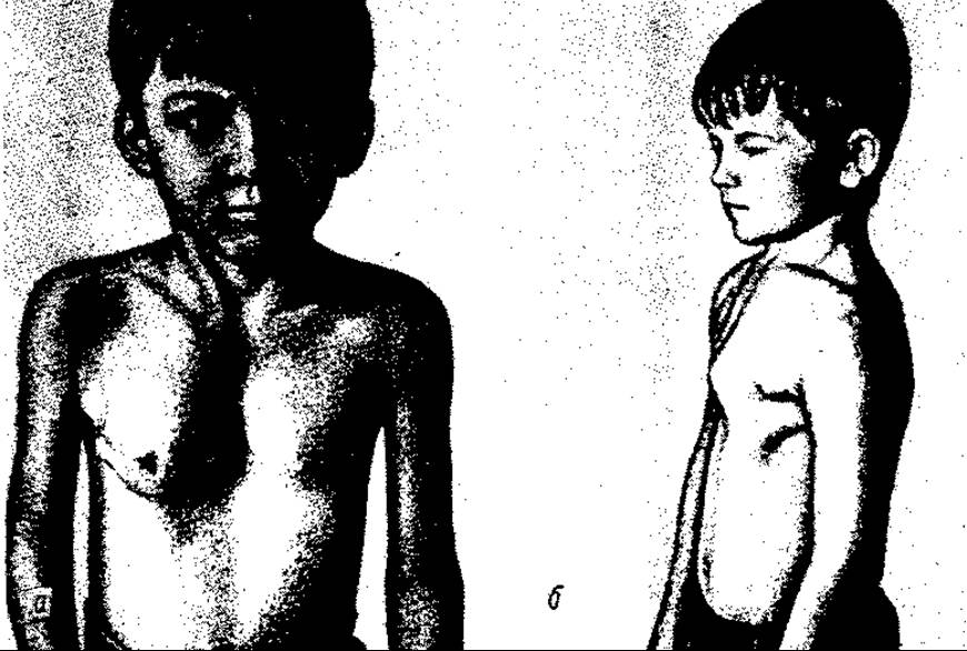
Fig. 122. Chest wall deformities: a — pectus excavatus; b — pectus carinatum (Pigeon chest)
If the hygroma extends to the face or Thorax, the main portion is excised while the remaining parts are fenestrated to prevent fluid accumulation and organ compression. Radical removal is usually unfeasible in such cases. To reduce pressure on the airways (preventing asphyxia), the hygroma is aspirated with a needle to drain the fluid. In cases of multichambered hygromas, multiple chambers are punctured separately.
CONGENITAL Anomalies of the CHEST
Medical practice encounters numerous malformations involving the Skeletal structure of the chest, mammary glands, and respiratory organs.
Pectus excavatus (funnel chest) is characterized by a broad depression of the sternal body along with the costal cartilages and xiphoid process (Fig. 122, a), which deforms the anterior chest wall. It is frequently associated with Kyphosis. Beyond the cosmetic defect, this anomaly exerts a negative impact on the heart by displacing it, potentially impairing both cardiac and pulmonary function. The developmental causes remain unknown. Some researchers consider the malformation to be a consequence of a growth rate disparity between the Diaphragm and the Sternum. Treatment for severe cases is surgical, involving plastic reconstruction.
Pectus carinatum (Fig. 122, b), also known as pigeon breast, involves a keel-like forward protrusion of the sternum and costal cartilages. Treatment consists of surgical correction.

Fig. 123. Supernumerary mammary gland (C.D. Haagensen)
Agenesis of one or both mammary glands (amastia) is extremely rare. More frequently observed conditions include juvenile breast hypertrophy and the development of one or two accessory mammary glands bilaterally (Fig. 123), typically localized in the anterior axillary fold (polymastia). The nipples are situated along the so-called milk lines, extending from the axilla to the Pubic Symphysis.
Treatment for accessory mammary glands involves surgical excision. In certain cases of breast hypertrophy, plastic surgery is also performed for cosmetic indications. Polymastia does not require treatment.
A rare chest wall malformation is sternal clefting. Mild forms present as a small longitudinal fissure, whereas severe forms are characterized by wide separation of the sternal segments and skin, with herniation of The Heart and Pericardium through the defect. This type of anomaly is treated surgically.
Even more complex is the so-called distal fissure, which typically occurs in combination with diaphragmatic and abdominal wall defects (omphalocele). Its management requires highly complex surgery.
CONGENITAL ANOMALIES OF THE ABDOMINAL WALL
The best-known and most prevalent congenital abdominal wall defects include embryonic hernia or omphalocele, umbilical cord hernia, funicular hernia, umbilical hernia, and inguinoscrotal hernia.
The most severe anomaly is omphalocele, which involves the Evisceration of abdominal viscera through a large defect in the abdominal wall into the embryonic amniotic sac within the umbilical cord region.
Between the 6th and 10th weeks of intrauterine development, the coelomic cavity shifts toward the umbilical stalk (cord), toward which intestinal loops are primarily directed. These loops grow faster than the cavity itself enlarges. After the 10th week, the abdominal cavity (its wall) grows at an accelerated rate, and the Internal Organs return into it. When desynchronization occurs between these processes, a situation may arise where the development of the abdominal wall (its tissues and skin) is delayed, creating a central defect in the wall through which abdominal organs herniate. They are covered solely by fused layers of peritoneum and the amniotic membrane, which is up to 1 mm thick and has a transparent, gelatinous appearance.
The second variant of a congenital abdominal hernia is a hernia that protrudes through the umbilical ring via a defect in the rectus abdominis muscles while the abdominal wall is otherwise normally developed. A congenital umbilical hernia is covered only by skin. This hernia is small in size and its clinical course differs radically from that of omphalocele. The skin protects the abdominal cavity from infection, whereas an omphalocele is covered only by a transparent gelatinous membrane through which infection penetrates rapidly, leading to Peritonitis. Therefore, surgical intervention for an omphalocele must be performed immediately after the child's birth.
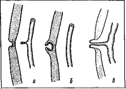
Fig. 124. Types of vitelline duct (ductus omphaloentericus) development: a — Meckel's diverticulum; b — blind fistula; c — complete persistence of the omphalomesenteric duct
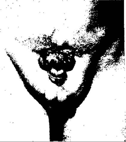
Fig. 125. Bladder exstrophy with epispadias
In cases of large omphaloceles, a non-radical operation is performed (covering the hernia with mobilized skin). Umbilical hernias can be operated on later, following attempts at conservative treatment.
Congenital inguinal or inguinal-scrotal hernias are pathogenetically associated with the descent of the Testes (descensus testiculorum) from the abdominal cavity into the inguinal region. This process is preceded by the formation of the so-called processus vaginalis of the peritoneum, into which the Testis descends (invaginating into it along with its posterior wall). After the descent of the testis, which occurs before birth, the cavity of the peritoneal process above it and extending upward to the very opening obliterates and closes.
If the peritoneal process fails to close by the time of delivery, it becomes filled with internal organs—primarily the intestines—under the influence of increased intra-abdominal pressure (which rises during the straining of the child's muscles, especially during crying and screaming), thus transforming into a hernia. Its diagnostic sign is the presence of abdominal organs in the hernial sac along with the testis. Since such hernias frequently become incarcerated, they are treated surgically at an early age.
In the region of the umbilical fossa, aside from hernias, malformations associated with the incomplete regression of embryonic omphalomesenteric and urachal ducts may be observed.
In cases of their complete failure to close, an opening typically forms in the umbilical fossa: an intestinal fistula (Fig. 124) when the omphalomesenteric duct remains open, and a urinary fistula when the urachal duct remains unclosed.
If only the distal part of the ducts remains unclosed, blind sinuses of these ducts open into the umbilical fossa, or simply a portion of the mucous membrane of the mesenteric duct (the so-called umbilical polyp), which secretes mucus. These incompletely obliterated ducts can be a source of abscess development in the abdominal wall and umbilicus. The treatment for these malformations is surgical.
Among external malformations of the urinary system, bladder exstrophy is the most severe (Fig. 125). The defect consists of underdevelopment of the anterior wall of the Urinary Bladder and the anterior abdominal wall directly above it, accompanied by diastasis of the pubic symphysis and prolapse of the bladder mucosa through the abdominal wall defect, frequently with the ureteral orifices visible on its surface.
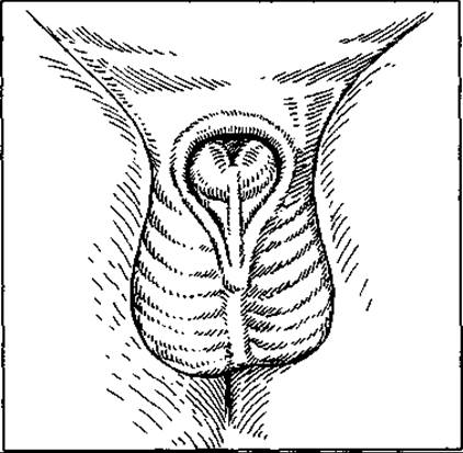
Fig. 126. Epispadias
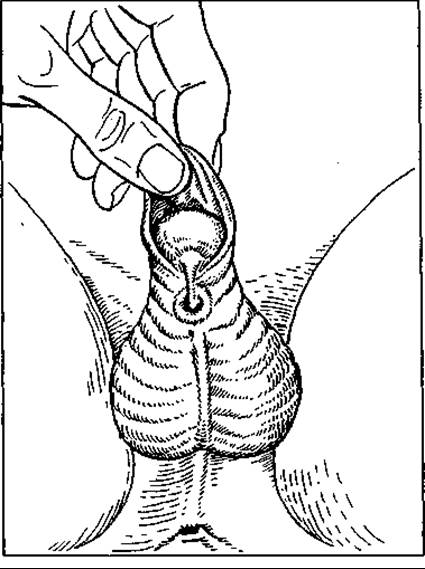
Fig. 127. Hypospadias
Due to the abnormal connection of the pubic bones, children with this anomaly walk with a waddling gait ("duck waddle"). This defect is frequently combined with other anomalies, notably cryptorchidism (undescended testes), etc.
Exstrophy occurs with a frequency of 1 in 100,000–120,000 boys and 1 in 500,000 girls.
The malformation is accompanied by continuous leakage of urine onto the skin, resulting in maceration and inflammation.
Treatment involves complex plastic reconstructive surgery of the urinary bladder and anterior abdominal wall. If correcting the defect by this method is impossible, transplantation of the bladder wall containing the ureteral orifices into the bowel is performed.
Urethral Anomalies include the ectopic placement of the urethral meatus on the anterior (dorsal) surface of the Penis (epispadias) or on its ventral surface (hypospadias).
Epispadias occurs rarely, but it belongs to the most severe pathologies when it cannot be corrected (Fig. 126). Its mildest form is the glandular (or balanic) type, in which the urethral opening is located on the glans penis; Urinary Incontinence is absent in this type. The second type is the penile (or corpus) type, where the urethra opens along the entire dorsal surface of the penis. Urinary incontinence may or may not be present in this type, depending on whether there is insufficiency of the bladder neck sphincter. The third type is the most common (penopubic). The defect spans the entire length of the penis and extends a short distance onto the abdominal wall, and is accompanied by failure of the pubic bones to fuse.
Epispadias occurs predominantly in boys, but it can also affect girls (clitoral, perineal, and complete types, which are accompanied by separation of the urethra, sphincter, and bladder). Surgery offers a more favorable prognosis in the first two cases, while the third requires ureteral transplantation (performed at 3–6 years of age).
Hypospadias (Fig. 127) occurs much more frequently than epispadias (20 times more often) and presents in three types: coronal (frenular), penile (mid-shaft), and penoscrotal or perineal. In the latter case, determining the child's sex can be difficult because the external genitalia are indistinctly differentiated. Hypospadias is invariably combined with various deformities, such as penile curvature and a rudimentary penis. Treatment is surgical.
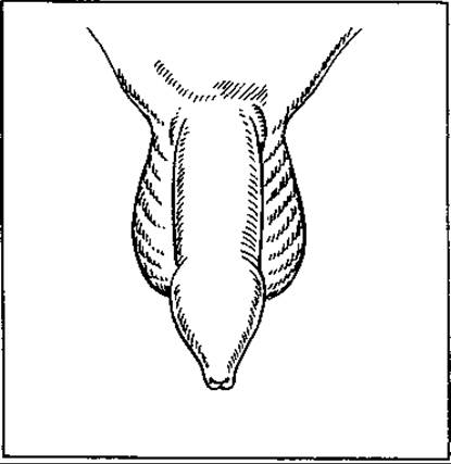
Fig. 128. Phimosis
Phimosis is a congenital or acquired condition characterized by severe narrowing of the prepuce (foreskin) opening, preventing it from retracting over the glans penis (Fig. 128). Urine and smegma accumulate beneath the foreskin (between it and the glans), leading to irritation and inflammation of both the foreskin and frequently the glans, thereby resulting in posthitis and balanitis. In adulthood, this anomaly can impair sexual function. Forcibly retracting the foreskin over the glans can cause paraphimosis, a condition where the trapped foreskin forms a tight band behind the glans, causing strangulation. The treatment for phimosis is surgical—specifically, excision of the distal part of the foreskin where the aperture is sufficiently wide.
Congenital Anomalies of the Anus and Rectum
Malformations of these segments of the digestive tract are among the most common, occurring slightly more frequently in boys. The most severe among them are anal atresia (atresia ani), rectal atresia (atresia recti), and agangliosis of the rectosigmoid colon, also known as Hirschsprung's disease. Anal atresia consists of the absence of an anal opening into the rectum (it remains covered by skin) due to a developmental arrest in embryogenesis—specifically, the failure of the anal membrane to perforate. Rectal atresia is characterized by a blind-ending rectum located some distance above the anal canal.
During early embryogenesis, when the embryo reaches a length of 7.5 mm, the cloaca is formed—a common chamber that unites the urogenital sinus and the hindgut.
Over time, this cavity is divided into separate cavities by the urogenital and anal membranes.
As a result of delayed or impaired separation of the urogenital sinus from the gut cavity by these membranes, along with a failure of their perforation, various malformations develop in this anatomical region (Fig. 129). These include: 1) anal stenosis, where the anal opening is present but markedly narrowed; 2) complete absence of the anal opening due to a failure of membrane perforation; 3) absence of the anal opening coupled with a blind-ending rectum situated at a distance from the closed skin surface; and 4) normally developed anus and anal canal, but with the rectum ending blindly above it, meaning a septum separates the rectum from the anal canal.
Malformations of the first three types are diagnosed within the first 24 hours of life, whereas the fourth type is typically identified only after several days due to acute intestinal obstruction. Consequently, the treatment outcomes for this latter type are consistently less favorable. In 70% of children with these anomalies, fistulous tracts connect the rectum to the Urogenital System or the Perineum.
The treatment for these anomalies is exclusively surgical. The simplest intervention is performed for the second type of defect, which involves a straightforward incision of the skin over the anal ring. For the 3rd and 4th types of defects, a staged approach is required, typically beginning with a sigmoid colostomy followed by radical corrective surgery within the first year of life.
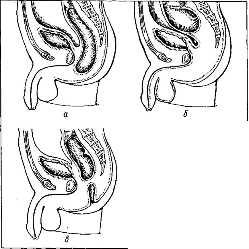
Fig. 129. Rectal atresia: a — anal atresia; b — atresia of both the anus and rectum; c — atresia of the rectum proper
CONGENITAL ANOMALIES OF THE BUTTOCKS, PERINEUM, AND MALE EXTERNAL GENITALIA
Dermoid cysts, fistulas, teratomas, duplicated Scrotum, and ectopic testes (located in the perineal region) can occur in the gluteal, sacrococcygeal, and perineal areas.
Congenital fistulas are primarily a component of rectal malformations, specifically rectal atresia, where the bowel lumen maintains an abnormal communication with the external environment (the skin surface) via a fistulous tract.
Dermoid cysts are predominantly localized anteriorly and slightly inferior to the coccyx, whereas teratomas typically develop in the sacrococcygeal and perineal regions. They often protrude markedly above the skin surface or extend deep into the surrounding tissues. Dermoid cysts contain skin appendage elements (sebaceous material and Hair). In addition to skin derivatives, teratomas frequently contain elements from germ layers other than the ectoderm, most notably endodermal tissues (such as bone or cartilage).
Cryptorchidism (from Greek *kryptos* — hidden; *orchis* — testicle) refers to the failure of one or both testes to descend into the scrotum, resulting in their arrest at various points along the normal descent pathway from the abdominal cavity into the scrotal sac.
This anomaly can be unilateral or bilateral. When the testes remain arrested within the ABDOMINAL CAVITY AND are absent from the Inguinal Canal, the condition is termed anorchism (or monorchism if only one testicle is missing). If a testicle is arrested within the inguinal canal or subcutaneously outside the scrotum, it is classified as cryptorchidism. Monorchism and cryptorchidism are the most frequently encountered forms. The undescended testicle may be located at various levels within the inguinal canal or just beyond its superficial ring, but it is typically anchored above the aponeurosis of the external oblique muscle by adhesions.
As a rule, the affected testicle is hypoplastic. Treatment is surgical. In cases of intra-abdominal location, surgical exploration of the abdominal cavity is required.
If the testicle exhibits severe hypoplasia, it is usually excised, as such tissue carries a significant risk of malignant transformation.
In cases of inguinal cryptorchidism, the testicle is surgically brought down and fixed in the scrotum (orchidopexy). This procedure should ideally be performed between the ages of 3 and 4 years, as irreversible degenerative changes begin to occur in the retained testicle thereafter.
ANOMALIES OF THE FEMALE EXTERNAL GENITALIA
Among the Congenital malformations of these organs, the most common are an imperforate hymen (*hymen femininus imperforatus*) and female pseudohermaphroditism.
An imperforate hymen, which completely seals the vaginal opening and obstructs the outflow from the uterus and upper Vagina, leads to the retention and accumulation of menstrual blood within the uterine cavity and vagina (hematocolpos), and potentially extending into the fallopian tubes and even the peritoneal cavity. This markedly stretches and enlarges the uterus (metromegalia / hydrometra) and causes the hymen to bulge outward. Such an anomaly can provoke severe intrauterine inflammation or even peritonitis.
Early diagnosis (utilizing colposcopy) and surgical incision (hymenotomy) are essential. Severe malformations of the FEMALE Reproductive System include pseudohermaphroditism, which is predominantly caused by congenital adrenal hyperplasia (resulting in excess adrenal androgens). This condition is characterized by underdevelopment of the vagina and uterus, accompanied by clitoral hypertrophy (Fig. 130).
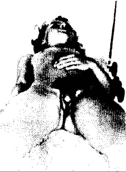
Fig. 130. Female pseudohermaphroditism with clitoromegaly
This defect is highly complex and requires precise diagnosis of both the genetic (chromosomal) sex and The Nature of the internal gonads. Treatment involving corrective surgery can be performed only after this.
Female pseudohermaphroditism may be combined with other malformations of the urogenital system and the rectum.
LIMB MALFORMATIONS
Limb malformations are numerous and diverse, ranging from minor defects to severe deformities. Commonly encountered anomalies include the fusion of fingers—syndactyly, an increased number of fingers—polydactyly (Fig. 131), or a decreased number—oligodactyly, an additional terminal (nail) phalanx (Fig. 132), as well as finger hypoplasia and deformities of the hands and feet.
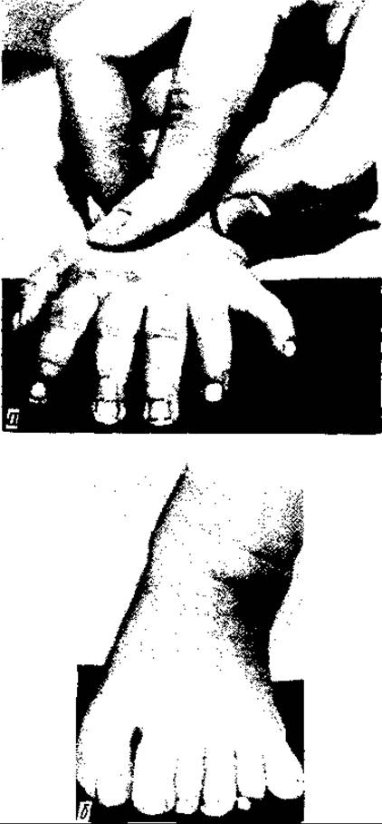
Fig. 131. Polydactyly of the upper (a) and lower (b) limbs
Rare anomalies include the absence of all limbs—amelia, the absence of the lower (apus) or upper limbs (abrachius); fusion of the lower limbs (simpus or sirena), absence of the humerus and BONES OF THE forearm, absence of the Femur and bones of the leg, and the attachment of hands or feet directly to the shoulder or pelvic girdles (phocomelia).
Underdevelopment of individual bones or limb segments, enlargement of individual fingers—Macrodactyly, congenital limb deformities in the form of constricting bands, and congenital types of elephantiasis are also observed. Congenital hip dislocation and Clubfoot occur relatively frequently.
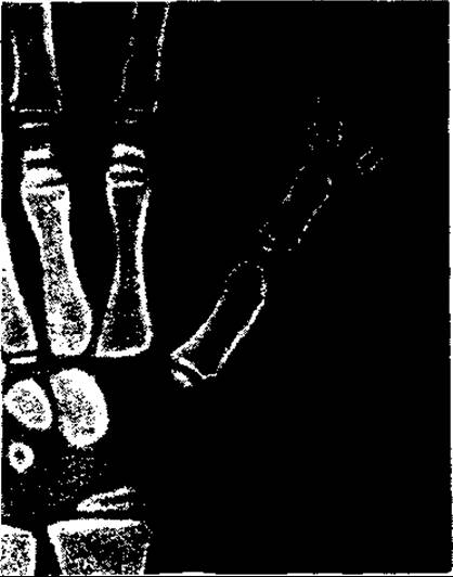
Fig. 132. Additional phalanx of the 1st digit of the upper limbs
Hip dislocation occurs predominantly in girls. Its signs include the following specific symptoms: the child begins walking much later and walks with a waddling gait; Trendelenburg's sign (the gluteal fold on the affected side is positioned higher than on the healthy side (Fig. 133)), and limited abduction and lateral rotation.
Treatment in preschool childhood is orthopedic, and at a later age, surgical.
Clubfoot (pes equinovarus) is a congenital FOOT deformity that occurs predominantly in boys and is caused by disorders in the development of the nervous system or muscles, as well as anomalies in the development of fetal membranes. The foot is turned inward with the plantar surface facing upward (supinated), flexed toward the sole, and the ligamentous connections between the Tarsal Bones and the bones of the leg are impaired. The Muscles of the Leg are atrophic, the thighs are conical, and the gait is awkward. Treatment is orthopedic and surgical.
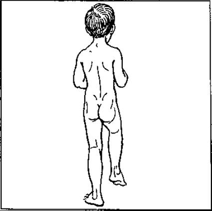
Fig. 133. Congenital hip dislocation — Trendelenburg sign
MALFORMATIONS OF INTERNAL ORGANS AND TISSUES
Malformations of internal organs and tissues, while not outwardly apparent, are accompanied by various functional disorders that are detected either immediately after birth or at various stages of life.
Some malformations may be completely incompatible with life or fatal without urgent surgical correction. In some cases, malformations may not cause any disorders in the patient's body and are discovered incidentally or when severe complications develop.
MALFORMATIONS OF THE DIGESTIVE TRACT AND ABDOMINAL ORGANS
Esophageal malformations. The most frequent esophageal malformations are stenosis and atresia. Anatomically, there are 6–7 types of defects. These include malformations of the esophagus itself (rarely) and combinations with tracheal malformations, characterized by the presence of a connection (fistula) between the underdeveloped or developed esophagus and the tracheal lumen (predominantly).
Among malformations of the esophagus itself, stenosis and atresia should be highlighted first. Stenosis is a defect consisting of a significant narrowing of the esophagus, mainly in its middle third. This is the mildest form of malformation. When the degree of stenosis is significant, it manifests in the first weeks or months of life, or later when semi-liquid or solid food is introduced.
A rare form of isolated esophageal malformation is its atresia at the level of the upper and middle thirds without a connection to the Trachea. The defect manifests within the first 2 days after birth and, without surgical intervention, is incompatible with life.
All Other forms of esophageal atresia are combined with tracheal malformations. Morphologically, up to 4–5 types of such malformations are distinguished. Among them, the most frequent (90%) is atresia of the upper segment of the esophagus combined with a connection between the upper end of the second, distal segment of the esophagus and the trachea above its bifurcation. This defect, like others accompanied by fistulas communicating with the trachea, clinically manifests in the first 2 days after birth (excessive salivation and hyperproduction of saliva, which does not pass from the oral cavity into the esophagus due to its obstruction, coughing, cyanosis, and wide-open eyes during breastfeeding).
The diagnosis is confirmed by esophageal radiography (2 ml of lipiodol is injected into the esophagus via a catheter). Treatment of this severe malformation is surgical. Without it, infants die from pulmonary complications (pneumonia) and the inability to feed.
Prior to 1939, all surgical approaches to congenital anomalies were unsuccessful. Today, many patients successfully undergo esophagoplasty and the repair of tracheal fistulas. Postoperative mortality ranges from 30 to 40%. Esophageal atresia is frequently associated with malformations of other organs, particularly cardiac and anorectal defects.
Malformations of the lower esophagus include cardiospasm—narrowing of the cardiac part of the esophagus (treated by dilation, mechanical esophageal expansion using bougies or pneumatic balloons)—as well as esophageal chalasia, which is the relaxation and gaping of the cardiac portion accompanied by mucosal prolapse into the gastric lumen.
CONGENITAL MALFORMATIONS OF The Stomach AND INTESTINES
Pylorospasm and hypertrophic pyloric stenosis. These congenital anomalies involve pathological spasm of the pylorus and hypertrophy of the circular smooth muscle of the pyloric sphincter, respectively. They occur predominantly in firstborn infants and may exhibit a familial predisposition.
Their Clinical presentation is nearly identical, manifesting as symptoms of high intestinal obstruction (metabolic disturbances, primarily water-electrolyte imbalance, and nutritional failure). Vomiting typically begins around the 10th day of life and rapidly leads to dehydration and weight loss. Treatment is primarily surgical.
In pylorospasm, the muscular layer and pyloric sphincter of the stomach are incised longitudinally down to the mucosa. In pyloric stenosis, a full-thickness longitudinal incision is made through the gastric wall in the pyloric region, combined with excision of the hypertrophied muscle ring.
Small bowel malformations include duodenal and ileal stenosis (rare), duplication of various segments, and Meckel's diverticulum. Duodenal stenosis can either occur independently (rarely) or develop secondary to an annular Pancreas that compresses the duodenum.
Meckel's diverticulum is a lateral pouch (of varying length) of the ileum located 50–70 cm proximal to the ileocecal valve. It represents a remnant of the unobliterated embryonic vitelline (omphalomesenteric) duct.
When the vitelline duct fails to close, it transforms into an intestinal fistula opening at the umbilicus.
Diagnosis of Meckel's diverticulum is usually incidental during small bowel X-ray examinations performed for other indications, as it remains asymptomatic until inflammation—diverticulitis—develops. The latter follows a clinical course similar to acute appendicitis, and the diverticulum is typically discovered during an appendectomy.
Congenital megacolon, or Hirschsprung's disease, is a developmental anomaly of the anorectal (rectosigmoid) segment of the Large Intestine characterized by marked narrowing, fibrotic changes, and, most importantly, the absence of peristalsis in this region.
This defect is associated with a deficiency or complete absence of ganglion cells within the intramural nerve plexuses of the affected colonic segment, which is why it is also referred to as colonic aganglionosis.
Severe functional and anatomical alterations in the rectosigmoid region lead to chronic intestinal obstruction, hypertrophy of the colonic wall proximal to the obstruction, subsequent bowel dilation, and fecal retention. The sigmoid and descending colons—and frequently other segments of the large bowel as well—become massively dilated (reaching 15–20 cm in diameter) and packed with impacted feces.
Children with congenital megacolon suffer from growth retardation and cachexia. The abdomen is markedly distended, and the abdominal musculature is underdeveloped (Fig. 134).
Treatment is surgical, involving the resection of the aganglionic rectosigmoid segment and the severely altered portion of the sigmoid colon, followed by the pull-through of the distal large bowel to the perineum. This operation has several modifications, all of which are performed with preservation of the anal sphincter.
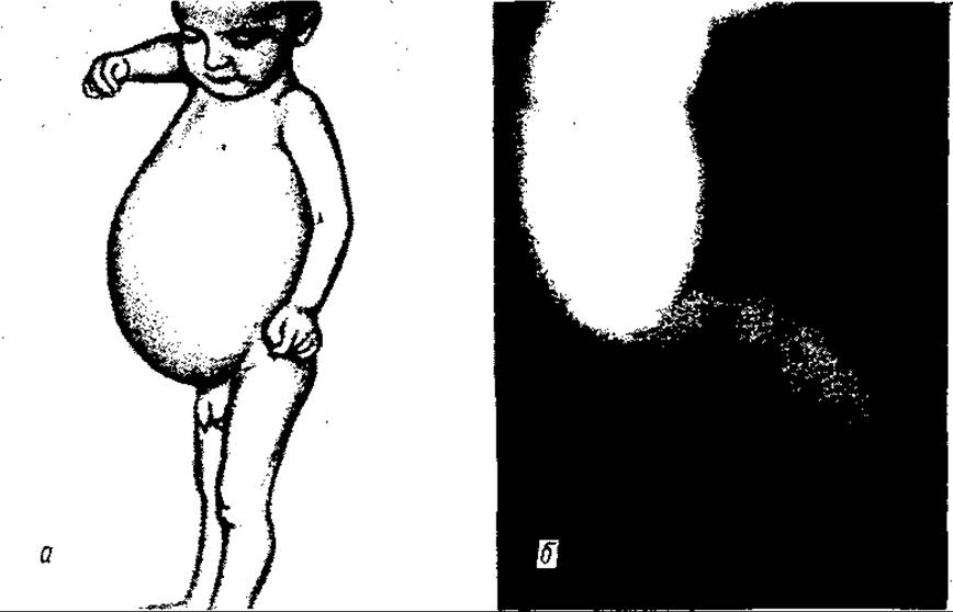
Fig. 134. Hirschsprung's disease:
a — general appearance of the patient; b — resected specimen of the rectum and sigmoid colon (M.B. Sitkovsky)
MALFORMATIONS OF THE BILIARY TRACT AND Liver
Congenital anomalies of the biliary tract and liver include atresia or agenesis of the common Bile duct, duplication of the Gallbladder, simple Liver cysts, and agenesis of the left hepatic lobe.
The most severe malformation is biliary atresia, which presents with jaundice and is fatal if left untreated by surgery.
ANOMALIES OF ABDOMINAL ORGAN POSITION, PERITONEAL FORMATION, AND THE OMENTUM
Abnormal positioning of abdominal organs includes situations such as situs inversus partialis (where the cecum and Appendix lie on the left side, the sigmoid colon on the right, etc.). Occasionally, this is accompanied by complete situs viscerum inversus, including dextrocardia.
Malformations related to the development of the peritoneum and omentum primarily involve the formation of deep peritoneal recesses at sites of organ transitions or reflections from the abdominal wall, as well as congenital apertures in the peritoneum and omentum. While these anomalies are mostly asymptomatic, they can occasionally cause acute or chronic intestinal obstruction due to the entrapment of bowel loops or omentum within these pockets and defects.
Peritoneal anomalies are most frequently localized in the duodenojejunal junction, where the peritoneum forms a deep recess prone to internal herniation of bowel loops (paraduodenal hernia or Treitz's hernia); in the ileocecal region (ileocecal fossae); and in the anterior peritoneal wall above the urinary bladder, where supravesical fossae may form, creating potential sites for internal herniation.
«Windows» are more commonly observed in the intestinal mesentery and the greater omentum (entrapment of intestinal loops can also occur in these defects).
A rare anomaly is the duplication of the peritoneum (where the entire intestine lies within a closed peritoneal sac inside a normally formed abdominal cavity).
URINARY SYSTEM ANOMALIES
These represent a wide range of defects, varying from minor to severe. Renal anomalies include underdevelopment (hypoplasia) of one or both Kidneys, their ectopia (dystopia—pelvic or iliac), fusion of the renal poles, most commonly the lower ones (Horseshoe Kidney), duplication of the kidney or Ureters, and Polycystic Kidney Disease, among others.
Many of these defects predispose to inflammatory and dystrophic processes, notably pyelitis, Pyelonephritis, and urolithiasis.
A duplicated Ureter, which may sometimes drain into the urethra rather than the urinary bladder, causes urinary incontinence. However, anomalies of the lower Urinary Tract are more frequent, particularly external defects of the bladder and urethra, as discussed previously.
ANOMALIES OF THE Internal female reproductive ORGANS
A large number of anomalies occur in the Internal Female Genitalia. Defects in the fusion of the Müllerian ducts can lead to the duplication of the uterus, fallopian tubes, and vagina. Common anomalies include hypoplasia, ranging up to Aplasia of the uterus, vagina, Ovaries, or fallopian tubes.
In some patients with secondary female sex characteristics, a testicle (ovotestis) is found instead of an Ovary. If located in the inguinal regions, it is removed due to its high malignant potential.
Respiratory system AND DIAPHRAGM ANOMALIES
Pulmonary anomalies include agenesis of a lung or its lobe; development of an accessory lobe with a bronchus; abnormal lobation of the Lungs; congenital Bronchiectasis; and pulmonary polycystic disease. Treatment is indicated only when the latter anomalies become complicated by suppurative processes.
Airway anomalies include atresia and stenosis of the larynx or trachea, which are either incompatible with life or require surgical intervention (in cases of stenosis).
Severe internal anomalies include diaphragmatic hernia, which involves an opening of varying size in the pleuroperitoneal membrane, up to the complete absence of the diaphragm.
This anomaly is accompanied by impaired function of both the cardiovascular and respiratory systems (palpitations, shortness of breath, hypoxemia) and the digestive organs due to their herniation (stomach, intestines) into the pleural cavity.
When the diaphragmatic openings are narrow, incarceration of the stomach and intestines can occur, leading to acute intestinal obstruction. Congenital diaphragmatic hernias are sometimes combined with other malformations, particularly external ones (fissure of the chest wall, cleft lip and palate, etc.). Treatment of diaphragmatic hernias is surgical.
Heart defects are observed in 0.6–0.8% of newborns, with nearly 100 Different types of cardiac anomalies identified. These include defects of the heart proper and the great vessels.
The most common are patent ductus arteriosus, pulmonary stenosis, coarctation of the aorta, atrial and ventricular septal defects (so-called septal defects), and combined heart and great vessel malformations (Tetralogy of Fallot).
Patent ductus arteriosus. This defect consists of the persistence of the fetal channel between the aorta and the pulmonary artery (the ductus arteriosus) remaining open after birth. It accounts for up to 15–20% of all congenital heart defects.
The anomaly clinically manifests primarily during school age, presenting with exertional dyspnea, growth retardation, low resistance to infections, etc. The defect is easily diagnosed by physical examination.
Its main hallmark is a loud systolic-diastolic (so-called machinery) murmur heard over the entire precordium, especially In the second intercostal space to the left of the sternum. The heart is moderately enlarged. Radiographic examination reveals dilation of the pulmonary trunk and «hilar dance».
This defect leads to elevated pressure in the pulmonary artery, Pulmonary Circulation Hypertension, hypertrophy of its vessels, and hypertrophy and dilation of the right ventricle (Eisenmenger syndrome). Eventually, pulmonary artery pressure may equal and even exceed aortic pressure due to increasing pulmonary hypertension and myocardial weakening. As blood flow reverses from right (pulmonary artery) to left (aorta), circulatory failure develops, resulting in fatal outcome during adolescence. With early diagnosis, this defect is surgically curable by ligating the duct at both ends and sometimes dividing it. The mortality rate is up to 1%. This procedure has become one of the simplest in cardiac surgery, with the first such operation performed in 1938.
Coarctation of the aorta is a malformation of the descending part of the aortic arch, characterized by its narrowing (Fig. 135). It is marked by a sharp increase in systolic pressure in the upper body (head, chest, upper extremities) with enhanced Blood supply to this region, alongside a sharp decrease in pressure and blood perfusion in the lower body (abdominal cavity and lower extremities).
The pulse in the lower extremities is weak, poorly filled, or absent, accompanied by pain in the legs. This blood redistribution leads to enhanced development of the shoulder girdle (broad shoulders, well-developed muscles) and underdevelopment of the lower body, particularly the lower extremities, which remain weak with poorly developed musculature.
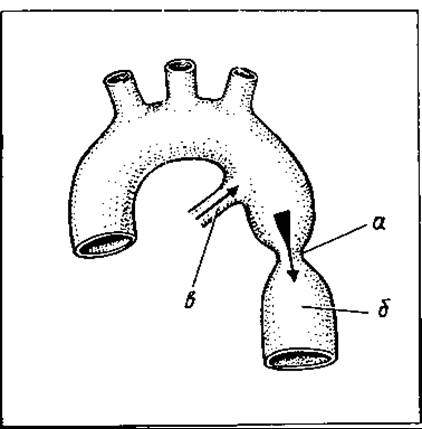
Fig. 135. Coarctation of the aorta: a — arterial duct; b — coarctation of the aorta; c — descending aorta
Patients complain of headaches, numbness in the lower extremities, and skin pallor. Physical examination reveals a harsh systolic murmur over the aorta and Left Ventricular Hypertrophy.
Many patients die from heart failure at an early age, while in adulthood death often results from hypertension complications.
With timely diagnosis, the condition is successfully treated surgically by resection of the narrowed aortic segment with a primary anastomosis (for a 1—2 cm stenosis) or by plastic repair. The procedure is performed without extracorporeal circulation (without a cardiopulmonary bypass machine).
A rare developmental anomaly of the aorta is a double aortic arch, which causes compression of the trachea and esophagus. It is treated surgically.
Atrial septal defect is a relatively common malformation (accounting for 5–25% of all congenital heart defects). It is typically classified as high (primum) or low (secundum).
This defect results in a left-to-right atrial shunt (due to the greater compliance of the right ventricle and the pulmonary Arterial System).
Small primary defects may remain clinically silent for a long time; however, with moderately large primary defects and all secondary defects, heart failure develops at an early age due to increased pulmonary blood flow and right ventricular overload. Patients experience dyspnea. Treatment involves surgical closure of the defect on an open heart under cardiopulmonary bypass.
Ventricular septal defect is somewhat more common than atrial septal defect and is located in either the membranous or muscular part of the septum. It varies widely in size, ranging from a small defect to complete absence of the septum (three-chambered heart). As with atrial septal defects, blood flows from left to right—from the left ventricle, where blood pressure is high, to the right ventricle, where pressure is lower. This leads to pulmonary hypertension and subsequent right ventricular failure due to overload.
A systolic murmur is auscultated in the third to fourth intercostal space to the left of the sternum. The heart is enlarged. The condition is accompanied by dyspnea (especially during physical exertion), palpitations, and rapid fatigue. Only small isolated defects do not require surgery. Conversely, large defects and those combined with other heart anomalies require surgical intervention at an early age (5–6 years), because pulmonary hypertension progresses rapidly. Once the pressure reaches 60 mm Hg, surgery becomes contraindicated, as closing the defect under such conditions places an excessive workload on the right ventricle, leading to its sudden failure.
Tetralogy of Fallot (first described by the French pathologist Fallot in 1885) is one of the most severe complex congenital heart defects of the "blue" or "cyanotic" type. Its morphological basis consists of pulmonary stenosis—specifically infundibular (funnel-shaped) stenosis of the right ventricular outflow tract—a high ventricular septal defect, dextroposition of the aorta, and right ventricular hypertrophy. Due to right ventricular outflow tract stenosis, very little blood reaches the lungs in this condition, resulting in hypoxemia. Furthermore, the ventricular septal defect allows venous and arterial blood to mix, which further exacerbates hypoxemia.
A high amount of reduced Hemoglobin accumulates in the blood (over 3% instead of the normal 0.3%), causing cyanosis. Children with this anomaly have bluish skin, rapid heart rate and breathing, delayed GROWTH AND DEVELOPMENT, and frequently suffer from so-called cyanotic spells (hypercyanotic spells) that can be fatal. Squatting helps patients breathe more easily.
A loud systolic murmur is heard in the third to fourth intercostal space to the left of the sternum, the chest is deformed, and clubbing of the fingers is present. On radiographs, the heart has a "woodden shoe" (coeur en sabot) appearance, characterized by concavity in the pulmonary artery segment and an elevated, rounded cardiac apex. Without surgical intervention, children typically die in adolescence.
Surgical correction of the defect is performed, often in two stages: The First stage involves creating an anastomosis between the pulmonary artery (distal to the stenosis) and one of the branches of the aorta (usually the Subclavian Artery), followed 1–2 years later by radical repair of all Components of the malformation.
DOUBLE AND MULTIPLE MALFORMATIONS
Double or multiple malformations result from developmental disorders within the uterine cavity involving two (or more) fetuses developing from multiple zygotes (normally resulting in dizygotic twins) or from a single zygote (normally resulting in monozygotic twins of the same sex).
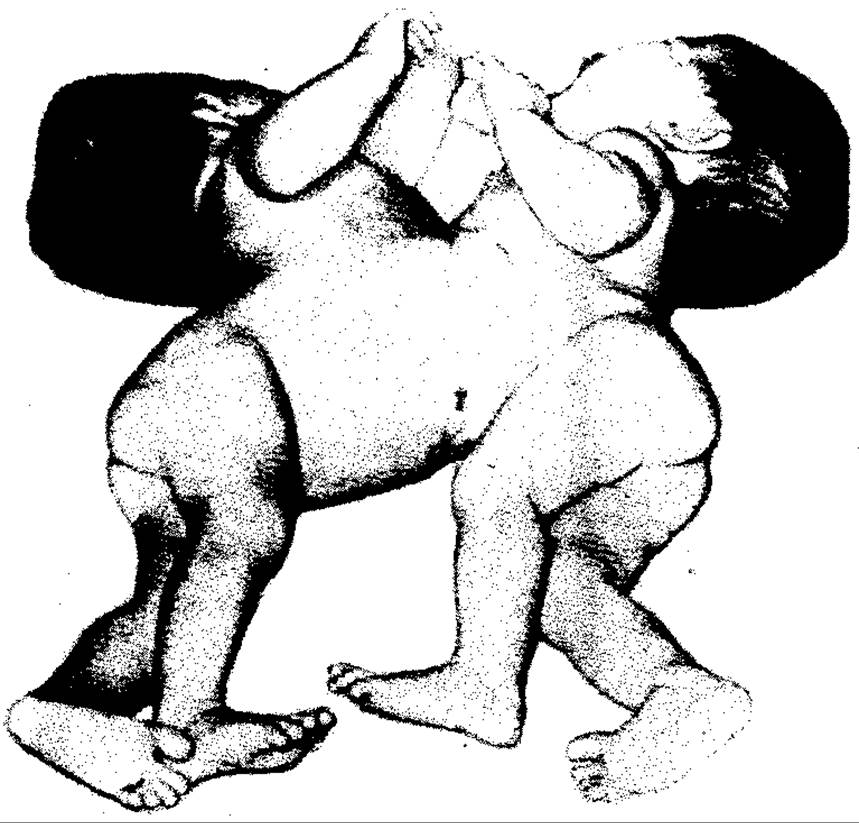
Fig. 136. Thoracopagus twins (K.L.Mooze)
Double malformations can be symmetrical or asymmetrical, separate or conjoined. Among asymmetrical separate twin fetuses, one may be normally developed while the other is malformed. The malformed twin may lack lower extremities or possess only a rudimentary heart and head—hence referred to as acardius or acephalus ("heartless," "headless")—or may consist solely of a head without a trunk and lower extremities (acardius acormus). Conjoined (attached) symmetrical double malformations represent equally developed organisms connected to each other by various PARTS OF THE body. They frequently share a Circulatory system (heart), brain, liver, other internal organs, or specific body regions.
Some of these organisms are nearly normally formed and joined only by skin or bones. Depending on the Topography of the union, they are classified as thoracopagi (Fig. 136; joined at the thorax), sternopagi (joined at the sternum), pygopagi (joined at the sacrum), craniopagi (joined at the skulls), etc. These are known as parallel double formations. Such twins can live for a very long time; a classic example is the Siamese twins (sternopagi), who lived for over 60 years.
Other variants include union of the upper part of the body with separately formed lower trunks and limbs (cephalopagi and thoracopagi), duplication restricted to the head region resulting in two faces or two heads (diprosopus and dicephalus), and fusion of the lower extremities. Craniopagi are joined only at their heads.
Recently, Canadian surgeons successfully separated such twins.
Asymmetrical double malformations consist of two unevenly developed organisms: one is nearly normal, while the other is malformed and attached to the first at various body sites. The developed twin is called the autosite, and the malformed one is the parasite.
Depending on the site of attachment of the parasite, they are termed: epignathus (a parasite attached to the jaw of the autosite), pygopagus, craniopagus, and thoracopagus (attached to the sacrum and perineum, skull, and chest, respectively).
Occasionally, a parasite is in an early stage of development inside a normal fetus (fetus in fetu). When the internal parasite appears merely as a amorphous tumor containing elements of various tissues or organs, it is termed a teratoma or organized teratoma.
Treatment of malformations and their separation is typically surgical. In symmetrical double malformations, this is feasible only if each of them possesses autonomous life-support organs.
Prevention OF CONGENITAL DEVELOPMENTAL DEFECTS
The prevention of developmental defects involves a comprehensive range of measures at individual, state, and global (planetary) levels.
Individual measures, which are easier to regulate, include maintaining a healthy lifestyle and work routine (proper Nutrition, eradication of bad habits), effective rest, and timely Genetic Counseling.
Genetic counseling is particularly essential for families with a history of children born with developmental defects.
Genetic counseling encompasses a general medical examination (case history, physical examination, dermatoglyphics—the study of palm and fingerprint patterns—along with genealogical, cytogenetic, and ultrasound evaluations, among others).
Modern diagnostic methods, particularly ultrasound and fetal karyotyping via amniocentesis, make it possible to diagnose fetal developmental defects before birth. Detecting a defect allows for timely notification of the family (mother) and, depending on the Nature of the defect and the parents' wishes, enables either termination or continuation of the Pregnancy.
Mass-scale preventive measures against congenital developmental defects comprise a wide range of national and international initiatives. The most critical among these include environmental health improvements (prevention and mitigation of radiation pollution in air, water, and soil; combating environmental contamination by pesticides, herbicides, and industrial toxic waste); strict adherence to regulations governing the Introduction of Pharmaceuticals through rigorous teratogenicity testing; occupational health protection for workers in hazardous industries; scientifically grounded and well-organized working and nutritional conditions; enhancement of public healthcare; and the elevation of the population's material well-being, cultural standards, and health literacy.
Last update: 08/08/2026
Editorial and Educational Adaptation: This material has been compiled based on the primary/original source text. The project team performed an editorial review, corrected technical inaccuracies, structured sections, and adapted the content for an educational format.
What was processed:
- elimination of formatting defects (OCR errors, structural breaks, corrupted characters);
- editorial organization of content;
- standardization of terminology in accordance with academic sources;
- verification of factual statements against the original source text.
All mentions of the author, publication year, and origin of the primary text have been preserved in accordance with the source.