Antibiotics (Properties, Application, Interaction) - M.P. Cherenko 1999
Features of examining a surgical patient. Diagnosis. Case history
Since ancient times, disease Diagnosis has been based on evaluating patient Complaints, medical history, and Objective Examination findings (inspection, Palpation, Percussion, Auscultation). In the 19th and early 20th centuries, these Methods formed the foundation of clinical practice. Years of experience contributed to their continuous refinement and The Development of semiology and Diagnostics for various conditions. Relying solely on data gathered through their own Senses, physicians of the past achieved a high level of diagnostic artistry and developed sharp logical clinical thinking, enabling them to make accurate diagnoses based on subtle, sometimes indirect, signs of illness.
However, the days when pure observation, experience, and The ability to interview a patient and uncover subtle symptoms were enough to unerringly recognize a disease and prescribe effective Treatment have passed. Today, physicians rely on laboratories and diagnostic departments to refine their diagnoses. Physical examination methods have taken a back seat.
Scientific and technological progress has led to the creation of diagnostic tools and devices that expand our cognitive horizons, allowing us to peer inside The Human Body, detect pathological changes in Internal Organs, and much more. This has significantly broadened the physician's diagnostic capabilities.
In recent years, there has been increasing Structure/133.html">Discussion about the dehumanization and even a crisis in the medical profession, often attributed to the scientific and technological revolution, narrow specialization, and an overreliance on laboratory and instrumental testing methods. Concerns have long been raised about the gradual marginalization of the classic clinician.
What is the solution? How can traditional, time-tested Research Methods be harmonized with modern ones? First, medical technological progress and the equipping of healthcare facilities with advanced tools do not exempt physicians from mastering Traditional Methods, including physical examination, and maintaining strong clinical reasoning skills. Second, we must foster a proper attitude toward diagnostic devices. The physician must remain the master, not an appendage or a soulless intermediary between the patient and the technology. We must remember the words of Norbert Wiener, the father of cybernetics: "To humanize what is human, and to compute what is computable."
The examination of a surgical patient differs little from that in other medical specialties. Yet, it possesses distinct features, often driven by the urgency required to confirm a diagnosis. In such cases, the surgeon must draw upon all their knowledge and experience to initiate treatment promptly. Examinations are necessary not only to establish an accurate diagnosis but also to monitor the course of the disease and the effectiveness of therapy.
The MAIN STAGES OF examining a surgical patient can be outlined as follows (see the diagram on p. 503).
Through a properly taken medical history and a systematic physical examination, surgical conditions can often be recognized quite quickly and accurately. Anamnesis and thorough examination must take precedence during the initial encounter between physician and patient. They form the foundation for any subsequent targeted use of technical diagnostic methods. The true nature of a surgical disease is captured more swiftly and accurately when the examination is structured rationally.
Class="center">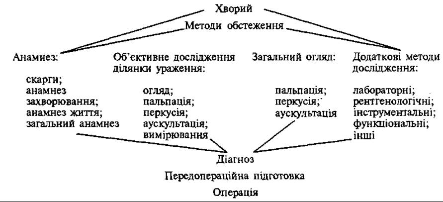
The encounter with the patient begins with taking the medical history (anamnesis). This encompasses the patient's complaints, The history of the present illness, personal and family history (social and past medical history), and a systems review (general anamnesis).
The word anamnesis originates from Greek and means recollection, or information regarding past states. This stage of patient examination relies entirely on the patient's self-assessment and is therefore considered subjective. In addition to demographic data (name, age, place of residence and employment), it includes the following sections: 1) patient complaints (querellae aegroti); 2) history of present illness (anamnesis morbi); 3) personal and family history (anamnesis vitae); 4) systems review (anamnesis communis).
Taking the medical history begins with the patient's complaints. Initially, the patient should be allowed to speak freely. Some individuals describe their symptoms fully and specifically, while others mention only certain troublesome manifestations, unconsciously omitting symptoms they deem secondary. Therefore, after the patient has shared their primary concerns, the surgeon should use targeted follow-up questions to uncover any other related complaints.
The METABOLISM/13.html">History of the present illness plays a crucial role in the examination process. It covers the timeline from the appearance of the very first symptoms until the patient's admission to the hospital. The patient should describe in detail when and with what signs the illness began. In certain acute surgical conditions, this history may be brief—particularly in cases of trauma—whereas in others, it spans a prolonged period. It is important to determine what the patient attributes the onset of the disease to, how it progressed (gradually or suddenly), what tests were performed (if results are available, they should be reviewed), what treatments the patient received, and what the outcomes were.
Since patients predominantly consult a doctor because of pain, special attention must be given to this symptom. It is essential to determine the Location OF THE pain, its onset, its character (constant or intermittent, dull, sharp, stabbing, boring, shooting, etc.), how it changes with body position, and its relation to the time of day.
When reviewing the personal history (anamnesis vitae), the physician should inquire about the patient's occupation (position, duties), bad habits (smoking, alcohol, substance use, prolonged medication intake), and past illnesses. It is vital to establish any sensitivities to certain medications (Antibiotics) or Blood products and plasma substitutes. In female patients, a gynecological history must be taken: age of menarche, past pregnancies, childbirths, abortions, and gynecological disorders. All this data may prove significant for assessing operability and predicting the postoperative course.
Family history holds an important place: family structure, the health of close relatives, living conditions, recreation, heredity, and past diseases among close relatives (Syphilis, Gonorrhea, Metabolic Disorders, etc.).
Beyond the history directly related to the condition that brought the patient to the surgeon, the physician must ascertain whether there are other complaints seemingly unrelated to the primary illness (systems review / general anamnesis). This, too, can influence the choice of treatment (conservative vs. surgical), preoperative preparation, anesthesia, surgical risk assessment, and postoperative management.
By questioning the patient, one can gain insight into the state of other organs and systems and detect comorbid conditions. Complaints are reviewed by system: nervous, respiratory, cardiovascular, digestive, urinary, musculoskeletal, and Sensory systems.
In emergency conditions, the time available for examination is generally limited, so it is sufficient to gather only the critical information required to perform surgical intervention.
If the patient is unconscious or unable to communicate (children, the deaf and mute, or cognitively impaired individuals), anamnestic information about their condition may be provided by relatives or witnesses to the trauma.
Taking a medical history is challenging when patients are experiencing severe pain at the time of the examination, as they may be reluctant to answer questions. In such cases, the patient should be placed in the most comfortable position possible.
Some patients intentionally exaggerate their pain (aggravation), while others fabricate non-existent symptoms (simulation). Additionally, some patients conceal certain symptoms out of fear of surgery (dissimulation). The physician must keep this in mind, though Conclusions regarding aggravation, simulation, or dissimulation should only be drawn after an objective examination and laboratory testing.
The Objective Examination of the patient involves assessing the body as a whole, including all its systems and organs (status praesens obiectius), as well as the local lesion (status localis morbi, locus morbi).
In therapeutic clinics, patient examination typically begins with investigating various body systems. In a surgical clinic, however, the examination begins with the lesion itself, followed by other organs and systems. This is especially true for emergency patients, where time is of the essence.
The site of the lesion may be a specific body region, an organ, a body cavity, or a combination thereof (e.g., a gluteal abscess, a torso hematoma, a fracture of the upper or lower extremities, Pleurisy or a lung tumor, appendicitis with Peritonitis, etc.). Objective examination, both general and local, usually begins with physical methods, supplemented by only basic instrumental investigations such as measuring body Temperature or blood pressure. Laboratory or advanced instrumental-apparatus tests (auxiliary methods), as well as specialist consultations, are ordered only after a preliminary diagnosis has been established.
The purpose of supplementary examinations is to refine the diagnosis and assess the degree of functional impairment in various organs and systems, as well as to evaluate the body's functional reserves in view of a potential surgical intervention (assessing surgical risk).
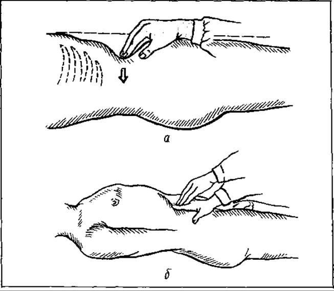
Fig. 112. One-handed palpation (a, b)
Examination of the affected area begins with inspection (inspectio). This allows for determining the shape of a specific body part, its position, volume, mobility, color, and transparency. Ideally, this should be performed in daylight, with the patient in both supine and standing positions (if the patient's condition permits). Inspection plays a particularly crucial role in superficially located pathological processes. If they are localized on one half of the body or on one of a pair of organs, their size and characteristics can be easily determined through comparison.
Inspection is followed by palpation (palpatio) of the affected area. It can be performed using one hand (Fig. 112), both hands, or in a specialized manner. This method helps determine local temperature, tenderness, consistency, size of the pathological formation, degree of Muscle tension, etc.
During bimanual palpation (Fig. 113), the hands move toward each other. The organ or pathological focus is positioned between both hands, with one hand preventing displacement while simultaneously guiding the examined organ or body area toward the second, palpating hand.
Specialized palpation also exists (Fig. 114). This refers to the examination of Body Cavities from the inside (Oral Cavity, rectum, Vagina).
Palpation must be performed gently and carefully, starting from regions distant from the affected area. Superficial palpation is conducted first, followed by deep palpation.
Palpation in various pathological processes has its own specific features.
Percussion (percusio) is thoroughly studied in clinical practice. It is used to detect Changes in the percussion note (from dull to tympanic, muffled to dull, tympanic to dull, pulmonary to tympanic, etc.). All of these indicate the presence of a pathological process.
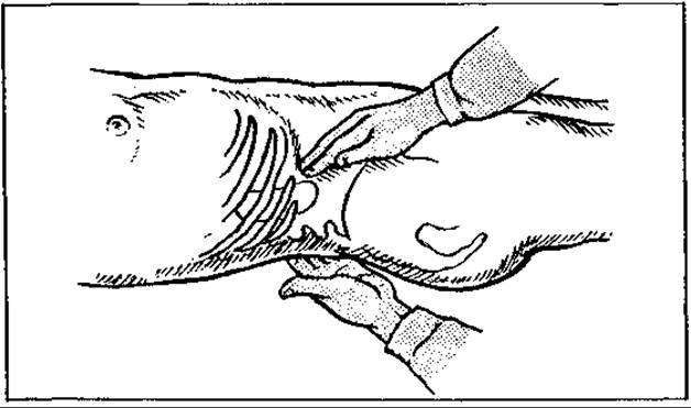
Fig. 113. Two-handed (bimanual) palpation
Auscultation (auscultatio) can be applied in the presence of a pathological process not only in the Lungs, but in any part of the body (abdomen, soft Tissues). Auscultation often plays a vital role in determining operability and diagnosing postoperative pulmonary or abdominal complications. It cannot always be replaced by radiological imaging methods.
Measurement. This investigative method is used in various pathological conditions. For instance, wounds, scars, or tumors are measured upon admission to the hospital, and their dimensions are recorded in the medical history. Periodic measurements help monitor the dynamics of the process. If a pathological focus is localized in the abdomen, periodic measurement of its girth may be necessary. To ensure measurements are taken at the same level, Skin markers are applied. A tape measure is used for these measurements, as well as for determining limb circumference. Palpable bony prominences serve as anatomical landmarks. The range of motion in joints is measured using a specialized goniometer.
General systemic examination of the patient. All organs and systems of the body are systematically examined using the same techniques applied during the investigation of the affected area (inspection, palpation, percussion, auscultation, measurement). These include the skin, Hair, Nails, subcutaneous tissue, Mammary Glands, Lymph Nodes, bones, joints, Thyroid Gland, respiratory, circulatory, digestive, urinary, and nervous systems, as well as external genitalia. Sensitivity and dermographism are also assessed.
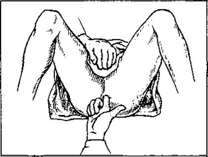
Fig. 114. Specialized palpation (rectal examination)
Inspection must be performed under adequate lighting, as yellow skin discoloration cannot be properly differentiated under artificial electric light. This helps form a general impression of the patient — their emotional state, facial expression (anxious, mask-like, surprised, or frightened), body position in bed (active, passive, forced), etc. Anthropometric parameters are determined: height, development of subcutaneous tissue, morphological constitution (asthenic, hypersthenic, normosthenic), facial complexion (pale, hyperemic with bright eyes), Lips (cyanotic), eye position (protruding — exophthalmos, sunken — enophthalmos), state of consciousness, sweating, presence of skin rashes or pigmentation changes, etc.
Attention is paid to the shape of individual anatomical regions or organs, configuration, and the enlargement or reduction of a part or the entire organ (HEAD, face, neck, chest contour, Abdominal cavity, limbs, etc.).
Palpation is of exceptional importance in the diagnosis of surgical diseases. Pulse palpation provides information on the state of Circulation, primarily cardiac function; palpation of the apex beat reflects heart activity, revealing painful points and changes in muscle tone. It also detects edema in various body areas, subcutaneous gas (crepitus) or fluid in joints, superficial and abdominal abscesses (rarely in the pleural cavity), and neoplasms in superficial organs or tissues as well as cavities (thyroid and mammary glands, superficial lymph node lesions, abdominal cavity, rectum, vagina), among others. Both superficial body areas and hollow or abdominal organs are palpated. The pulse, apex beat, and state of superficial vessel walls are assessed. This investigative method helps detect various organ disorders, including inflammatory, traumatic, and neoplastic conditions (lymphadenitis, thrombophlebitis, dislocations and fractures, goiter, and breast or rectal tumors), painful zones, and the accumulation of fluid or gas in tissues or cavities. Palpation plays a particularly major role in diagnosing abdominal conditions. Abdominal palpation is rightfully considered the most critical method for examining this region. The classic technique for palpating abdominal organs was developed by Ukrainian therapeutic scientists V. P. Obraztsov and M. D. Strazhesko. This method is known as gliding methodical palpation. The key principles of this palpation are maximum relaxation of the anterior abdominal wall Muscles and painlessness. It is performed with warm hands using gliding Movements of the fingers (with the hand positioned at a very sharp angle to the skin surface of the abdominal wall) over the abdominal organ. The fingers should be oriented perpendicular to the long axis of the organ. It is called methodical palpation because it is performed sequentially: in the absence of abdominal pain complaints, it begins with palpation of the sigmoid colon, whereas if there is pain in the left half of the abdomen, it starts on the right side (in the cecum region). In other words, palpation should begin in the area furthest away from the zone of tenderness and gradually approach it.
Palpation determines: zones of tenderness in the abdominal, retroperitoneal, and other regions, as well as muscle changes (protective muscle guarding or atrophy); temperature variations in the palpated area and its configuration; the presence of gas in tissues and fluid; the presence of a tumor or tumor-like formation in the abdominal cavity or its wall (tumor, inflammatory infiltrate, enlarged or distended hollow organ that is not normally palpable), etc. Palpation should be performed in both supine and standing positions. For instance, a displaced (floating) Kidney is sometimes more readily identified in the upright position than in the supine position. Palpating the mammary gland in the supine position in cases of mastopathy (dyshormonal breast hyperplasia) yields a completely different impression of the pathology compared to palpation in the vertical position. Therefore, palpation in both positions holds differential diagnostic value. Abdominal palpation is performed using either one hand or both hands placed on top of each other. In the latter case, the upper hand presses down on the lower one, while the lower hand glides (along with the abdominal wall) over the organ's surface. Each phenomenon is diagnosed using specific palpation maneuvers: for example, a quick tap on the abdominal wall detects the splashing sound of fluid in a hollow organ (Stomach, intestine), while a similar lateral push on one side of a distended abdomen (with a stationary hand placed on the opposite side) indicates the presence of fluid in the abdominal cavity (ascites, peritonitis). To correctly evaluate data obtained via palpation, one should always palpate the symmetrical area or healthy paired organ alongside the affected area or organ (comparative palpation).
Percussion allows for the detection of sound changes over The surface of various organs and cavities. It is of particular significance in diagnosing lung and pleural diseases (Pneumonia, exudative pleurisy, etc.), abdominal processes (tympanitis — intestinal obstruction, dullness — ascites, etc.), and heart conditions (enlarged cardiac dullness due to changes in chamber volume, muscle mass, or fluid in the Pericardium). Like palpation, percussion should be performed concurrently over symmetrical areas when examining paired organs and cavities.
Auscultation aims to detect various natural or abnormal sounds, tones, or murmurs. This method is especially valuable for diagnosing Diseases of the lungs (pneumonia, tuberculosis, pleurisy), heart (congenital or acquired defects), as well as certain pathological states of the abdominal cavity (intestinal obstruction), among others. A thorough knowledge of the normal acoustic range is essential for the accurate evaluation of sound characteristics.
Often, the objective examination methods described above are sufficient to establish a diagnosis, but in certain cases, additional methods become necessary. These include laboratory, radiological, endoscopic, ultrasound, functional, instrumental, and hardware-based investigative methods.
General clinical supplementary methods of investigation include: complete blood count and urinalysis, blood glucose testing, and chest fluoroscopy (or radiography). Treatment of the patient cannot proceed without these fundamental diagnostic methods.
Sometimes urinalysis AIDS in diagnosis. For instance, glycosuria may indicate Diabetes Mellitus. Microscopic examination of urine helps diagnose Pyelonephritis (presence of leukocytes, epithelium) and Renal Colic (presence of erythrocytes). Based on these findings, a Differential diagnosis can be made to rule out acute abdominal conditions (such as acute appendicitis). Assessing urine specific gravity allows clinicians to differentiate acute tubular dysfunction from inadequate renal perfusion caused by hypovolemia.
To determine certain substances (creatinine, hydrocorticosteroids, calcium, Porphyrins, amylase, electrolytes), a 24-hour urine collection is analyzed.
Blood tests determine Hemoglobin levels, erythrocyte and leukocyte counts, and the differential WHITE BLOOD Cell count (the appearance of immature neutrophil forms, or a left shift in the leukocyte formula, may indicate an active inflammatory process), as well as the hematocrit and platelet count.
Blood and urine tests are performed not only for diagnostic purposes but also during treatment to monitor its efficacy.
Every patient's blood type and Rh factor must be determined, regardless of whether these tests were performed previously.
In some cases, a biochemical blood panel is ordered to assess electrolyte and bilirubin levels, acid-base balance indicators, residual nitrogen, creatinine, and coagulation profile.
Additional, sometimes complex, diagnostic Procedures are often required. Their scope depends on the localization and Nature of the pathological process. Such investigations include analyses of gastric juice, duodenal contents, blood hormone levels, stool, and sputum, as well as immunological assays and microbiological tests. Stool analysis focuses on consistency and the presence of mucus or blood. Microscopic stool examination, particularly in cases of diarrhea, helps detect pus Cells, helminths, and Bacteria. In A number of cases, stool culture is indicated.
Analysis of gastric juice and duodenal contents allows for the assessment of gastric secretion, the detection of blood in gastric juice, and the evaluation of individual Bile fractions.
Instrumental and device-based diagnostic methods play a crucial role in diagnosing surgical pathologies.
Among them, thermometry and blood pressure measurement take precedence. These general status indicators are assessed first. In addition to measuring axillary body temperature, rectal temperature is also measured in many surgical patients, particularly those with abdominal conditions. This has significant diagnostic value in acute inflammatory processes within the abdominal cavity (such as acute appendicitis, peritonitis, and salpingo-oophoritis).
In critically ill patients, especially those in Shock, central venous pressure (CVP) is measured—that is, the pressure in the Veins that directly return blood to The Heart, namely the SUPERIOR VENA CAVA or brachiocephalic trunk. CVP is measured by puncturing a peripheral vein in the neck, chest, or arm and inserting a thin catheter into the central veins, which is connected to a Water manometer. During CVP measurement, the patient is positioned so that The Heart and central veins are at the same level.
Spirometry is of utmost importance, as it determines key pulmonary ventilation parameters (such as tidal volume, vital capacity, etc.). All respiratory parameters are graphically recorded on paper as curves (spirography).
X-ray Examination is used for Pathologies of the lungs, heart, stomach, intestines, bones, joints, and Skull. It can involve plain radiography or fluoroscopy, as well as more advanced methods such as tomography, kymography, xeroradiography, and contrast-enhanced radiological procedures. These include cholecystography (examination of the Gallbladder), intravenous and retrograde urography (examination of the Urinary Tract), retroperumoperitoneography (examination of the retroperitoneal space via oxygen insufflation), pneumomediastinography (examination of the Mediastinum following oxygen administration), pneumothyroidography (examination of The Thyroid Gland), angiography (examination of Blood and Lymphatic vessels after contrast injection), and bronchography (examination of the bronchial tree), among others.
In recent years, computed tomography and multi-slice radiography have become widely adopted. Computed axial tomography is a sophisticated imaging technique that evaluates a narrow section (segment) of the human body along its transverse axis. X-rays are directed at specific points along an arc to generate an image of the targeted body segment. Tomography can detect pathological foci (such as tumors, abscesses, or cysts) as small as 1 cm in diameter.
Fig. 115. Radionuclide evaluation of a tumor in the ROOT of the Tongue. Nuclide uptake at the level of the Mandible (a). Verified lingual thyroid ectopia (b)
Radiation exposure during this Procedure does not exceed that of a conventional X-ray examination. Computed tomography is particularly valuable for diagnosing pathological lesions in fixed anatomical regions (such as The Skull and brain), although it is also used to great effect in other areas, including the thoracic and abdominal cavities.
Radiological imaging involves the intravenous or oral administration of radioactive nuclides that are selectively taken up by specific organs (e.g., 99Tc sodium pertechnetate by the thyroid and Parathyroid glands, and 131I and 125I by bones). Their accumulation in these organs helps determine the location of the pathological process and assess the functional status of the affected organ (Fig. 115).
Thermography is based on the principle that any object with a temperature above absolute zero emits infrared rays, which are recorded as a thermogram. Temperature differentials within tissues generate an electrical current that can be captured on an oscilloscope and photographed. Areas of the body characterized by increased vascularity (where warm blood flows) or elevated metabolism emit more radiation, making them "warmer" than surrounding tissues. This diagnostic method is used to identify various diseases and injuries, as well as to pinpoint the localization of inflammatory processes.
Ultrasound examination. A major limitation of conventional X-ray methods, particularly scanning, is patient radiation exposure. Consequently, the development of diagnostic ultrasound technology two decades ago—which lacks the adverse effects associated with X-rays and radioactive isotopes—marked a major breakthrough in clinical diagnostics. Ultrasound is completely safe and can surpass computed tomography in diagnostic accuracy, particularly when evaluating moving structures (such as the heart and blood flow).
Echocardiography and echocardiotomography enable the detection of intracardiac defects and the diagnosis of arterial and venous disorders.
Ultrasound imaging relies on the varying ability of tissues to absorb sound. As a sound wave travels between two media with different acoustic impedances, a portion of the energy is reflected back into the first medium as an ultrasonic echo. This echo is captured by a detector aligned with the path of the ultrasound beam and can be displayed on an oscilloscope screen as a one-dimensional wave.
In surgical practice, ultrasound is used to assess tissue consistency (whether solid or cystic), diagnose abdominal tumors, detect gallstones and kidney stones, and evaluate goiters and thyroid tumors.
Endoscopy allows direct visualization of the interior of hollow organs and cavities. Laryngoscopy, bronchoscopy, mediastinoscopy, and laparoscopy are used to examine the abdominal cavity; thoracoscopy evaluates the pleural cavity; esophagoscopy, fibrogastroduodenoscopy, and proctoscopy examine the rectum; sigmoidoscopy evaluates the lower Large Intestine; colonoscopy assesses the entire large intestine; and cystoscopy is used for bladder examination.
The Introduction of flexible fiber-optic endoscopes into surgical practice—where images are transmitted through thousands of microscopic Glass fibers with an outer frosted surface—has significantly expanded the diagnostic capabilities of these instruments.
To determine the functional state of certain organs, a series of functional tests are performed (assessment of microcirculation, blood rheological properties, bioelectrical activity of the brain and heart, oscillography of the Vessels of the lower extremities, etc.). All of these make it possible to determine the degree of risk associated with surgical intervention and to predict the course of the postoperative period.
Naturally, not all patients require all of the aforementioned supplementary examination methods. The Scope of examination depends on The Nature of the disease, the presence of concomitant pathology, the scale of the surgical intervention, etc.
For many patients, examination and diagnosis take 1–2 days. Some tests can be performed on an outpatient basis. However, in rare cases, even all available examination methods make it difficult to establish a diagnosis.
In such cases, the surgeon resorts to various diagnostic procedures, notably biopsies. This is particularly true for the diagnosis of malignant tumors.
Several types of biopsies are distinguished.
Aspiration biopsy involves using a needle to puncture the tissue and aspirating small fragments of cellular material into a syringe for subsequent Cytological examination.
Needle core biopsy is most commonly used to obtain biopsy specimens from the Liver, kidney, muscles, bones, and Spinal Cord. Using a specialized needle equipped with a cutting tip and a tissue-sampling mechanism, a tissue fragment is harvested for histopathological examination.
Drill biopsy is performed using a specialized drilling device consisting of a small, sharp needle and a high-speed pneumatic drill. A tissue fragment is collected for analysis (most commonly from bone).
Simple open (excisional) biopsy. A tissue fragment is obtained via surgery for histological examination.
Extended open biopsy. During surgery, all abnormal tissue is removed with subsequent histological examination.
Other diagnostic procedures include diagnostic laparotomy or thoracotomy (opening of the abdominal or thoracic cavity). In some cases, a diagnostic procedure may turn into a therapeutic one.
The purpose of examining a patient is to establish a diagnosis. The process of establishing a diagnosis (from Greek diagnosis — recognition) is called diagnostics. Diagnostics is a complex process that can be conditionally divided into 4 stages:
1) gathering facts or data;
2) evaluating facts, comprehending them, and interpreting them based on Anatomical and physiological knowledge and experience;
3) formulating probable hypotheses regarding the nature of the disease (diagnosis);
4) choosing among the hypotheses (differential analysis).
DIAGNOSIS
A diagnosis is a precisely formulated definition of a patient's disease, based on the results of the examination, in accordance with the state nomenclature of diseases.
Starting from the initial encounter with the patient and the collection of medical history, the physician simultaneously puts forward a tentative hypothesis (or hypotheses) regarding the diagnosis alongside the examination. This hypothesis is either supported by the results of further investigation or discarded and replaced by a new one. Thus, diagnostics is a complex process of investigation and intensive analytical-synthetic mental effort on the part of the physician. In most cases, after a physical examination, the physician develops a probable working diagnosis, which, through differentiation with other conditions, transforms into an accurate, verified diagnosis. However, an accurate verified diagnosis cannot always be established, especially in acute conditions. This may be due to the patient's severe condition, which precludes auxiliary testing, and a lack of time to conduct complex and time-consuming studies due to urgent indications for surgery.
In cases of complex conditions involving occult diseases, a so-called exploratory surgery may be performed as the final act of diagnostics and, simultaneously, as a therapeutic intervention.
In patients with acute surgical conditions where an exact diagnosis cannot be established for various reasons, the therapeutic strategy—particularly the indications for surgery—must instead be determined based on a general diagnosis (a severe, life-threatening complication of a disease or trauma) without wasting time on establishing a specific disease diagnosis, which plays no significant role. For instance, in a patient with abdominal trauma, the primary objective is to detect intra-abdominal Hemorrhage or peritonitis—that is, injury to internal organs—rather than determining which specific organ caused the peritonitis or hemorrhage. The same tactical approach applies to acute abdominal conditions. Diagnosing peritonitis or hemorrhage is more important than identifying the specific disease that caused this complication.
MEDICAL HISTORY (CASE HISTORY)
All examination results, as well as the physician's reasoning regarding the Diagnosis and treatment, are recorded by the physician in the primary statutory document for an inpatient—the medical history (case history). For outpatients, the medical history is maintained as an outpatient medical record.
The medical history, containing an accurate or probable diagnosis, must be documented (completed) on the day the patient is hospitalized, and in cases of emergency (acute) conditions, even within the first hours of admission. Naturally, within such a limited timeframe, all the investigations necessary to establish an exact diagnosis (sometimes quite numerous) cannot always be performed. Therefore, the preliminary (initial) diagnosis recorded by the physician in the medical history is far from always exact; frequently, it is merely probable and based primarily on the results of physical examination (inspection, palpation, percussion, and auscultation) with a minimal amount of instrumental tests, primarily blood pressure measurement and perhaps a few others, such as ultrasound or gastric endoscopy. More complex laboratory and hardware diagnostic methods are performed as prescribed by the physician only after the preliminary diagnosis has been established.
Consequently, the structural flowchart of the medical history—reflecting the physician's sequential actions in examining the patient (establishing a diagnosis) and managing their treatment—will look as follows:
1. General patient information.
2. Patient complaints.
3. History of present illness (including general and systemic review).
4. Past medical history (previous illnesses, harmful habits, living and working conditions, heredity, etc.).
5. Examination of the affected area.
6. Data from the objective general physical examination.
7. Provisional diagnosis (definite, probable, or suspected).
8. Supplementary diagnostic tests to confirm or rule out the diagnosis, including consultations with relevant specialists.
9. Differential diagnosis.
10. Final or definitive diagnosis of the primary disease and any comorbid conditions.
Once the diagnosis is established, the management strategy is documented in the medical record. If Surgical treatment is required, its rationale must be justified, and the scope of preoperative preparation (its content and expected duration) determined. For chronically ill patients with significant homeostatic imbalances and impaired Functions of various systems and organs, the efficacy of preoperative measures is monitored through ongoing tests, while the patient's clinical dynamics during preparation are recorded in the daily progress notes. In acute conditions, such preparation is conducted within a condensed timeframe and restricted to essential measures.
Following a comprehensive evaluation and preoperative preparation, the physician writes a so-called preoperative epicrisis immediately prior to the surgery. This document briefly outlines the clinical diagnosis, the rationale for surgery, procedural risks, the extent of preparation completed, and the anticipated nature and scope of the intervention. Patient consent for surgery is mandatory; for minors under 16 years of age, consent must be provided by their parents. The patient is informed about the nature of the procedure, its necessity and rationale, as well as expected risks and outcomes. Postoperatively, a refined, detailed diagnosis is added to the medical record, and the procedure itself is described in a separate surgical report (detailing the names of the surgeon and assistants, anesthesiologist, and circulating nurse, type of anesthesia, exact start time and duration, name of the procedure, and The Nature and scope of the intervention).
Following the histological Analysis of the tissue specimen removed during surgery, the postoperative diagnosis may be significantly modified or refined.
Postoperative care is documented daily in the medical record, noting the clinical course and the recovery dynamics. Treatment concludes with discharge from the inpatient facility, accompanied by the compilation and recording of a discharge epicrisis. In the event of a patient's death, a fatal (postmortem) epicrisis is entered into the record, detailing the clinical course, treatment modalities including the type of surgery, complications of both the procedure and the disease, and the full clinical diagnosis of the primary condition, comorbidities, and complications. Following the autopsy, the pathologist enters the pathomorphological and histological diagnoses into the medical record. The medical record is then transferred through the administration to the archive, where it is retained for 25 years. Every case of inpatient mortality (both surgical and non-surgical) is reviewed at joint meetings of the surgical and pathological departments, attended by the administration as well as invited physicians from other departments and expert reviewers.
The medical record is a vital medical, scientific, and legal document. It serves to record comprehensive examination and treatment data, functioning as an operational document for clinical practice. Its scientific value lies in the fact that medical records enable researchers to draw conclusions regarding disease prevalence, the validity of etiological concepts during a specific period, and the outcomes of various therapeutic methods or techniques. This, in turn, aids in training subsequent generations of physicians in diagnosis and treatment.
The medical record serves as an important legal document in cases of unfavorable treatment outcomes or misunderstandings and conflicts between the patient or their family and the physician. It can act both as a defense for the physician against groundless complaints from the patient or relatives, and as evidence against them—even when treatment was managed correctly, if this was inadequately documented, and especially if the physician failed to provide standard-of-care treatment.
The exceptionally significant and multifunctional role of the medical record demands from physicians not only a conscientious approach to patient care, diagnosis, and treatment, but also prompt and thorough documentation of examination and treatment findings, disease progression dynamics, and therapeutic measures performed. All changes in the patient's condition, particularly complications arising from the disease or treatment, must be recorded in a timely, accurate, and truthful manner, properly interpreted, and accompanied by a record of the therapeutic actions taken.
SURGICAL PROCEDURE. PREOPERATIVE AND POSTOPERATIVE PERIODS
A surgical operation, or surgical intervention (derived from the Latin operatio — an action), refers to a mechanical (rarely thermal) intervention on tissues and organs for therapeutic or diagnostic purposes.
Surgical intervention as a therapeutic modality undoubtedly preceded Conservative methods of treatment. Amidst a hostile natural environment, even prehistoric humans attempted surgical procedures, such as removing foreign bodies from tissues or arresting wound hemorrhage.
With Human Development and the progress of civilization, primitive interventions were refined, and new, more complex techniques based on evolving knowledge of anatomy, physiology, and other sciences were developed and implemented. However, all surgical operations in the past—up until the second half of the 19th century—were limited to the body surface and were mainly performed for trauma and certain congenital anomalies. Even these superficial surgical interventions were far from consistently effective and carried high risks due to various complications, predominantly of an infectious nature. The cause of these complications remained unknown until the latter half of the 19th century, leaving major internal surgery without a solid theoretical foundation.
Surgical procedures were performed by practical craftsmen who belonged to the guilds of barbers and bonesetters and were not part of the university-trained medical community.
It was not until the 17th century that French surgeons were granted equal rights with physicians.
Following major breakthroughs in physiology, microbiology, chemistry, and technology, surgery from the late 19th century and particularly throughout the 20th century—especially its second half—developed to such an extent that it became one of the leading branches of medicine. Today, surgery not only corrects traumatic and pathological Tissue and organ damage and Congenital Malformations, but has also paved the way for organ restoration and replacement through transplantation.
Surgical operations are classified as bloodless (less common) or bloody (involving disruption of the integrity of the skin or mucous membranes). Examples of bloodless procedures include the reduction of a joint dislocation, bone fragment alignment in closed fractures, and obstetric maneuvers in complicated deliveries. Bloody interventions may be confined to the integument and superficial soft Tissues of the body, but they are much more frequently associated with gaining surgical access to a diseased organ or tissue. Examples of superficial bloody operations include the incision of an abscess or Phlegmon, removal of a superficial tumor, and wound skin grafting. Examples of bloody operations on internal organs include appendectomy, cholecystectomy, and lung resection.
Until recently (the 1980s), surgical procedures were invariably open, meaning they involved extensive tissue dissection or cavity exposure to ensure optimal access to the pathological site. Today, endoscopic surgeries have become commonplace, where tissue incisions are made solely to introduce endoscopic equipment (laparoscopes, thoracoscopes, manipulators, video cameras, and suction devices) into the target area. Typically, such procedures involve several 1—3 cm skin incisions.
Just a few years ago, endoscopic surgeons performed only a limited range of procedures using endoscopic techniques: cholecystectomy for Chronic Calculous Cholecystitis, appendectomy for chronic appendicitis, cystovariectomy, and the like. Today, with the advent of tissue staplers, clips, and surgical meshes, this list has expanded considerably. Modern surgery now addresses benign and malignant tumors of the intestines and Ovaries, inguinal hernias, gastric and duodenal ulcers, and many other conditions.
Depending on the timing of the intervention, surgeries are classified as non-urgent (elective) or urgent.
Elective surgeries are performed at an arbitrary time in a Setting favorable to the patient. Such procedures are scheduled for patients with chronic, long-standing conditions that cannot be resolved through conservative medical management (e.g., surgeries for a reducible inguinal hernia, VARICOSE VEINS OF the lower extremities, cosmetic procedures, etc.).
Urgent surgeries are procedures that cannot be postponed for long due to disease progression (e.g., for malignant tumors diagnosed at a stage amenable to radical surgery; gastric and duodenal ulcers complicated by subcompensated or decompensated stenosis; gastric ulcers suspected of malignant transformation; chronic calculous cholecystitis, etc.).
In addition to the chronological Classification based on urgency, operations are also categorized as emergency and immediate.
Emergency operations are performed very shortly after patient admission—typically within 1—3 hours—following diagnostic clarification and conservative preoperative preparation. Such procedures are indicated for patients presenting with acute abdomen symptoms (intestinal obstruction, acute cholecystitis, acute appendicitis, peritonitis, acute thrombosis of major lower-limb vessels, etc.).
Immediate operations are performed without delay, concurrently with resuscitation efforts against shock or even clinical death. These are primarily procedures for severe (profuse) hemorrhages of various origins (e.g., traumatic rupture of the Spleen or liver, lacerations of major mesenteric vessels, cardiac wounds, bleeding from a gastric or duodenal ulcer, etc.) or, for instance, acute laryngeal obstruction. These operations may also be termed resuscitative, as their primary goal is to preserve the patient's life.
Depending on its impact on the disease, an operation may be classified as radical or palliative. A radical operation is one that achieves the optimal therapeutic goal—typically, a complete cure and the elimination of any possibility of disease recurrence.
THE CONCEPT OF radicality is somewhat relative, as the true efficacy of a surgical intervention can often only be assessed years later. When benign tumors (fibromas, lipomas, osteomas) are excised, a complete cure is achieved, and recurrence is not observed. However, when benign tumors of The Stomach or intestines (most commonly polyps) or Ovarian Cysts are removed while preserving organ tissue, recurrence remains a possibility, even though these procedures are considered radical. Similarly, surgeries for certain chronic gastric and duodenal conditions are only conditionally radical—for example, the standard surgical treatment for duodenal ulcers (resection of 2/3 of the stomach) or selective proximal vagotomy, the latter of which has a 5—10% recurrence rate.
In the surgery of malignant tumors, a radical operation is defined as the complete removal of the entire tumor along with the affected tissues, the organ (or part of it), and the zone of potential regional metastasis. It can only be performed in the absence of distant metastases. However, even in these cases, reliable criteria for radicality do not exist; the radicality of Cancer surgery is relative and can only be determined by long-term outcomes.
Malignant tumor surgeries are further distinguished into typical, extended, and combined radical operations. Typical operations involve the en-bloc resection of the tumor-affected organ (or part of it) along with regional lymphatic vessels. Extended radical operations additionally involve the removal of lymphatic collecting basins. Combined operations are performed when the oncological process affects two or more adjacent organs, which are then excised or resected together with the regional lymphatic apparatus.
A palliative (symptomatic) operation is one that does not cure the patient's underlying disease, but alleviates suffering and prolongs life. For instance, in cases of inoperable esophageal cancer complicated by obstruction, a gastrostomy is performed—creating a stoma in the stomach either via an abdominal incision or endoscopically, and inserting a wide tube for enteral feeding. In advanced stages of rectal cancer with bowel obstruction, segments of the large intestine, particularly the sigmoid colon (less frequently the transverse colon), are brought out to the skin surface to create an artificial anus (anus praeter-naturalis) to allow bowel evacuation.
The vast majority of operations are performed in a single stage, known as single-stage operations. However, due to various factors (poor patient condition, the nature of the surgery, or technical constraints), some interventions are carried out in two or more stages—hence two-stage, three-stage, or multi-stage operations. For example, the Surgical Treatment of an adenoma or hypertrophied prostate in elderly patients sometimes begins with a high cystostomy (secto alta) to insert a tube for urinary diversion and bladder irrigation, aimed at resolving or reducing urinary tract inflammation. The Second Stage involves the actual prostatectomy. Multi-stage procedures also include reconstructive surgeries utilizing skin, intestinal segments, or tissue complexes, as well as the closure of extensive skin defects resulting from severe Burns.
Operations range from minor and technically simple to major and complex. Examples of the former include the excision of small superficial tumors or the drainage of abscesses. Major, complex procedures include partial or total gastrectomy (gastric resection and total gastrectomy, respectively), esophagectomy, pulmonary segmentectomy or lobectomy, open-heart surgeries for congenital or acquired defects, and organ transplantation.
The nature of the disease determines the necessity for surgery. Indications for surgery are divided into absolute and relative. Absolute indications arise in conditions that pose an immediate threat to the patient's life. These include trauma, acute surgical abdominal emergencies (such as a perforated gastric or duodenal ulcer, profuse ulcer or bleeding from another pathological source, intestinal obstruction, esophageal stricture, and many others) for which practically no alternative treatments exist. Relative indications encompass various conditions that do not currently threaten life directly, but may become life-threatening due to progression if conservative therapy fails (e.g., treatment failure in gastric or duodenal ulcers) or due to the unpredictable risk of impending complications (e.g., a reducible oblique inguinal hernia always carries the risk of sudden, unpredictable strangulation, leading to intestinal obstruction and peritonitis); chronic conditions lacking alternative treatments (e.g., benign tumors).
Patients with absolute indications for surgery present a simpler management and tactical challenge for surgeons, as neither patients nor physicians have a choice—without surgery, the outcome is inevitably tragic. Emotionally, however, these procedures are naturally more stressful and technically demanding. Naturally, not all patients with emergency conditions can be operated on immediately: if a patient is in agonal shock not caused by hemorrhage, mechanical asphyxia, or cardiac tamponade, resuscitation must be attempted first, followed by surgery. If resuscitation efforts fail, the patient is deemed inoperable.
More complex psychological dilemmas arise for surgeons when proposing operations with relative indications. In such cases, one must weigh the risks of conservative management against the risks of surgery itself, as well as compare the expected quality of life under both approaches. Surgeons must always evaluate their own skill level objectively, avoiding overconfidence, and ask themselves: "Would I recommend or perform this operation for my loved ones or myself?"
Today, numerous risk assessment scoring systems exist, most of which factor in a complex set of parameters. The majority of these systems classify surgical risk into four grades, with grades III and IV representing the highest threat levels.
Among risk factors, The most significant are: age over 70; Heart Failure and coronary artery disease; cerebral atherosclerosis; severe stable Hypertension; chronic pulmonary and respiratory failure; class III—IV obesity; diabetes mellitus, particularly type 1; Chronic Kidney Disease; chronic hepatitis and liver cirrhosis; hemorrhagic diathesis; cachexia (anemia, hypoproteinemia); malignant tumors; immunological deficiency; thromboembolic syndrome.
While these risk factors are not equivalent in weight, each is extremely serious. Their combination (or heart failure on its own) is particularly hazardous and constitutes a grade III—IV risk.
Accurately assessing the degree of surgical risk is the cornerstone of successful surgical treatment. Optimal risk evaluation is achieved through the surgeon's professional training, thorough preoperative investigation, and comprehensive preparation of the patient.
An operation is a complex undertaking performed by a surgeon leading a team of surgical specialists and nursing staff. The primary operating surgeon is always assisted by a team, typically including a first and sometimes a second assistant, as well as a scrub nurse (rarely two). Additionally, the team includes an anesthesiologist and an anesthesia nurse if the procedure is performed under general anesthesia or advanced regional techniques (spinal or epidural). A surgeon is a specialist who possesses a profound understanding of the underlying pathology and absolute mastery of the surgical technique required.
If a surgeon encounters intraoperative complications or unexpected findings that necessitate a more complex procedure than anticipated—one for which they feel insufficiently prepared—they must seek assistance from a more qualified colleague.
Operations are performed in operating rooms equipped with sophisticated, automatically or mechanically controlled operating tables that allow the patient to be positioned in any required posture (supine, prone, lateral, Trendelenburg, reverse Trendelenburg, etc.). During most procedures, the surgeon stands to the patient's right, with the first assistant directly opposite. The second assistant typically stands on the left, more caudally than the first. The scrub nurse generally stands near the FOOT end of the table with a dedicated instrument and supply trolley.
However, during surgeries on the lower half of the abdominal cavity or pelvic organs, the surgeon stands to the left of the patient, as this position offers greater convenience. For procedures involving the Thorax and thoracic organs, the surgeon should stand on the affected side. All members of the surgical team act under the operating surgeon's guidance, executing their plan and intent silently while understanding the surgeon's gestures and movements. Only the surgeon—and the first assistant when necessary—has the right to speak, issue commands, or give instructions. Everyone else must work in silence. However, this does not mean that team members should be passive executors. Silent operation is a hallmark of sound work Organization and high professional skill, and most importantly, critical evidence of a rigorous aseptic regimen, which is essential for preventing postoperative infectious complications.
PREOPERATIVE PERIOD
The preoperative period is the timeframe from the patient's hospitalization until the decision to perform surgery is made and executed. Its duration depends on the nature of the disease (acute or chronic), the scope of the operation, the patient's condition, and their physiological reserves. Surgeries performed in acute states that directly threaten the patient's life are generally carried out after a minimal amount of preoperative preparation.
For elective surgeries, the preoperative period ranges from a few hours (more commonly one day) to several days, less frequently a week, and very rarely weeks.
From a medical and economic perspective, the preoperative period should be kept as short as possible: the less time the patient spends in the ward prior to surgery, the lower the risk of hospital-acquired infection. In the surgical ward, contact between patients awaiting surgery and those with purulent infections must be prevented. Therefore, a vital approach in combating nosocomial infections today is maximizing the completeness of patient examination and preparation in outpatient settings. Patients who cannot be adequately prepared within a short timeframe on an outpatient basis are prepared in specialized therapeutic departments (cardiology, endocrinology, etc.).
With comprehensive outpatient examination and preparation, the preoperative period remains short. During this stage in the surgical inpatient unit, the surgeon is introduced to the patient and their test results, physical and auxiliary examinations upon admission are conducted, and the diagnosis and indications for surgery are substantiated. If deficiencies in prior examinations or discrepancies between the diagnosis and test indicators are found, the surgeon orders auxiliary tests to clarify the diagnosis or its details. When a patient is hospitalized without prior outpatient workup, they must be examined accordingly in the hospital. In such cases, hospital physical examination always takes precedence.
For relatively minor, particularly superficial interventions (surface benign tumors and tumor-like lesions, abdominal wall hernias, etc.) in patients who, apart from the pathology requiring surgery, show no significant deviations from the norm during physical examination, auxiliary tests can be kept to a minimum: blood typing, blood and urine tests, blood sugar levels, chest fluoroscopy or radiography (unless contraindicated or performed within the past 1–2 months), and electrocardiography for patients over 40–50 years of age.
In patients who present with certain disorders (identified either through medical history or objectively) in organs or systems unrelated to the primary condition requiring surgery, these organs and systems—as well as those closely functionally linked to them—must be investigated more thoroughly, regardless of the nature and scope of the procedure. Physical examination is supplemented by assessing their functional reserves using various auxiliary methods: radiological, endoscopic, ultrasonic, radioactive, laboratory-biochemical, immunological, etc. For example, if a patient has a history of rheumatism, it is necessary to evaluate not only the functional capacity of the myocardium and valve apparatus but also The activity of the rheumatic process. For liver and kidney diseases, a full series of biochemical tests is performed, including blood bilirubin, blood and urine Proteins, Enzymes such as transaminases, creatinine, electrolytes, etc.
Major surgical interventions, regardless of physical findings during the patient's physical examination, accordingly require a greater number of auxiliary tests to detect even minor functional deviations in major systems and organs that could become life-threatening postoperatively under the stress of surgical trauma and anesthetic procedures.
In every patient prior to surgery—particularly those operated on under local anesthesia (novocaine block)—sensitivity to novocaine must be determined via an intradermal injection of 0.25% solution in a dose of 0.1 ml. If hypersensitivity is indicated in the medical history, it should be tested even by cutaneous application. The same precautions apply to antibiotics, especially those of the penicillin group.
Once the definitive diagnosis is established and necessary examinations are completed, preoperative preparation is carried out. During this period, the anesthesiologist meets the patient, determines the type of future anesthesia, and provides recommendations regarding surgical preparation and premedication.
First and foremost, informed consent for surgery is obtained from the patient, or from their parents if the patient is under 16 years of age. Only unconscious patients in urgent need of immediate surgical intervention are operated on by the decision of the physicians alone.
All abnormalities in the patient's body must be either corrected or mitigated to a safe level using all available therapeutic means in the shortest possible time. If this cannot be achieved, the patient should be transferred for preparation to a specialized therapeutic or outpatient department, or—in cases of a poor prognosis regarding the correction of severe concomitant organ dysfunction—surgery should even be temporarily refused.
Nowadays, many patients are prepared for surgery in specialized therapeutic departments—cardiology, pulmonology, endocrinology, etc.—and are operated on only after achieving compensation for disorders in other organs and systems.
The scope and content of preoperative preparation are determined by the patient's condition, the state of their organs and systems, and the nature of the surgery itself. For minor surgical interventions in patients without notable deviations in their major systems and metabolism, preoperative preparation can be limited to psychological and sanitary-hygienic preparation, supplemented by sedatives and hypnotics if Nervous system disturbances are present.
For patients scheduled for major internal organ surgeries and those with significant bodily changes associated with disease and age, preparation must be multidisciplinary and considerably more complex.
Preoperative preparation consists of general preparation (required for all patients without exception) and individualized preparation of specific organs and systems where various disorders have been detected. The latter involves both standard and specific measures.
General preparation. Regardless of the nature of the surgery, psychological preparation is administered to the patient. Surgery constitutes traumatic aggression, despite its therapeutic purpose. Patients associate it with a range of unpleasant sensations, such as the anticipation of pain and bleeding. This triggers stress in the nervous and endocrine systems—the so-called alarm reaction with psychoemotional arousal and sympathoadrenal activation. The intensity of these reactions varies individually depending on the typological features of Higher Nervous Activity.
Therefore, the first element of preoperative preparation should be a reassuring, supportive conversation with the patient (tailored to their level of education) regarding the surgery, its essence, expected positive outcomes, and potential complications if the disease is left untreated. The surgeon must honestly and soberly weigh the risks of the surgery itself against the risks of potential complications without it, discuss alternative treatment methods with the patient, and objectively evaluate the latter based not only on personal experience but also on national scientific and statistical data.
For instance, surgery for DIFFUSE TOXIC GOITER in a patient with no contraindications to radioactive iodine therapy is more hazardous than the disease itself, especially if the surgeon lacks sufficient experience. Possible alterations in the patient's body image post-surgery (such as mastectomy, limb amputation, creation of an anus praeternaturalis, etc.) require particularly careful discussion.
Conversations with the patient should maintain an optimistic tone and reference examples of previously operated patients. However, excessive optimism, the absolute denial of surgical risks, or concealing them is unacceptable—especially when speaking with patients scheduled for complex procedures or elderly individuals with significant systemic changes, as any postoperative complications would give the patient and their relatives valid grounds to challenge the surgeon's actions. To suppress negative nervous system responses (agitation, anxiety, insomnia), sedatives and hypnotics are prescribed prior to surgery: valerian infusion with bromine or motherwort (25–30 drops 3 times daily); trioxazine, diazepam, seduxen; and at bedtime—barbamil, phenobarbital, noxyron, etc.
Many patients with surgical pathologies are malnourished or debilitated. They exhibit deficits in body weight, plasma volume, blood proteins, water, and electrolytes, alongside anemia and other conditions. Anemia is most commonly the result of malignant tumor growth, chronic infectious processes (particularly in the gastrointestinal tract, such as Ulcerative Colitis, Chronic gastritis, and pancreatitis), chronic hemorrhoids, PEPTIC ULCER DISEASE, etc.
Deficits in Blood Plasma volume, specifically water (dehydration), can result from dyspeptic disorders (vomiting in pyloric stenosis complicating ulcer disease; diarrhea in chronic colitis, polyposis, and colorectal tumors, etc.) or, less frequently, tissue edema, which involves the accumulation of water in the extracellular space.
Chronic, mild anemia is sometimes accompanied by lowered blood pressure and tachycardia. Only significant anemia, particularly of an acute type (circulating blood volume deficit exceeding 15%, hemoglobin level of 100 g/L or lower), requires replacement preoperative transfusion of whole blood or packed red Blood Cells (in chronic anemia).
Moderate anemia (110 g/L) is virtually not a contraindication even for major surgeries. Compensating for protein and fluid-electrolyte losses demands significantly greater attention. Deficits in these blood components must be replenished through the administration of solutions (albumin, Amino Acids, electrolytes) under dynamic laboratory monitoring (total protein, its fractions—albumins and globulins, blood Na, K, Ca, and chlorides). To assess the correction of fluid deficits, hourly diuresis is measured (normally 40–60 ml per hour).
Emaciation is usually caused by a low intake of proteins, Vitamins, and other essential nutrients in the diet. Therefore, such patients require high-calorie Nutrition (including tube feeding) and the parenteral administration of energy substrates (glucose with Insulin, amino acids, protein, and vitamin supplements). Obesity increases the risk of cardiac, pulmonary, infectious, and thromboembolic complications, not to mention poor surgical access, unclear anatomical contours, and so forth. Consequently, whenever possible, patients with grade III–IV obesity are advised to reduce their body weight to a reasonable level before surgery.
The Blood Coagulation SYSTEM is closely monitored and corrected in cases of a history of thromboembolism, hemorrhagic elements (spontaneous hemorrhages and skin rashes), chronic liver diseases—particularly with elevated levels of bile pigments and bile acids in the blood—as well as following recent cytotoxic or Radiation therapy, or The Use of medications that affect coagulation (vikasol antagonists, oral contraceptives).
If clotting capacity is heightened, attempts are made to normalize the fluid balance, increase physical activity, and administer anticoagulants.
In cases of decreased blood clotting, vitamin preparations are prescribed (such as vikasol and ascorbic acid) alongside blood products—antihemophilic globulin (factor VIII) or cryoprecipitate (factor IX), platelet concentrate, proteolysis inhibitors, and others.
Hygienic preparation of the patient for surgery, along with a shortened preoperative preparation period, reduces the risk of nosocomial infection. This involves the patient taking a shower on the eve of the surgery (the evening before) and changing into clean underwear and bed linen. The skin in specific areas (groin, skin folds, thighs, feet, etc.) is treated with an antiseptic (such as a furatsilin solution, chlorhexidine, or similar agents). The last meal is permitted in the evening; however, patients undergoing abdominal surgery are restricted to liquid foods only (broth, kefir, kissel). On the morning of the surgery, patients do not eat, except for those scheduled for a midday operation, who are allowed a cup of tea in the morning. Most patients, particularly those undergoing abdominal procedures, receive a cleansing enema in the evening (and occasionally on the morning of surgery).
Half an hour prior to surgery (sometimes immediately before the procedure), the skin in the operative area is shaved.
PREPARATION OF SYSTEMS AND ORGANS
Cardiovascular system. Examination of and attention to this system must be exceptional. Its physical examination involves determining pulse rate and characteristics, and blood pressure; detecting signs of right ventricular failure (venous distension, edema, hepatomegaly); and auscultation and percussion findings. In young people, these methods help identify congenital heart or vascular defects (such as a ventricular septal defect or aortic coarctation), whereas in middle-aged and elderly individuals, they reveal diseases primarily caused by atherosclerosis (coronary artery disease, Atherosclerotic Cardiosclerosis, hypertension, etc.). All patients presenting with cardiovascular abnormalities, as well as elderly and senile patients, routinely undergo supplementary instrumental examinations of this system to gain a clearer picture of its functional state (such as ECG and ultrasound scanning).
Impaired cardiac function resulting from myocardial damage, rhythm disturbances, or obstacles to blood flow—such as those caused by hypertension—if left uncorrected prior to surgery, can lead to a sharp decline in tissue perfusion and oxygenation, including that of the myocardium itself, resulting in critical disruptions of metabolism and vital bodily functions.
The risk of cardiac operative and postoperative complications is particularly high in the presence of the following preoperative disorders: 1) a history of myocardial infarction less than 6 months prior; 2) atrial fibrillation or other rhythm disorders (non-sinus rhythm); 3) jugular venous distension or gallop rhythm; 4) age over 70 years; 5) significant aortic stenosis; 6) marked blood gas imbalances (partial pressure of oxygen below 60 mmHg and partial pressure of CO2 exceeding 50 mmHg); and 7) low potassium levels (below 3 mEq/L) and HCO3 (below 20 mEq/L).
Among the factors listed above, recent myocardial infarction, gallop veins, atrial fibrillation (electrocardiographic rhythm changes), and extrasystole (more than 5 premature ventricular contractions per minute) carry the highest risk.
Therapeutic measures to prepare patients with concomitant heart disease for surgery are carried out in collaboration with a cardiologist. In cases of myocardial weakness, particularly in elderly patients, cardiac Glycosides, antiarrhythmic agents (amiodarone, propranolol, phenoptin, verapamil, novocainamide, quinidine, etc.), coronary vasodilators (papaverine, no-shpa, nitroglycerin), and beta-blockers (propranolol, obsidan, reserpine, etc.) are prescribed.
If arterial hypertension is detected prior to surgery, its cause and nature must be determined, and blood pressure should be lowered to the patient's habitual ("working") level rather than to normal values. Diastolic pressure should be brought down to no higher than 100 mmHg (7.5 kPa).
To ensure adequate tissue oxygen perfusion, the partial pressure of oxygen in arterial blood should be 90 mmHg, and in venous blood, at least 40 mmHg. This can be achieved given normal cardiac function, optimal hemoglobin levels, and the absence of hypovolemia. Both low hemoglobin concentration and hypovolemia are frequently the result of hemorrhage.
If cardiovascular dysfunctions cannot be fully corrected during the patient's preoperative preparation, caution must be exercised when employing anesthetic techniques that impair myocardial oxygen supply, reduce coronary blood flow, or fail to increase ventricular and diastolic pressure.
Respiratory system. Respiratory disorders vary widely—from relatively mild conditions (such as chronic smoker's Bronchitis) to severe pathologies (tuberculosis, pneumosclerosis, Bronchial Asthma, etc.) accompanied by respiratory or even pulmonary failure. All these conditions require preoperative therapeutic measures aimed at combating lung and airway infections, improving bronchial drainage, facilitating mucus expectoration, suppressing coughs, reducing respiratory failure, and increasing vital capacity. Heavy smokers should quit smoking several weeks before surgery or, at the very least, drastically reduce the number of cigarettes smoked. It should be noted that respiratory complications can occur both in patients with chronic lung and bronchial diseases and in those with entirely normal respiratory function.
The correction of respiratory disorders and preoperative preparation are carried out using a wide array of methods. These include breathing exercises with prolonged expiration, vibratory and manual chest massage, postural drainage, inhalation of bronchodilator, anti-inflammatory, and expectorant agents, as well as the application of mustard plasters and pepper patches. Antimicrobial agents, particularly antibiotics, are used for purulent and chronic specific infections, while antiasthmatic drugs (theofedrine, alupent, euphylline, isadrin, prednisolone) are indicated for bronchial asthma and asthmatic bronchitis.
Smokers, elderly individuals, and obese patients also require heightened attention. An important criterion for adequate preparation is an increase in vital capacity. In patients scheduled for major surgery, it should not be less than 50% of the predicted value. The match-blowing test can be used to assess lung function; the patient must extinguish a flame from a distance of 25 cm using only their breath, without pursing their lips. However, the most objective methods for monitoring the dynamics of respiratory function are spirography and blood gas analysis—specifically measuring the partial pressures of O2 and CO2 in the blood.
Urinary System. Patients with healthy Kidneys and urinary tracts rarely experience postoperative complications involving this system, and therefore, evaluations are usually limited to routine urinalysis. Only when pathological elements are detected in the urine are blood nitrogen, urea, and creatinine levels measured. Acute or chronic inflammatory processes in the urinary system are treated using diet and antibiotics.
Patients with chronic renal failure are operated on exclusively for vital indications (perforated ulcer, peritonitis, hemorrhage, etc.) and require postoperative management in intensive care units.
Liver. Among surgical patients, There is a high prevalence of individuals with chronic liver diseases (hepatitis, cirrhosis) and biliary tract disorders (cholelithiasis, chronic cholecystitis). Patients with hepatitis or cirrhosis require preoperative preparation aimed at improving liver function, particularly its antitoxic and protein-synthesizing capacities. To achieve this, anti-inflammatory agents such as corticosteroids (prednisolone), detoxification solutions, infusions of glucose with insulin, albumin, vitamin preparations (B, C, K, PP, etc.), Diuretics, and hepatoprotective agents (Essentiale) are employed. Although reliable tests for assessing liver function are lacking, a general idea can be obtained by measuring blood proteins (total and fractions), bilirubin and other pigments, enzymes such as transaminases, the Quick test, and others. In jaundice, the primary threat is cholemia and the associated coagulation disorders (decreased clotting and increased bleeding tendencies due to vitamin K deficiency). Consequently, the mainstays of preparation for such patients are saturating the body with vitamins K, C, and B, alongside the administration of fibrinogen and proteolysis inhibitors.
Stomach and Intestines. Operations on the digestive tract and abdominal wall constitute the largest group. Diseases of the Digestive System (dystrophic, inflammatory, neoplastic, vascular, etc.), by impairing nutritional function, frequently lead to metabolic disorders, weight loss, anemia, and exhaustion (hypoproteinemia, dehydration, fluid-electrolyte imbalances, hyponatremia and hypokalemia, acid-base disturbances, etc.). Often, these conditions are accompanied by (or result from) impaired propulsive function of the digestive tract (pyloric stenosis, adhesive intestinal obstruction), leading to stasis in suprastenotic bowel segments accompanied by compensatory dyspeptic and intestinal disorders. All metabolic, intestinal, and dyspeptic disorders are subject to preoperative treatment and correction using various modalities—diet therapy (sometimes including transnasal enteral nutrition and infusions of amino acids, electrolyte solutions, and glucose), anti-inflammatory and antispasmodic drugs (antibiotics, no-shpa), antacids and H2-receptor blockers (for peptic ulcer disease), gastric lavage and drainage (in cases of stenosis), enzymatic preparations (for Chronic Pancreatitis), cleansing enemas (for adhesive chronic intestinal obstruction, which frequently accompanies large postoperative ventral hernias), therapeutic enemas and suppositories (for certain Diseases of the Colon and rectum), and physical therapy procedures such as diathermy, novocain and antispasmodic/anti-inflammatory iontophoresis, UHF therapy, intestinal electrical stimulation, and gymnastic exercises (for chronic duodenal obstruction, reflux gastritis, chronic pancreatitis, etc.).
Prior to surgery, these patients are prescribed a low-fiber diet (broth, lean meat in the form of meatballs, fish, juices, kissel).
On the eve of the surgery, patients with gastric outlet stenosis undergo gastric lavage, and on the morning of the procedure, a gastric tube is inserted and left in place throughout the entire operation. For all other patients, a cleansing enema is administered in the evening, and in some cases, early on the morning of the surgery. For surgeries involving the large intestine, patients often undergo total bowel irrigation the day before. The patient is seated on a commode chair with an opening positioned over a bucket; a tube is introduced into the stomach through which up to 8–10 liters of boiled, cooled water—or preferably isotonic sodium chloride solution—is gradually poured to wash the bowel until the effluent is clear. Alternatively, the colon can be thoroughly washed using a siphon enema or a glycerin enema. Colon irrigation significantly reduces the risk of postoperative putrefactive (anaerobic) infection, which frequently complicates such surgeries. The risk of infection during colon operations is also reduced by the oral administration of antibiotics active against the intestinal microflora (levomycetin, synthomycin, kanamycin, metronidazole, tienam, etc.) 2–3 days prior to surgery.
In recent years, instead of such intestinal sterilization, a daily dose of a broad-spectrum antibiotic is administered intravenously or (more rarely) intramuscularly 1–2 hours before surgery.
Specific preparation. Surgical intervention for certain diseases requires specific preoperative preparation. For example, surgical treatment of toxic goiter or goiter with hypothyroidism involves preparation with antithyroid drugs (such as mercazolil, thiamazole, 6-methylthiouracil) or thyroid drugs (thyroidin or thyroxine), respectively.
For adrenal tumors (pheochromocytoma or paraganglioma) that produce large amounts of catecholamines (adrenaline and noradrenaline), ganglionic blockers (such as the alpha-blocker phenoxybenzamine) are used in the preoperative period and during surgery to lower and stabilize blood pressure.
Patients with immune system disorders are prescribed immunostimulating and immunomodulating agents (such as levamisole, thymalin, taktivin, thymogen) before and after surgery, while organ and allotissue transplantation requires immunosuppressants (corticosteroids, cyclosporine A, imuran, etc.).
Premedication. The final element of immediate preoperative and anesthetic preparation is premedication—the administration, 15–30 minutes before surgery, of nervous system depressants, non-narcotic analgesics (1% promedol solution – 1 ml, less frequently 1% omnopon solution – 1 ml, etc.), parasympatholytics (primarily atropine sulfate – 0.1–0.5% solution – 1 ml) to suppress vagal Reflexes on the heart (reducing heart rate) and agents that prevent laryngospasm and bronchospasm, inhibit mucus secretion by the mucous membranes of the digestive tract and respiratory airways, as well as neuroleptics and antihistamines.
Atropine sulfate is not administered to patients with glaucoma (characterized by elevated intraocular pressure) because it increases intraocular pressure.
Monitoring of functional changes in the patient's body during surgery is carried out by the surgeon and anesthesiologist. When surgery is performed under local infiltration anesthesia, the surgeon, typically with assistants, monitors the patient's condition and reactions to surgical trauma. If the operation is performed under general anesthesia or complex forms of local anesthesia, such as spinal or epidural anesthesia, bodily functions are monitored jointly by the anesthesiologist and the surgeon.
The surgeon must not delegate the monitoring of the patient's intraoperative condition entirely to the anesthesiologist.
Often, the first signs of changes in Cardiac Activity and hemodynamics are noticed by the surgeon through the color of blood and tissues, changes in the intensity and character of bleeding, and so on. Deviations from the norm, particularly in The Cardiovascular System during surgery, are primarily caused by insufficient depth of anesthesia, and less frequently by excessive intraoperative bleeding and respiratory impairment. Therefore, both the anesthesiologist and the surgeon must closely monitor the level of anesthesia and patient oxygenation, as well as maintain circulating blood volume (CBV).
The latter is achieved by continuous, controlled intravenous infusion (via a vein accessed before anesthesia by puncture or cutdown, and through an indwelling venous catheter) of therapeutic solutions (5–10% glucose, electrolytes, blood protein products if blood loss reaches or exceeds 15% of CBV, i.e., 750 ml, etc.). The anesthesiologist also administers other intravenous drugs to stabilize hemodynamics and normalize neuroendocrine responses.
Special attention during surgery, particularly during anesthesia with muscle relaxants, must be paid to respiration, since under this type of anesthesia the patient's spontaneous breathing is replaced by artificial mechanical ventilation.
POSTOPERATIVE PERIOD AND POSTOPERATIVE CARE
The period from the completion of surgery until the patient's discharge from the hospital is called the postoperative period. The first 2–5 days following surgery constitute the early postoperative period. Most severe complications, such as traumatic and hemorrhagic shock, peritonitis, failure (dehiscence) of intestinal anastomoses, pleurisy, and others, predominantly develop during this early period.
All patients undergo systematic dynamic monitoring in the postoperative period, both through observation and physical examination, and, when necessary, using instrumental, device-based, laboratory, and Biochemical Methods.
Patients who undergo surgery under general anesthesia are kept in intensive care and resuscitation units during the early postoperative period, whereas those operated on under local anesthesia are usually cared for in the surgical ward. All postoperative patients have their pulse, blood pressure, body temperature, and diuresis measured, and the state of their skin (perspiration, dryness, and color) and sclera (jaundice) is monitored. For patients in the ICU, these checks are frequently accompanied by complex instrumental Methods for Assessing hemodynamic and respiratory parameters, often determining central venous pressure, blood gas levels, hourly diuresis via a urinary catheter, and specific biochemical markers (blood bilirubin, glucose, urea and creatinine levels, transaminases, etc.).
Since patients operated on under local anesthesia generally have a low risk of postoperative complications, postoperative monitoring in most cases is limited to the aforementioned physical examinations. However, if an uncomplicated course occurs in ICU patients, only routine physical and instrumental-laboratory methods are likewise applied. Conversely, if serious complications arise in patients who underwent surgery under local anesthesia in the general ward, the scope of examinations and medications is expanded, and such patients are frequently transferred to the intensive care unit. Regardless of the nature of the surgery, all postoperative patients must be monitored for both hemodynamic and respiratory indicators (auscultation, percussion, and, if necessary, fluoroscopy and radiography).
Patients who have undergone abdominal surgery must undergo systematic abdominal examinations (palpation, percussion, and auscultation). Evaluating the reflex sphere in neurosurgical patients (motor and sensory functions of peripheral nerves), as well as peripheral vascular patency (pulse), is of utmost importance.
In patients undergoing surgery on neck vessels and tissues, as well as elderly patients, obese individuals, and those with diabetes mellitus, hypertension, or heart disease regardless of the surgical site, the pulsation and patency of peripheral and regional vessels must be monitored.
Typically, the local status of the operative site, the condition of the body cavity or region, the wound itself (presence or absence of bleeding, hematomas), the condition and function of drains, and the nature and volume of drainage output, especially During the first days after surgery, must remain the focus of attention.
Postoperative treatment primarily involves relieving postoperative pain (one of the causes of complications) and correcting fluid and electrolyte deficits in the vascular and perivascular body compartments.
Pain relief is achieved by administering analgesics every 6–12 hours, or significantly more often in many cases (50% analgin solution, diphenhydramine, baralgin, less frequently opiates such as promedol or omnopon). It should be remembered that opiates severely depress the respiratory center; therefore, they must be used with caution in elderly patients and administered in small doses.
Adequate analgesia is a vital factor in preventing respiratory complications, particularly in patients operated on for thoracic and upper abdominal conditions, because pain causes splinting of the chest wall and Diaphragm, leading to inadequate lung expansion, Atelectasis, and pneumonia. To prevent respiratory complications, it is important to encourage an active in-bed regimen, early ambulation, breathing exercises, and vibratory and mechanical chest massage. Patients with coughing and inadequate sputum clearance should receive inhalations of bronchodilator and mucolytic agents.
An essential measure for preventing cardiovascular and urinary system complications is ensuring an adequate supply of fluids and electrolytes. In patients undergoing superficial surgery, particularly under local anesthesia, this fluid and electrolyte requirement is met naturally through oral intake (mineral water, boiled water, tea, compote, etc.).
Patients who have undergone abdominal, thoracic, brain, or other major surgeries cannot drink or eat adequately in the first days postoperatively, both due to the nature of the operations (e.g., abdominal surgery) and their severe general condition, as well as the residual effects of anesthesia. Therefore, bodily Water and Electrolyte deficits are replenished via parenteral fluid administration (intravenous, rectal, or subcutaneous).
The Need for and volume of fluid and electrolyte infusion are determined based on physical parameters of cardiovascular and renal function (pulse rate and characteristics, blood pressure, hourly diuresis, vein filling, skin turgor, and, in severe cases, central venous pressure measurements).
For major surgeries without massive blood loss or shock, the fluid volume averages 1.5–3 liters per day (approximately 30–40 ml per 1 kg of body weight, or 1.5 liters per 1 m2 of body surface area).
In severe patient conditions, the volume of infused fluid may be increased to 5—6 L per day. However, it must be borne in mind that systemic fluid overload (hyperhydration) is more dangerous than dehydration. Hyperhydration can not only cause circulatory overload and acute heart failure, but also promotes brain edema and anastomotic leakage following surgeries on the brain and digestive tract, respectively, ultimately leading to impaired function of the operated organs.
When calculating fluid requirements, allowances must be made for ongoing losses through potential fistulas, gastric tube drainage, vomiting, perspiration, and respiration.
Only a comprehensive assessment of all potential fluid losses can provide reliable guidelines for restoring fluid and electrolyte balance through infusion therapy.
Infusion therapy is indicated in various clinical scenarios, the primary ones being: 1) prolonged surgery; 2) procedures accompanied by massive blood loss; 3) surgeries complicated by infection; 4) fluid deficits due to acute illness; 5) advanced age; 6) diabetes mellitus; 7) surgeries involving the creation of intestinal fistulas.
Infusion therapy should be discontinued only once full enteral intake of fluids and nutrition becomes feasible.
Special attention must be paid to monitoring potassium and sodium cation concentrations. Potassium-containing solutions should only be administered once normal diuresis is established. Notably, postoperative urinary retention is observed predominantly in patients with a history of urinary tract disorders. In other patients, postoperative urinary hesitancy is usually reflex-mediated (occurring mainly in male patients undergoing surgery for abdominal wall hernias and lower abdominal organs). Postoperative oliguria in patients without preoperative signs of renal failure is primarily associated with dehydration and hypovolemia.
Anti-inflammatory and anti-infective postoperative therapy. Surgical trauma triggers an inflammatory response, comprising both aseptic and septic (infectious) processes. The latter frequently develops despite preoperative and postoperative precautions, driven by factors such as the opening of the bowel and other hollow organs colonized by pathogenic microflora, foci of infection, prolonged operative time, breaches of asepsis, advanced or geriatric age, atherosclerosis and reduced tissue vascularization, poor resistance to infection, diabetes mellitus, the use of xenomaterials, and impaired immunological reactivity.
A significant role in suppressing the inflammatory response within the surgical area and preventing its spread is played by measures that improve tissue perfusion (active in-bed mobilization, massage and wiping of the back and sacral region with camphor or ethyl alcohol, hygienic gymnastics) combined with anti-inflammatory agents—such as shortwave diathermy (UHF), alcohol compresses on the wound, and occasionally short courses of low-dose (30—50 mg) prednisolone over several days. Postoperative anti-infective prophylaxis following procedures complicated by pathogenic contamination is carried out primarily using broad-spectrum antibiotics, among which ampicillin, gentamicin, third-generation Cephalosporins, and tienam are the most effective, alongside metronidazole as a potent agent against non-spore-forming anaerobic microflora.
When infectious complications develop, targeted antibiotic therapy is routinely employed, utilizing agents to which the isolated pathogens are sensitive and which exhibit minimal toxicity, as is characteristic of the penicillin group.
In addition to antibiotics, quinolone derivatives such as ciprofloxacin have found widespread application in combating infection.
Enzymes and Enzyme Inhibitors play a crucial role in the postoperative management of patients. Enzymes, particularly proteases (Trypsin, Chymotrypsin, Wobenzym, etc.), are widely utilized as anti-inflammatory, necrolytic, and antimicrobial agents in the treatment of wound infections.
Recently, enzymes have also been employed for tissue bonding—such as biological glues used in wound closure, fistula sealing, and tissue defect repair.
Protease inhibitors are widely used to treat edematous forms of acute pancreatitis and purulent wounds once they have been debrided (during the granulation and proliferation phases). Prostaglandin inhibitors, notably acetylsalicylic acid (at a daily dose of 0.25–0.325 g), also possess anti-inflammatory and antithrombotic effects.
Prevention of postoperative thromboembolic complications. Thromboembolic events following surgery are relatively infrequent yet carry severe, sometimes fatal consequences. Although preventing these complications remains one of surgery's major challenges, understanding their risk factors is essential for implementing clinical prophylactic measures. Risk factors for thromboembolic complications include: circulatory insufficiency, particularly atrial fibrillation; chronic lower extremity venous insufficiency with a history of phlebothrombosis and thrombophlebitis; chronic infections, foremost rheumatic disease accompanied by acquired heart valve defects (such as mitral stenosis); atherosclerosis, diabetes mellitus, obesity, and hypertension (frequently combined with atherosclerosis); hypercoagulability of diverse etiologies; polycythemia; and prolonged surgeries involving temporary disruption of blood flow, for example in the lower limbs (abdominal aortic aneurysm repair), as well as major orthopedic procedures on long tubular bones.
The mainstay of thromboembolic prophylaxis includes the prompt correction of fluid and blood losses, early patient mobilization and active in-bed limb exercises, respiratory and general hygiene gymnastics, the application of compression stockings in patients with varicose veins prior to surgery and maintaining them postoperatively, preoperative correction of hyperlipidemia (specifically lowering blood Cholesterol in obese and hypertensive patients scheduled for major surgeries), and the administration of either direct anticoagulants (heparin, calcium heparin, nadroparin) or indirect anticoagulants (hydroxycoumarin derivatives such as dicumarol, phenprocoumon, ethyl biscoumacetate, phenindione, etc.), alongside antiplatelet agents like acetylsalicylic acid (at a dose of 0.25–0.3 g) in the absence of contraindications (such as peptic ulcer disease), administered to elderly postoperative patients, those with arrhythmias or coronary atherosclerosis, diabetic patients, and individuals with a prior history of thromboembolism.
For patients exhibiting impaired blood clotting (jaundice, cholemia), menadione (sodium bisulfite), ascorbic acid, calcium preparations, and blood coagulation factors—including native blood—are prescribed.
Postoperative nutritional support. The heightened catabolic state in the postoperative patient must be compensated for by supplying adequate energy substrates in the form of natural foods or high-energy nutrient solutions. Patients undergoing minor superficial procedures, particularly under local anesthesia, meet their energy demands through a normal diet.
Meals should be high-calorie and easily digestible. Patients undergoing major gastrointestinal surgeries who cannot tolerate oral feeding for 3–5 days (due to intestinal paresis and the risk of anastomotic leakage), as well as those recovering from extensive operations on the brain, heart, or organ transplantation, receive predominantly parenteral nutrition during the first few days (3–5 days) until gastrointestinal and systemic functions recover.
Parenteral nutrition primarily utilizes a 10% glucose solution with insulin (1 IU per 4 g of glucose, not exceeding 3 g of glucose per kg of body weight), alongside xylitol, sorbitol, and fructose, which are metabolized independently of insulin. Protein requirements are met through amino acid infusions. Fat emulsions are used less frequently. Critically ill patients are occasionally administered a 20% ethanol solution (30 g), which has a very high caloric value.
Parenteral nutrition in patients with normal gastrointestinal function is supplemented with enteral nutrition from the first postoperative days. This may be administered orally or, in surgeries involving the Esophagus or stomach, via a nasogastric tube placed into the Small Intestine or stomach. Liquid high-calorie nutritional formulas (broths, egg-and-milk glucose mixtures, etc.) are delivered into the stomach or bowel via the tube. Meeting the body's Vitamin Requirements—particularly vitamins C, B-complex, and A—constitutes an essential element of postoperative nutritional care.
However, parenteral nutrition cannot normalize a patient's nitrogen balance; therefore, clinicians should strive to initiate enteral nutrition as early as possible. Indications for enteral nutrition include the normalization of intestinal motility (resolution of paresis) and the resumption of spontaneous bowel movements.
Proper postoperative nursing care is a prerequisite for favorable surgical outcomes. An empathetic attitude toward the patient, combined with the prompt fulfillment of their needs and requests, ensures the psychological and physical tranquility essential for an optimal recovery process. Maintaining optimal Sanitary and hygienic conditions (fresh air, appropriate room temperature, etc.), good nutrition, personal hygiene care (washing, cleansing of the oral cavity, Nose, and eyes), timely linen changes, and assistance with physiological functions all constitute vital elements in preventing postoperative complications. General nursing care, alongside meticulous execution of physician orders by nursing staff, monitoring of dressings, surgical wounds, and drains, timely removal of drains and suture removal, informing the patient's family about their postoperative status, and the surgeon's discharge counseling regarding surgical outcomes and future recommendations, collectively form a harmonious system of postoperative treatment and patient rehabilitation.
Adjuvant and complementary therapies in postoperative management. In addition to the aforementioned postoperative treatments, many patients receive Various Forms of adjunctive therapy starting prior to discharge and continuing through outpatient or spa-based medical rehabilitation. These therapeutic modalities enhance The impact of surgery on the underlying disease process and mitigate the effects of surgical trauma. Foremost among these is radiation therapy for oncological patients—including X-ray therapy, y-ray therapy, and radioisotope therapy—as well as other physical modalities for inflammatory processes (UHF diathermy, UV irradiation, etc.).
X-ray and y-ray therapy are typically administered after the patient has been discharged from the surgical ward, since tissue edema in the region of the surgical wound (scar) generally persists during their inpatient stay. Radiation therapy is primarily indicated for patients with superficial malignancies, such as breast and thyroid cancer, limb and trunk sarcomas, and skin cancers. Radiotherapy may also be applied preoperatively, mainly in advanced cancer cases, to induce tumor necrobiosis and limit its dissemination following surgery, as well as to treat certain malignancies, particularly of the skin.
Radionuclides, such as 131I, are also employed as sources of radiation therapy. Radioiodine is administered following the resection of follicular thyroid carcinoma to eradicate residual tumor cells and metastases (as follicular carcinoma, unlike other histological variants of thyroid cancer, actively takes up radioactive iodine nuclides in the majority of cases).
Radiation therapy can be administered immediately following surgery. Radionuclide therapy is carried out in specialized inpatient facilities.
Various types of physical therapy—such as alcohol compresses, warm soaks, UHF therapy, UV radiation, Sollux lamp treatments, as well as vibration and conventional massage—are widely used during the postoperative period as adjunctive treatments.
In addition to radiation therapy and physical therapy, hormone therapy is frequently prescribed. For numerous forms of hormone-dependent malignant tumors (such as differentiated thyroid cancer, breast cancer, and prostate cancer), hormonal agents are utilized: thyroxine for thyroid cancer patients, testosterone and estrogen receptor inhibitors (triphenylamine-tamoxifen) for breast cancer, and estrogens for prostate cancer, among others. In premenopausal women with breast cancer, if the tumor tests positive for estrogen receptors, hormone therapy is typically preceded by an oophorectomy.
For the treatment of patients with stage III and IV malignant tumors, chemotherapeutic agents are additionally employed, including 5-fluorouracil, cyclophosphamide, methotrexate, cisplatin, vincristine, doxorubicin, prednisolone, and others. In this context, Chemotherapy in cancer patients with regional metastases is considered adjuvant, whereas chemotherapy in patients with distant metastases is referred to as supplemental. Chemotherapeutic drugs are most often administered in combinations with agents of varying Mechanisms of action to reduce toxicity and enhance efficacy. Immunotherapy is also prescribed during the postoperative period, particularly for patients with immunodeficiencies and those who have undergone organ transplantation (immunosuppressive therapy).
Last update: 08/08/2026
Editorial and Educational Adaptation: This material has been compiled based on the primary/original source text. The project team performed an editorial review, corrected technical inaccuracies, structured sections, and adapted the content for an educational format.
What was processed:
- elimination of formatting defects (OCR errors, structural breaks, corrupted characters);
- editorial organization of content;
- standardization of terminology in accordance with academic sources;
- verification of factual statements against the original source text.
All mentions of the author, publication year, and origin of the primary text have been preserved in accordance with the source.