Medical Radiology - Lazar A.P. 2008
Radiation Examination of the Digestive System
Diseases of the Colon
The Large Intestine consists of the cecum, colon, and rectum. The colon is framed within the Abdominal cavity in the shape of an arch and is subdivided into the ascending colon, hepatic flexure, transverse colon, splenic flexure, descending colon, and sigmoid colon. In a living human, the length of the large intestine is approximately 1.5 m. The luminal width decreases from 6–8 cm in the cecum and ascending colon to 2–5 cm in the sigmoid colon and rectum. The rectum terminates in the anal canal, which is 1.5–5.0 cm long and pierces the pelvic Diaphragm.
The relief of the large intestinal mucosa is formed by transverse semilunar folds (Fig. 190). Thehaustra (haustral indentations) of the colon are most densely spaced in the transverse and descending colons. In the distal sigmoid colon and the rectum, haustra are absent, whereas longitudinal folds may be visible. Radiologically, the contours of the large intestine appear smooth throughout its length, with well-defined haustration. The large intestine fills with contrast medium According to the following timeline: the cecum fills 4–5 hours after ingestion of the barium suspension, the hepatic flexure at 6–7 hours, the splenic flexure at 12 hours, the sigmoid colon at 18 hours, and the entire colon at 24 hours. Complete evacuation of the contrast medium from the large intestine occurs within 24–48 hours.
Class="center">
Fig. 191. Multiple diverticula of the descending colon on a double-contrast barium enema radiograph.
The primary radiological Methods for examining the large intestine are irrigoscopy and irrigography, performed via retrograde administration of a contrast agent using an enema or a Bobrov apparatus. Irrigoscopy allows for the assessment of contrast passage through the intestine, whereas irrigography provides detailed visualization and documentation of identified abnormalities. The Use of pharmacological modifiers during the radiological examination increases the sensitivity of the method. Standard techniques include conventional radiography of the colon with full contrast filling, post-evacuation mucosal study, and double-contrast barium enema. Other Diagnostic Imaging Modalities (computed tomography, Magnetic Resonance imaging, Ultrasonography) are primarily used to determine the extent of the pathological process, including involvement of the lymphatic pathways. Recently, endoscopy—specifically fibrocolonoscopy—has been increasingly employed, yielding a higher rate of positive diagnoses for colonic diseases.
Colonic diverticula occur frequently, especially in elderly and senile individuals. They are predominantly pulsion, pseudodiverticulums, measuring up to 1 cm. Typically, multiple diverticula are observed in the large intestine, most commonly in the sigmoid colon (Fig. 191). Radiologically, diverticulosis manifests against the Background of spastic colitis—characterized by segmental narrowing and asymmetric haustration—appearing as outpouches along the bowel wall.
Chronic colitis is an inflammatory disease of the large intestine accompanied by morphological and functional alterations. It may involve the entire large intestine or only a portion of it (sigmoiditis, proctitis, transversitis).

Fig. 192. Ulcerative Colitis on a double-contrast barium enema radiograph.
1 – the affected sigmoid colon is slightly dilated, haustration is absent, and the mucosal relief exhibits a honeycomb pattern;
2 – the descending colon shows no pathological changes.
In chronic colitis, the caliber of the large intestinal lumen is uneven: areas of normal diameter alternate with segments of spastic narrowing, sometimes to the point of compartmentalization. Depending on the Clinical presentation—whether dominated by diarrhea or constipation—radiological examination reveals either abnormally rapid or markedly delayed transit of the contrast medium. The normal symmetric arrangement and height of the haustra are altered, with their depth unevenly reduced. Existing haustra are deformed and vary in size. Due to mucosal irritation and hypersecretion, fluid levels and small gas bubbles are visualized within the dilated segments of the bowel.
When the large intestine is filled with a contrast enema, the mass passes through the entire bowel very rapidly. The most irritated areas remain in a state of prolonged spastic contraction. In the presence of pronounced mucosal edema, the colon fails to dilate normally, retaining the appearance of a narrow, featureless tube devoid of haustration. Following evacuation of the contrast enema, the altered mucosal relief becomes apparent: the number of folds is reduced, the interfold spaces are narrowed, while the folds themselves are widened and deformed. In some areas, the mucosal folds are so edematous that they merge together, rendering the mucosal surface featureless and smooth.
Ulcerative colitis is a fairly common condition predominantly affecting young adults. Patients present with abdominal pain and frequent bowel movements containing Blood and mucus. The distal large intestine is most frequently involved. The main radiological signs include the absence of haustration, hazy and serrated bowel contours due to multiple tiny ulcers, and the alternation of healthy and affected mucosal patterns creating a honeycomb appearance (Fig. 192), alongside narrowing of the affected bowel segment resembling a narrow ribbon (the "lead pipe" sign). Fibrocolonoscopy is the most appropriate method for assessing the extent and severity of colonic involvement. For Differential Diagnosis from Crohn's disease, fibrocolonoscopy with biopsy and subsequent histological examination is performed.

Fig. 193. Acute appendicitis on CT (A) and US (B).
Thickening of the appendiceal wall (large arrows) and periappendiceal edema (small arrows).
Appendicitis, or inflammation of the vermiform Appendix, is best detected sonographically based on characteristic signs: appendiceal wall thickening greater than 6 mm (visualization itself indicates inflammation), non-compressibility of the appendix, appendicolith, echogenic areas of inflamed omentum, and fluid accumulation in the ileocecal region and other peritoneal recesses. Computed tomography is utilized when ultrasound findings are negative or inconclusive, providing superior visualization of a thickened appendix, appendicoliths, and appendiceal abscesses (Fig. 193). Radiographic signs of acute appendicitis are non-specific and include gaseous distension of the ileum and cecum with fluid levels, alongside thickening and rigidity of the cecal mucosal folds.
Benign tumors of the large intestine. Polyps are the most common neoplasms. A solitary polyp appears radiologically as a regular-shaped filling defect with smooth, well-defined contours; the surrounding mucosal relief and bowel lumen remain unaltered, and bowel mobility is preserved. In cases of multiple polyps (diffuse polyposis), numerous regularly shaped radiolucencies with clear contours are observed throughout the colon (Fig. 194). Polyps are also readily detected using spiral computed tomography as soft-tissue mass lesions within the bowel lumen; 3D volume-rendering capabilities help differentiate polyps from fecal residues. The malignant transformation risk for colonic polyps is 2.5% at 5 years and exceeds 20% at 20 years. Every second polyp measuring 2 cm3 or more already represents a cancerous tumor; therefore, surgical removal of polyps is recommended.
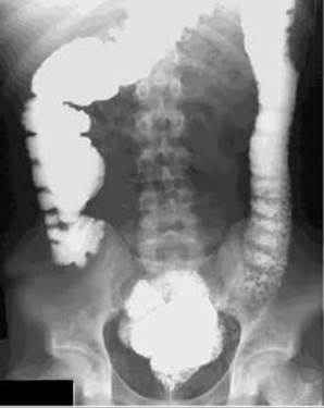
Fig. 194. Colonic polyposis (irrigography). Numerous rounded filling defects in the left half of the large intestine.
Colorectal Cancer. In Ukraine, among oncological diseases, colorectal cancer ranks third in incidence after lung and Stomach cancer. The global incidence of colorectal cancer has been rising recently, which can be attributed to the prolonged retention of fecal matter in the large intestine when the diet lacks sufficient plant fiber.
The majority of colorectal cancer patients are between 40 and 60 years of age.
Early detection of this condition is hampered by a prolonged asymptomatic course. Clinical manifestations depend on the tumor's Location and the stage of the disease. Characteristic signs include bloating, localized abdominal pain, melena, partial bowel obstruction, anemia, Skin pallor, fatigue, alternating constipation and diarrhea, and blood in the stool.
Colorectal cancer is most commonly classified into two main morphological forms: exophytic and endophytic. According to numerous observations, the endophytic form of cancer occurs more frequently in the left half of the large intestine, whereas the exophytic form predominates in the right half.
Radiological signs depend on the growth pattern of the neoplasm and the stage of the process. The following radiological signs of colorectal cancer can be identified during irrigoscopy: 1) a filling defect (marginal, central, circular); 2) an atypical mucosal relief; 3) wall rigidity; 4) an additional shadow against the background of the gas-distended intestinal lumen; 5) a superficial mucosal defect in the form of an irregularly shaped barium suspension pool with blurred, uneven contours; 6) a sharp demarcation of the tumor from healthy tissue – the "step-off" sign (Fig. 195); 7) intestinal deformity; 8) narrowing of the intestinal lumen, with significant narrowing revealing the "intestinal amputation" sign; 9) supra- or infrastenotic dilatation of the colon; 10) absence of haustration.

Fig. 195. Cancer of the sigmoid colon (irrigography). Narrowing of the bowel with uneven contours and undermined, stepped edges (arrow).
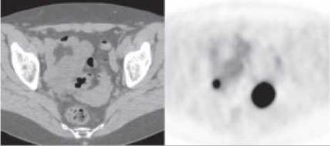
Fig. 196. Sigmoid colon cancer on CT and PET.
CT, MRI, ultrasound, and PET make it possible to establish reliable radiological signs of cancer, clarify the depth of tumor invasion into the colon and adjacent Organs, as well as assess the extent of the process and the state of regional Lymph Nodes (Fig. 196).
Last update: 08/08/2026
Editorial and Educational Adaptation: This material has been compiled based on the primary/original source text. The project team performed an editorial review, corrected technical inaccuracies, structured sections, and adapted the content for an educational format.
What was processed:
- elimination of formatting defects (OCR errors, structural breaks, corrupted characters);
- editorial organization of content;
- standardization of terminology in accordance with academic sources;
- verification of factual statements against the original source text.
All mentions of the author, publication year, and origin of the primary text have been preserved in accordance with the source.