Medical Radiology - Lazar A.P. 2008
Radiological Examination of the Digestive System
Emergency Diagnostic Radiology
A special role is played by Diagnostic imaging Methods in emergency conditions, such as "acute abdomen", intestinal obstruction, abdominal organ injuries, and the Diagnosis of intra-abdominal abscesses.
Acute intestinal obstruction. It is classified into mechanical and dynamic (functional) intestinal obstruction. Mechanical obstruction results from an organic narrowing of the intestinal lumen (tumor, adhesions, scar tissue, volvulus, intussusception). Dynamic obstruction occurs reflexively in various intra-abdominal conditions (appendicitis, pancreatitis, biliary colic, Peritonitis).
When an obstruction occurs in the intestinal lumen, defecation is delayed, triggering Fermentation and putrefaction processes. This leads to excessive gas production and severe irritation of the intestinal mucosa with significant fluid secretion. Proximal to the obstruction, the intestine becomes markedly distended, forming large gas-fluid levels with horizontal planes, the so-called "Kloiber's cups" (or gas-fluid levels), which are clearly visible on plain abdominal radiography or fluoroscopy (Fig. 197).
Class="center">
Fig. 197. Intestinal obstruction on a radiograph (diagram).
A - upright position reveals "Kloiber's cups";
B - supine position reveals the pneumorelief of the intestine.
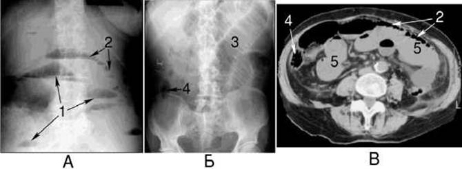
Fig. 198. Small bowel obstruction on radiographs in the upright (A) and supine (B) positions, and on a computed tomography scan (C).
1 - "Kloiber's cups"; 2 - "string of pearls" sign; 3 - gas-distended small bowel; 4 - unaffected large bowel; 5 - fluid-filled small bowel.
Intestinal obstruction caused by a malignant tumor typically develops in the late Stages of the disease. The obstruction progresses gradually, and only rarely presents suddenly and acutely due to secondary inflammation in the affected area or smooth Muscle spasm.
Direct radiographic signs of obstruction appear within 2–3 hours of onset, differing slightly between small and large bowel obstructions. Specifically, in small bowel obstruction, Kloiber's cups are predominantly located in the central abdomen, and the horizontal fluid level is wider than the height of the gas bubble above it. In contrast, in large bowel obstruction, they are located mainly in the flanks, and the width of the fluid level is less than the height of the gas bubble. Furthermore, obstructed small bowel loops often exhibit a sequence of small gas bubbles—the "string of pearls" sign (Fig. 198).
Based on the Location of Kloiber's cups, the radiologist can also infer the site of the obstruction, which lies just proximal to the lowest cup. However, to pinpoint a large bowel obstruction, a contrast enema is performed. The segments of the intestine distal to the obstruction appear normal.
To differentiate between mechanical and dynamic (paralytic) ileus, the patient is given a Glass of cold Water to drink; in mechanical obstruction, this causes an increase in the number of horizontal fluid levels.

Fig. 199. Inguinal hernia on a series of CT scans.
For rapid and precise diagnosis during the progression of acute intestinal obstruction, when severe obstruction is suspected and the surgeon considers emergency surgery, computed tomography (CT) is indicated. CT not only establishes the diagnosis of obstruction but also directly visualizes the site of bowel blockage. Oral contrast follow-through studies are contraindicated in this Setting due to the time required and low diagnostic yield, as the contrast gets severely diluted by intestinal contents and blocked by dilated bowel loops. Moreover, filling the bowel with barium sulfate prior to surgery is highly discouraged. CT has a higher Specificity than conventional radiography: the absence of a visible cause of obstruction on CT, such as a mass, makes an adhesive Etiology highly likely.
When CT is unavailable, abdominal radiography should be complemented by Ultrasonography (ultrasound). Ultrasound helps detect small bowel obstruction when radiographic signs are equivocal, demonstrating fluid-filled small bowel loops and mucosal edema. Unlike CT, ultrasound also provides functional information—such as the absence of peristalsis in paralytic ileus or bowel wall necrosis. However, ultrasound has low diagnostic value in large bowel obstruction due to marked gas distension.
One of the causes of intestinal obstruction can be the incarceration of a bowel loop within a hernia. The presence of bowel within a hernia sac is readily detected on CT (Fig. 199).
The most common cause of intestinal obstruction in children is intussusception, which predominantly occurs between the ages of 3 and 12 months. Types include ileocolic, colocolic, and enteroenteric intussusception, with the ileocecal region being the most frequent site. Contributing factors include intestinal tumors, a long mesentery, a mobile cecum, and dyspepsia. The onset is sudden. The child is restless, cries out in pain, vomits, and passes bloody stools. Later, passage of gas and feces ceases, leading to bowel distension and increasing abdominal wall rigidity that hinders Palpation.

Fig. 200. Perforated duodenal ulcer.
A - plain abdominal radiograph;
B - computed tomography scan.
Free air (1) and fluid (2) are visible within the peritoneal cavity.
To diagnose and treat intussusception, colonic air insufflation is employed, which frequently yields positive results. This Procedure begins with abdominal fluoroscopy or a plain abdominal radiograph. Occasionally, the gas pattern reveals an oval or rounded intussusception shadow. If this shadow is insufficiently defined, a contrast enema examination of the Large Intestine is performed by introducing air rectally using a Bobrov apparatus. Under fluoroscopic guidance, the colon is insufflated, and the air becomes trapped at the site of the intussusception, which appears as a dense, rounded shadow. In cases of small bowel intussusception, following colonic air insufflation, the child is given a radiopaque contrast agent to drink, and its progression toward the intussusception is monitored. Peritonitis may develop by the second day or later following the onset of the disease. In such cases, radiological examination reveals horizontal fluid levels within the Abdominal cavity.
Abdominal trauma and injuries to Internal Organs result from physical trauma and gunshot wounds. Transmural injury to a hollow viscus may also occur As a result of ulcer perforation. In most cases, patient survival depends on early detection and prompt surgical intervention.
Diagnostic imaging plays a pivotal role in identifying injuries by evaluating the patient in various positions, which reveals the characteristic changes, site of injury, and foreign bodies. The primary radiological and CT/ ultrasound signs of gastrointestinal tract injury include the presence of free air in the peritoneal cavity beneath the Diaphragm, fluid in the ABDOMINAL CAVITY AND pelvic space, and localized meteorism, which can be detected via radiography, computed tomography, and ultrasound (Fig. 200). The volume of gas depends on the size of the perforation.
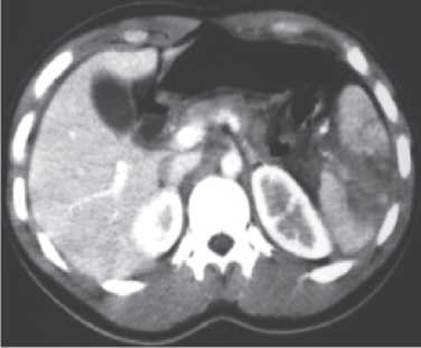
Fig. 201. Traumatic rupture of the Spleen on a contrast-enhanced computed tomography scan of the abdominal cavity.
Diaphragmatic injuries are radiologically manifested by elevation of the diaphragmatic dome and restricted mobility. Injuries to The Liver and spleen are more clearly visualized using CT (Fig. 201) and are frequently accompanied by rupture of a hollow gastrointestinal organ.
Intra-abdominal suppuration. The Development of intra-abdominal abscesses is typically caused by surgical interventions, ulcer perforation, gunshot wounds, and various other conditions requiring urgent surgical management. Intra-abdominal purulent collections are most frequently localized beneath the diaphragmatic dome, in the hepatic region, near the gastric stump, and within the lesser sac.
Diagnostic imaging occupies a prominent place in the evaluation of intra-abdominal suppuration, enabling the detection of both Direct and Indirect signs. Direct signs include the visualization of an abscess cavity containing gas and fluid, whereas indirect signs comprise morphological alterations and functional disorders of surrounding organs (displacement, and reactive Changes in the diaphragm and Pleura, among others). Abscess sizes vary and may reach 10 cm or more.
Abscesses are best visualized using CT and ultrasound. On CT and sonographic scans, an abscess appears as a rounded mass with irregular, indistinct margins and heterogeneous density (echogenicity), mimicking a tumor. Differential diagnosis is facilitated by identifying a thick, hyperdense (hyperechoic) abscess wall, a gas bubble at the upper pole of the abscess, and clinical signs of an acute inflammatory process. To confirm the diagnosis, CT-, ultrasound-, or MRI-guided needle biopsy of the abscess is performed (Fig. 202).

Fig. 202. CT-guided anterior abdominal wall percutaneous needle biopsy of a pelvic abscess.
Last update: 08/08/2026
Editorial and Educational Adaptation: This material has been compiled based on the primary/original source text. The project team performed an editorial review, corrected technical inaccuracies, structured sections, and adapted the content for an educational format.
What was processed:
- elimination of formatting defects (OCR errors, structural breaks, corrupted characters);
- editorial organization of content;
- standardization of terminology in accordance with academic sources;
- verification of factual statements against the original source text.
All mentions of the author, publication year, and origin of the primary text have been preserved in accordance with the source.