Medical Radiology - Lazar A.P. 2008
Radiological Examination of the Digestive System
Diseases of the Small Intestine
The Small Intestine consists of three parts: the duodenum, the jejunum, and the ileum. The length of the entire small intestine in a living person varies individually, ranging from 3 to 10 m.
The first and most distinct part of the small intestine is the duodenum. The length of the duodenum is about 25-30 cm. On radiographs, it has a horseshoe shape embracing the HEAD of the Pancreas. The duodenum comprises the superior, descending, horizontal (inferior), and ascending parts. Its superior part is expanded, bulb-shaped, and directed from the pylorus upward, backward, and to the right.
Class="center">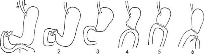
Fig. 185. Diagrams of Stomach and duodenal surgeries and their radiographic complications (diagram).
1 - gastroenterostomy; 2 - pyloroplasty; 3 - Billroth I gastric resection; 4 - Billroth II gastric resection with Finsterer modification; 5 - Billroth II gastric resection with Polya modification; 6 - peptic ulcer of the efferent loop.

Fig. 186. Stomach and small intestine on a barium sulfate contrast radiograph.
The proximal 2-3 cm of the duodenum, like The Stomach, are located intraperitoneally, while the remaining part is extraperitoneal.
Pathological Changes in the duodenum are better visualized during artificial hypotonia (relaxational duodenography). Hypotonia is induced by the parenteral administration of anticholinergic drugs (metacin, atropine, calcium gluconate, etc.) or the enteral administration of local anesthetics (novocaine, xylocaine, etc.). After 10-20 minutes, duodenel hypotonia develops and lasts for 15-25 minutes. At this time, the duodenum is filled with a barium suspension through a pre-inserted duodenal tube, and a radiograph is taken.
The jejunum and ileum are collectively referred to as the "mesenteric part" of the small intestine; the division between these segments is quite conventional, based primarily on their Location within the Abdominal cavity: the jejunum lies in the upper left quadrant, and the ileum in the lower right. The luminal width of the small intestine, as determined by imaging modalities, decreases distally from 2.5 cm to 3.0 cm. Circular folds (Kerckring folds), about 2 mm thick, occur at a frequency of 6-12 per 5 cm of length, producing a feathery mucosal relief pattern during contrast radiography (Fig. 186). The serration of the contours and the thickness of the folds decrease slightly in the distal small intestine. Under normal conditions, food mass transit through the small intestine takes 6-12 hours, depending on the COMPOSITION OF THE meal. On an empty stomach, the barium contrast mixture passes through the small intestine in 4-8 hours. To accelerate the transit of the contrast medium through the intestine, the patient is given cold Water to drink. As a result, the contrast mass reaches the ileocecal valve in just 30-40 minutes.
Duodenal ulcers are very similar to gastric ulcers. They are predominantly localized in the duodenal bulb (Fig. 187), and less frequently in the upper half of the descending part of the duodenum. In approximately 10% of cases, multiple ulcers are detected; about half of these are situated on the anterior wall and are difficult to visualize, necessitating a multi-projection examination using lateral and oblique views.
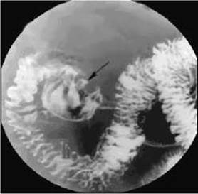
Fig. 187. Duodenal bulb ulcer. The arrow points to the ulcer niche.
The gold standard method for detecting duodenal and gastric ulcers is instrumental examination via fibrogastroduodenoscopy.
Duodenal diverticula are most commonly found in the middle third of the descending part of the intestine along its medial border. During CT and ultrasound examinations, such a diverticulum can be mistaken for a pancreatic cyst. Due to stasis within the diverticulum, inflammation—diverticulitis—may develop, presenting with clinical symptoms mimicking a peptic ulcer, cholecystitis, or pancreatitis. Duodenal diverticula are usually solitary. Multiple small bowel diverticula, or diverticulosis, are more frequently observed in elderly individuals. Diverticula of the jejunum and ileum are structurally identical to those of the duodenum.
On radiographs, CT, and MRI scans, diverticula appear as additional rounded cavities communicating with the intestinal lumen. Characteristic features include the presence of fluid and gas within the diverticular cavity, contour irregularity, and thickening of the mucosal folds surrounding the diverticulum.
Enteritis is an inflammatory condition of the small intestine that, in a chronic course, leads to mucosal atrophy. Patients complain of abdominal pain, gurgling, bloating, and weight loss. Radiographic examination reveals accelerated transit of intestinal contents—instead of the usual 6-8 hours, the contrast medium fills a significant portion of the small intestine within just 2 hours. Secretion is increased due to irritation of the inflamed mucosa.
During contrast radiography, the delicate feathery pattern disappears, and the mucosal folds of the small intestine become edematous and thickened, causing the entire wall to be hypomobile with altered tone: decreased in some areas and increased in others. Certain segments of the small intestine are moderately dilated, containing gas collections and horizontal fluid levels, while other segments are spastically shortened. In some places, the internal surface of the intestine appears smoothed and featureless, with uneven filling in separated, segmented areas (segmentation sign). Frequently, the contrast medium is scattered throughout the small intestine in the form of small flakes—the flocculation sign (Fig. 188).

Fig. 188. Enteritis on a radiograph.
1 - segmentation;
2 - flocculation.
Terminal ileitis (Crohn's disease), or granulomatous colitis, is most frequently localized in the small intestine. In 1936, Crohn reported that this disease affects the terminal segment of the ileum, hence naming it terminal ileitis. Subsequently, observations emerged describing similar lesions in other PARTS OF THE digestive tract, ranging from the Pharynx to the rectum.
Terminal ileitis is more common in men aged 25-40 years. The disease begins with involvement of The Lymphatic system, followed by the extension of the inflammatory process to all layers of the intestinal wall, leading to granulomatous inflammation. Numerous small ulcers appear in the mucosa. Over time, the scarring of these ulcers results in intestinal stenosis.
The radiographic presentation depends on the phase of the disease. Initially, thickening, flattening, and disappearance of the mucosal folds, along with numerous ulcers, are observed. Characteristic features include segmental involvement, unevenness of the intestinal lumen, mucosal edema, and a cobblestone-like mucosal relief pattern. Later, areas of marked ribbon-like narrowing of the small intestine, ranging from 1 to 20 cm in length, become apparent (Fig. 189), with drastically slowed peristalsis and evacuation.
Tuberculosis of the small intestine is most commonly localized in the ileocecal region and the distal ileum within 20–40 cm of the ileocecal valve, as well as in the duodenum. It generally occurs secondarily, in the presence of a primary specific lesion in the Lungs, Kidneys, bones, etc. Ulcerative, tumor-like, stenosing, and mixed forms of tuberculous involvement are distinguished. The affected segment is deformed and presents jagged, irregular contours. Inflammatory infiltration of the ileocecal valve is observed, and the ileocecal junction appears patulous. The folds are thickened, occasionally flattened, and exhibit polypoid elevations in certain areas, giving the mucosal pattern a mottled appearance.

Fig. 189. Terminal ileitis on a radiograph.
1 - stomach;
2 - transverse colon;
3 - jejunum;
4 - narrowed terminal portion of the ileum;
5 - cecum.
The intestine is rigid with limited mobility.
Malignant tumors of the small intestine are uncommon at any age. Clinical manifestations depend on the LOCATION OF THE process.
In the duodenum, malignant tumors may originate in the bowel wall itself or its papillae. The primary radiological diagnostic method is hypotonic duodenography.
Exophytic growth is characterized by wall rigidity, deformation of the mucosal relief, the presence of a filling defect, impaired evacuation from the suprastenotically dilated intestinal segment, tenderness upon Palpation, and limited mobility.
Endophytic growth presents with narrowing of the intestinal lumen accompanied by suprastenotic dilation, absent peristalsis, rigidity of the walls and folds, deformation of the mucosal relief, and asymmetrical narrowing of the small bowel lumen in a specific area.
A characteristic radiological sign of Cancer of the major duodenal papilla is a filling defect with clear margins and distinct demarcation from healthy tissue. As it grows, the tumor causes stenosis of the major duodenal papilla and involvement of the Bile duct. Radiological differentiation from carcinoma of the head of the pancreas invading the duodenum is impossible. Findings from fiberoptic esophagogastroduodenoscopy with biopsy, intraoperative biopsy, CT, and ultrasound should be taken into account.
Within the mesenteric part of the small intestine, tumors are more frequently localized in the proximal jejunum and the distal ileum. Exophytic growth is marked by prominent wall rigidity, atypical mucosal relief, and a filling defect. When the tumor invades the mesentery or retroperitoneum, radiographs reveal a fixed pattern of motionless small bowel loops.

Fig. 190. Barium enema (diagram)
A - dense filling with contrast medium;
B - after evacuation (emptying) of the contrast medium.
Benign and malignant tumors of the small bowel mesentery are rare and remain clinically silent for a long period. Radiographs reveal Displacement of the small intestine loops by the tumor, creating an area devoid of opacified bowel loops.
Currently, the Diagnosis of small intestinal tumors increasingly relies on findings from MSCT and ultrasound, which allow for the detection of the tumor and the identification of regional Lymph node hyperplasia.
Last update: 08/08/2026
Editorial and Educational Adaptation: This material has been compiled based on the primary/original source text. The project team performed an editorial review, corrected technical inaccuracies, structured sections, and adapted the content for an educational format.
What was processed:
- elimination of formatting defects (OCR errors, structural breaks, corrupted characters);
- editorial organization of content;
- standardization of terminology in accordance with academic sources;
- verification of factual statements against the original source text.
All mentions of the author, publication year, and origin of the primary text have been preserved in accordance with the source.