Medical Radiology - Lazar A.P. 2008
Radiation examination of the digestive system
Diseases of the stomach
On an X-ray image taken with the patient standing in the anteroposterior projection, The Stomach is positioned vertically to the left of the spine; only its outlet crosses the spine and partially extends to the right. In the supine position, the stomach shifts upward and assumes an oblique orientation. The size and position of the stomach largely depend on the individual's constitutional type and body build.
In most people, the stomach typically has a J-shape (fishhook shape); in tall, thin individuals of a dolichomorphic type, it is elongated like a stocking; and in short, stocky individuals of a brachymorphic type, it resembles a bull's horn, lying more horizontally and higher up (Fig. 172).
Class="center">
Fig. 172. Morphological types of stomach shape on an anteroposterior radiograph (diagram).
A - horn;
B - hook;
C - stocking;
D - cascading.
The stomach changes its shape and size depending on the degree of filling, wall tone, condition of surrounding Organs, patient's posture, and respiratory phase. An empty stomach does not Touch the anterior abdominal wall, with the transverse colon lying anterior to it. When fully distended, the stomach capacity increases up to 4 L, and it descends to the level of the navel or even lower (typically, the lower border of the greater curvature projects 3 cm above the navel).
Anatomically, the following PARTS OF THE stomach are distinguished: the fundus (or fornix, located beneath the left dome of the Diaphragm); the cardia (surrounding the cardiac orifice); the body of the stomach; and the pyloric part, the cavity of which consists of the pyloric antrum (antral section) and the pyloric canal. The pylorus is the narrowed terminal part of the stomach connecting it to the duodenum. In radiology, the subcardiac region (located below the level of the cardiac orifice) and the prepyloric region (situated 2-3 cm proximal to the pylorus) are also distinguished (Fig. 173).
Radiation imaging Methods reveal a certain amount of air in the stomach, which is swallowed by the person during eating and talking. In the upright position, this air forms a gas bubble located in the Cytology/practical/108.html">Fundus of the stomach. The gastric gas bubble is a permanent Structure that appears at the moment of birth. In forensic medicine, its presence is considered a sign of a live-born infant, whereas its absence in a patient is often indicative of cancerous involvement of the stomach.
In the lateral projection, the retrogastric space is visualized posterior to the stomach, which normally does not exceed the width of the lumbar vertebra (Fig. 174). It may increase with weight gain, in hypersthenic individuals, and in space-occupying lesions of the Pancreas.

Fig. 173. Parts of the stomach.
1 - fundus; 2 - cardia; 3 - subcardiac region; 4 - body of the stomach; 5 - angular notch; 6 - pyloric part; 7 - antral region; 8 - prepyloric region; 9 - pylorus.
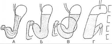
Fig. 174. Radiographs of a contrast-filled stomach in the standing position (diagram):
A - with a thin layer of contrast medium, allowing visualization of the mucosal relief; B - in the anteroposterior projection; C - in the right oblique projection with compression to study the angular notch; D - in the lateral projection.
The normal thickness of the gastric wall is 0.3-0.5 cm. In the body of the stomach, 4-5 longitudinal folds are visible, running parallel to the lesser curvature (Fig. 175). Oblique folds extending from the anterior to the posterior wall cause serration of the greater curvature. The highest and widest folds are found in the fundus and the lower part of the body. When the stomach is distended, the folds flatten and disappear, allowing the fine mucosal relief—gastric areas (areolae)—to be traced on X-rays as rounded elevations 2-3 mm in size. The gastric gas bubble is normally rounded, fluid and mucus are absent in the fasting state, and 2-3 peristaltic waves can be simultaneously observed along the gastric contour, starting from the middle of the body and fading out near the pylorus.
Functional Disorders of the stomach can occur in pathological processes affecting either the organ itself or other abdominal viscera. Functional disorders are classified into hypertonic (observed during the acute phase of Inflammatory Diseases), hypotonic (characteristic of the remission period of chronic diseases), and mixed types (manifesting during exacerbations of chronic inflammation).
Hypertonic functional disorders are characterized by the following radiological signs: gastric tone is increased, the stomach is positioned high, obliquely or horizontally, the gastric angle is increased, the sinus is smoothed out, the gas bubble is oriented horizontally, its size may be reduced, and the pyloric part may be rotated. In a hypertonic organ, due to its reduced size, the mucosal folds become higher and the sulci between them deeper, resulting in a more distinct Differentiation of the mucosal relief. Secretion is characterized by the presence of fluid and mucus in the stomach prior to the examination, with a significant increase in their volume during the Procedure. Spasms, whether total or local, are most intense in the outlet region of the stomach and the pylorus. A characteristic feature is a local spasm at the site of the pathological process, for example, at the ulcer site in PEPTIC ULCER DISEASE. Pronounced spasms can cause kinks in various parts of the stomach. Peristaltic waves start high, are deep and frequent, and segmenting waves may occur.

Fig. 175. Mucosal relief on a radiograph (diagram).
A - stomach; B - duodenum; C - Base of the duodenal bulb; D - Formation of the relief pattern;
1 - posterior wall; 2 - anterior wall; 3 - fold; 4 - sulcus.

Fig. 176. Chronic antral gastritis on a double-contrast radiogram (A) and computed tomography scan with oral and intravenous contrast enhancement (B). Thickened mucosal folds and stomach wall (arrow).
Hypotonic functional disorders are associated with decreased gastric tone. X-ray Examination reveals an enlarged stomach with an elongated body, an acute angle, a stretched lower body, and a pyloric region directed upward and positioned obliquely or vertically. Due to gastric distension, the mucosal folds appear flattened and stretched, while the interfold spaces are shallower, making the mucosal relief less distinct. Peristalsis is weakened, with a reduction in both the depth and propagation speed of peristaltic waves.
Mixed-type functional disorders are characterized by a gastric tone that depends on the patient's condition prior to the exacerbation (i.e., during remission), determining whether the stomach will be hyper-, normo-, or hypotonic. Gastric evacuation function may vary throughout a single examination, shifting from accelerated to delayed.
Chronic gastritis. Endoscopy plays a primary role in the Diagnosis of chronic gastritis. Radiographic diagnosis of Various Forms of chronic gastritis is based on the most typical morphological and functional features. The wall and folds of the stomach become thicker. Fold thickening is accompanied by an increase in their height and length (Fig. 176). Thickening of the folds leads to narrowing of the grooves between them. As the folds elongate, their course becomes more tortuous. Areae gastricae acquire an irregular shape and expand up to 5 mm or more. Further disease progression leads to the atrophy of folds and a weakening of the honeycomb pattern of the gastric areas (atrophic gastritis) or the appearance of multiple erosions (erosive gastritis).
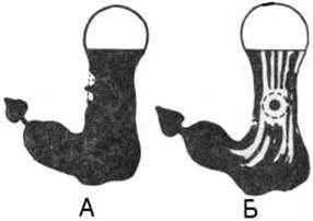
Fig. 177. Peptic ulcer niche on a gastric radiograph (diagram).
A – on the contour; B – on the relief.
Functional Changes in the stomach during chronic gastritis present as a hypertonic type, specifically: hypersecretion, presence of fasting fluid in the stomach, increased peristalsis, and elevated gastric tone, sometimes progressing to prolonged spasms, particularly in the antral region.
Peptic ulcer of the stomach. Gastric ulcers are predominantly localized along the lesser curvature, most frequently in the subcardiac region and the middle third of the gastric body. Both acute and chronic gastric ulcers are distinguished. An Acute gastric ulcer manifests as a rounded mucosal wall defect penetrating to varying depths. A direct radiological sign of an ulcer is a "niche" in the wall. When viewed in profile, the niche appears as a protrusion on the gastric contour (contour niche), whereas when viewed en face, it appears as a rounded accumulation of contrast medium surrounded by a radiolucent halo (relief niche) (Fig. 177).
The contour niche of an acute ulcer has a funnel-shaped appearance. Pronounced inflammation surrounds the ulcer, projecting above the mucosal surface as a ridge. The inflammatory process occasionally extends a considerable distance from the ulcer, manifesting as thickening and edema of the mucosal folds (ulcerative gastritis).
A Chronic Gastric Ulcer is characterized by The Development of Connective Tissue around the ulcer crater, which is why it is often referred to as a calloused or indurated ulcer. In chronic ulcers, inflammatory changes in the mucosa result in a characteristic deformation of the folds, manifesting as their radial convergence toward the niche (Fig. 178), which is a favorable prognostic sign as it is more frequently observed during ulcer healing. The contour niche of a chronic ulcer features a narrowed base and a flat or rounded apex (Fig. 179).

Fig. 178. Chronic ulcer on a contrast-enhanced gastric radiograph (A) and targeted radiograph (B).
1 – ulcer relief niche; 2 – thickened folds.

Fig. 179. Chronic ulcer along the lesser curvature on a targeted gastric radiograph (A). The arrow indicates the contour niche.
Indirect radiological signs of an ulcer include hypertonic-type functional disorders of the stomach. Regional spasm at the ulcer level causes an inward retraction of the gastric wall contour on the side opposite the ulcer—known as the "pointing finger" sign.
Complications of an ulcer may include penetration, perforation, malignant transformation, Hemorrhage, and pyloric stenosis.
When an ulcer penetrates into an adjacent organ, it is termed a penetrating ulcer. A penetrating ulcer is characterized by three distinct layers within its crater when the patient is in a vertical position: a layer of barium suspension, a layer of fluid above the suspension, and a layer of air above the fluid. Depending on the ulcer's Location in the gastric wall, one can infer which organ has been penetrated: when located on the lesser curvature, penetration most commonly occurs into the hepatogastric ligament; when located on the posterior wall, into the pancreas; and when located on the anterior wall, into the left lobe of the Liver.
The extension of an ulcer into the peritoneal cavity is termed perforation. Free gas in the Abdominal cavity occupies the highest position (under the diaphragm in the upright position; in the contralateral paracolic gutter when the patient is in lateral decubitus). Free gas is easily detected radiologically (as a radiolucent strip), as well as via CT, MRI, and ultrasound.
Malignant transformation of an ulcer occurs in 10–12% of chronic ulcer patients. The regular shape of the ulcer crater is disrupted, and its contours become indistinct and serrated. The inflammatory ridge transitions into unaffected wall tissue as a protrusion. Malignant degeneration of an ulcer cannot always be precisely determined; gastroscopy with biopsy helps clarify the diagnosis.
Hemorrhage from an ulcer is not a contraindication for a contrast X-ray examination, although it may be hindered during this period due to functional gastric spasms.
Pyloric stenosis develops As a result of scarring from a pyloric ulcer or due to tumor growth narrowing the pyloric channel. Compensated, subcompensated, and decompensated stages of stenosis are distinguished. In compensated stenosis, the stomach is slightly hypotonic, contains a small amount of fluid and mucus, evacuation occurs in small portions, and the stomach empties of the contrast medium within 24 hours. In subcompensated stenosis, the stomach is hypotonic, contains a large amount of fluid and mucus mixed with food residues, and up to 1/3 of the administered contrast volume remains in the stomach after 24 hours. In cases of decompensated stenosis, the stomach is hypotonic, significantly enlarged, contains abundant fluid, mucus, and food residues, and more than half of the contrast volume remains in the stomach after 24 hours.

Fig. 180. Gastric polyposis on a radiograph. Numerous filling defects are visible.
Gastric diverticula are frequently mistaken for ulcers. Gastric diverticula are true diverticula. In 3/4 of cases, they are located on the posteromedial wall in the subcardiac region of the stomach. Diverticulum shape is typically regular and rounded, with smooth, arched contours and mucosal folds that extend into them from the stomach wall.
Gastric tumors. Gastric tumors are classified as benign or malignant. Based on their histological structure, benign tumors are divided into epithelial and non-epithelial. Epithelial tumors include polyps, Papillomas, and adenomas, whereas non-epithelial tumors include leiomyomas, fibromas, lipomas, neuromas, vascular tumors, and others.
Epithelial tumors originate from the mucous membrane and consist of parenchyma and stroma. When parenchymal proliferation, specifically of the glandular apparatus, predominates, the tumor is classified as an adenoma. When stromal proliferation, i.e., connective tissue, predominates, the tumor is referred to as a papilloma or villous polyp. Epithelial tumors can be solitary or multiple, located in any part of the stomach, though most frequently in the antrum. Their diameter ranges from 2 mm to 2.5 cm. Polyps and adenomas are frequently asymptomatic. Radiologically, a solitary polyp presents as a rounded filling defect with clear, smooth contours. Polyps often feature a stalk, which may be short, long, wide, or narrow. When multiple polyps are present, numerous filling defects are observed (Fig. 180). Peristalsis and gastric emptying remain unimpaired.
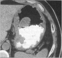
Fig. 181. Polyposis and Gastric Cancer on a CT scan. Irregular thickening of the gastric wall containing a contrast agent.
Non-epithelial tumors develop within the submucosal and muscular layers of the stomach. The radiological signs of non-epithelial tumors include a filling defect, restructuring of the mucosal relief, and an additional mass during double contrast imaging. The filling defect exhibits a regular shape and smooth, well-defined contours. The mucosal folds overlying non-epithelial tumors are flattened or displaced. In patients with small tumors, the shape, tone, and peristalsis of the stomach remain unaltered.
According to the majority of oncologists, all benign tumors of the gastrointestinal tract—especially large ones—are considered precancerous conditions due to their high propensity for malignant transformation.
Gastric cancer. Cancer is the most prevalent malignant disease of the stomach. It predominantly affects men aged 40–60 years. Gastric cancer is most commonly localized in the pyloric region. Clinical manifestations of gastric cancer most frequently include dull, aching pain unrelated to food intake, nausea, vomiting, and weight loss. Depending on the direction of growth, two main forms of gastric cancer are distinguished: exophytic and endophytic (Fig. 181).

Fig. 182. Radiological forms of gastric cancer (diagram).
1, 2 - diffuse-infiltrative form (localized wall rigidity);
3 - diffuse-infiltrative form (wall infiltration with straightening of the contour);
4 - ulcer-infiltrative form;
5 - exophytic form;
6 - narrowing of the body;
7 - narrowing of the antrum;
8 - pyloric stenosis;
9 - malignant ulcer of the angular notch;
10 - diffuse-infiltrative form smoothing out the angular notch.
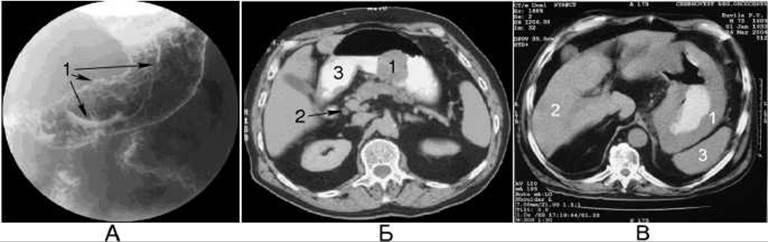
Fig. 183. Gastric cancer. A double-contrast gastric radiograph (A) reveals a filling defect (1). A computed tomography scan (B) demonstrates the exophytic form of cancer: 1 - tumor; 2 - Lymph node; 3 - contrast agent in the stomach. A computed tomography scan (C) demonstrates the endophytic form of cancer: 1 - tumor; 2 - liver; 3 - Spleen.
Exophytic cancer grows primarily into the gastric lumen and may present as polypoid or crater-like (cup-shaped). Polypoid tumors are more commonly found in the lesser curvature and the cardia. The cup-shaped form of cancer is characterized by elevated edges and central necrosis (ulceration), developing more frequently along the greater curvature and the gastric fundus. The radiological signs of the exophytic form of cancer include a filling defect with irregular contours within the contrasted stomach (Figs. 182, 183). On the surface of the cup-shaped cancer, directly in its center, lies a large ulcer, which appears as a pool of contrast medium. The mucosal folds terminate abruptly at the edge of the filling defect. Wall rigidity and the absence of peristalsis are best visualized when the tumor is located along the outlining contour.
The endophytic form of cancer grows primarily intramurally and can be ulcer-infiltrative or diffuse (flat-infiltrative, without ulceration). Ulcer-infiltrative cancer is most frequently localized in the prepyloric region and along the lesser curvature of the stomach. Diffuse-infiltrative cancer is primarily localized in the outlet region and may involve the entire stomach. The development of an endophytic cancerous tumor in the stomach is characterized by the following radiological signs: the appearance of a rigid area with straightened mucosal folds (atypical relief), abrupt termination of mucosal folds at the edge of the affected area, straightening of the organ contour and absence of peristalsis in the affected zone, and organ deformation. Circular spread of endophytic cancer is accompanied by narrowing of the gastric lumen.
For endophytic cancer, X-ray Computed Tomography is the optimal diagnostic imaging modality, enabling the detection of the tumor as asymmetrical, irregular thickening of the gastric wall with heterogeneous enhancement of densitometric density following intravenous contrast administration.

Fig. 184. Gastric cancer (endophytic form) on a sonogram.
1 - tumor; 2 - fluid in the stomach.
To determine the stage of gastric cancer, CT, MRI, and ultrasound are performed, which help assess the thickness of the stomach wall (Fig. 184), the degree of involvement of adjacent organs, enlargement of regional and retroperitoneal Lymph Nodes, and the presence of metastases.
Malignant tumors and gastric ulcers are often treated surgically. The effectiveness of the surgery, the condition of the operated stomach, and potential complications are evaluated radiologically (Fig. 185).
Last update: 08/08/2026
Editorial and Educational Adaptation: This material has been compiled based on the primary/original source text. The project team performed an editorial review, corrected technical inaccuracies, structured sections, and adapted the content for an educational format.
What was processed:
- elimination of formatting defects (OCR errors, structural breaks, corrupted characters);
- editorial organization of content;
- standardization of terminology in accordance with academic sources;
- verification of factual statements against the original source text.
All mentions of the author, publication year, and origin of the primary text have been preserved in accordance with the source.