Medical Radiology - Lazar A.P. 2008
Methods of Radiation Diagnostics
X-ray Computed Tomography
In 1963, the Journal of Applied Physics published an article by A. Cormack, a then-little-known physicist from South Africa, in which he proposed a mathematical method for reconstructing Brain images using a narrowly collimated X-ray beam. Only seven years later did this publication catch the attention of a team specializing in electronic music production, led by a then-unknown engineer, G. Hounsfield. In a short span, the scientists managed to develop a novel type of apparatus. Scanning the first object (a formalin-preserved brain) took 9 hours. However, as early as 1972, the first tomographic scan was performed on a woman with a brain tumor. The advantages of the new technique were so compelling that many major electronics companies shifted their focus to manufacturing these devices. They eventually came to be known as computed tomographers (CT, or CAT scanners). The first CT scanners were designed exclusively for HEAD examinations, but whole-body scanners soon followed. Today, CT can be used to visualize any part of the body (Fig. 9).
The core principle of X-ray computed tomography is the digital reconstruction of cross-sectional tomographic slices of The Human Body, obtained by computer Analysis of the attenuation degree of a narrow X-ray beam. Much like conventional radiography, the method relies on the property of Tissues differing in density and atomic weight to absorb X-ray radiation unequally.
Class="center">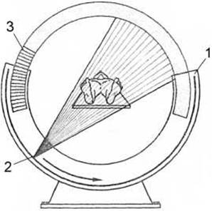
Fig. 10. Diagram of an X-ray computed tomograph.
1 - gantry frame; 2 - X-ray tube; 3 - detectors.
The X-ray tube, which emits a narrow X-ray beam via a system of slit collimators, rotates in a circle around the object, completing a full 3600 revolution (Fig. 10). A circular array of detectors mounted within the gantry frame converts the radiation energy into electrical signals, which are then mathematically processed and displayed visually on a monitor screen as a black-and-white image. This image can be saved digitally or printed onto photographic film.
Dense body areas that cause significant X-ray absorption appear bright on the computed tomogram, whereas low-density areas appear dark. The densitometric density of various human body tissues is measured in Hounsfield units (HU) (Fig. 11). The density of Water is defined as 0 HU, bone density as +1000 HU, and air density as -1000 HU. Most human body tissues have a densitometric density ranging from -100 to +200 HU. To better distinguish between adjacent anatomical structures with different densitometric densities, examinations are performed within a specific electronic window set by the operator on the computer, for example, from 0 to +200 HU. Reducing the width of the electronic window increases image contrast.
Computed tomography offers several advantages over conventional radiography: a) it features high sensitivity, allowing for the differentiation of individual Organs and tissues by density within 1%, whereas standard radiographs achieve only about 10%; b) unlike conventional tomography, CT provides a clear image of organs and pathological foci strictly within the plane of the scanned slice without the superimposition of overlying and underlying structures; c) it provides precise information on the size and density of individual organs, tissues, and pathological formations; d) it allows for the evaluation of the spatial relationship between a pathological process and surrounding organs and tissues.
Typically, computed tomographs acquire up to 10 horizontal slices (also referred to as axial slices) with a thickness of 5-10 mm, spaced 5-10 mm apart. Acquiring thinner slices (down to 1 mm) requires increasing the patient's radiation dose. Through computer reconstruction of the acquired slices, images can be generated not only in the horizontal plane but also in the coronal and sagittal planes, albeit with somewhat lower quality. These slices are used to study organ Structure, densitometric density, topographic Location, and their relationships to adjacent anatomical structures.
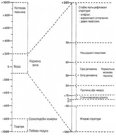
Fig. 11. Hounsfield absorption scale (at the head level).
In modern spiral computed tomographs, the simultaneous longitudinal movement of the patient table occurs during the circular Rotation of the X-ray tube within the gantry frame (Fig. 12). Consequently, the tube moves in a helical path around the patient's body, making it possible to acquire a series of slices of a specific body region quite rapidly—within 20-30 seconds—unlike conventional (sequential) tomographers, where acquiring a series of tomographic slices takes 10-15 minutes. The advent of spiral CT scanners has enabled three-dimensional (3D) organ volume rendering, computed angiography, and virtual endoscopy.
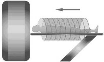
Fig. 12. Operating principle of a spiral computed tomograph.
Intravenous contrast enhancement with iodine-based compounds improves the visualization of individual organs and pathologically altered tissues during contrast-enhanced computed tomography. Oral contrast administration during abdominal CT is used to differentiate various segments of the gastrointestinal tract from adjacent structures.
CT guidance is utilized to perform punctures and targeted biopsies of various organs and pathological lesions. CT plays an extraordinarily vital role in monitoring both conservative and surgical patient Treatment. Furthermore, CT is an invaluable method for the precise localization of tumor neoplasms and for targeting the ionizing radiation source to the lesion during radiotherapy planning for malignant tumors.
Last update: 08/08/2026
Editorial and Educational Adaptation: This material has been compiled based on the primary/original source text. The project team performed an editorial review, corrected technical inaccuracies, structured sections, and adapted the content for an educational format.
What was processed:
- elimination of formatting defects (OCR errors, structural breaks, corrupted characters);
- editorial organization of content;
- standardization of terminology in accordance with academic sources;
- verification of factual statements against the original source text.
All mentions of the author, publication year, and origin of the primary text have been preserved in accordance with the source.