Antibiotics (Properties, Application, Interactions) - M.P. Cherenko 1999
Fundamentals of Transplantology. Plastic and Reconstructive Surgery
Organ and tissue TRANSPLANTATION
Plastic and reconstructive surgery aims to restore the integrity of human body parts affected by acquired or congenital defects, as well as to recover the anatomical form and function of a damaged or lost organ.
Plastic and reconstructive surgery has a long history. It is well established that certain tissue transplantation Procedures were performed in antiquity. Historical documents indicate that as early as 8000 BC, ancient Egyptians utilized plastic surgery techniques to reconstruct nasal tissues.
The Book of Life by the prominent Indian physician Sushruta, written around 1000 BC, describes rhinoplasty using Skin flaps from the Cheeks and forehead. Ancient Indians also performed free skin grafting from the buttocks. This art was practiced by a lower caste of priests or potters, as well as executioners who severed noses.
In 1450, the Sicilian physician Branca and his son Antonio developed a pedicle flap technique for nasal reconstruction, harvesting skin from the upper arm. This method became known as the Italian method.
Plastic surgery achieved major Milestones in the 19th century, associated with the names of Ukrainian surgeons (Y. Shimanovsky, V. Karavaiev), Russian surgeons (N. Pirogov, E. Pelikan), German surgeons (Graefe, Dieffenbach), French surgeons (Lisfranc), and English surgeons (Lane).
In 1865, in his book Operations on the Surface of The Human Body, Y. Shimanovsky summarized the plastic surgery techniques existing at the time and provided a detailed Overview of local tissue plastics and pedicled skin flaps.
On December 9, 1835, at the St. Petersburg Academy of Sciences, N. Pirogov delivered a lecture entitled "On Plastic Operations in General and Rhinoplasty in Particular," in which he thoroughly analyzed transplantation and outlined its future development. With this work, N. Pirogov essentially laid the foundations for a new branch of medicine: transplantology.
In the 1860s, new transplantation Methods were introduced: free bone grafting (the French physician Ollier) and free skin grafting (the French physician Jacques Reverdin).
By the late 19th century, surgeons began transitioning from tissue transplantation to organ transplantation.
A major breakthrough in The Development of transplantology was the discovery by the French surgeon Carrel, who developed an effective technique for vascular anastomosis. This ensured that the transplanted organ received an adequate supply of Blood and nutrients at the new site.
In the late 1930s, surgeons performed clinical transplants of only certain tissues (bone, skin, cornea), while organ transplantations were restricted to animal experiments. Skeptics grew increasingly vocal, questioning the very feasibility of organ transplantation.
It was then that a historic event for transplantology took place. On April 3, 1933, the young Ukrainian surgeon Yuriy Voronoy performed the world's first human Kidney transplant. The case is described as follows.
“...On the evening of March 31, 1933, a 24-year-old woman was admitted to the hospital in a state of severe poisoning. Earlier that day, she had ingested 4 g of corrosive sublimate in an attempted suicide. The patient was in a semicomatose state, interrupted by cramping abdominal and lumbar pain. Despite intensive therapeutic measures, her condition did not improve, and the symptoms of intoxication progressed.
At that point, Dr. Yu. Voronoy decided to take a chance that medicine had not yet explored: he dared to perform a kidney transplant from a deceased donor. The donor was a 60-year-old man admitted to the hospital with a Skull base fracture, who had died in the emergency department. Yu. Voronoy transplanted the kidney not to its anatomical site, but to the thigh. Such a Procedure is known as a heterotopic transplant, as opposed to an orthotopic transplant, where an organ is placed in its natural Location. However, the patient's body was unable to overcome the severe, fatal poisoning, and she survived for 48 hours with the transplanted kidney...”.
Much time has passed since then. In December 1971, a team of scientists led by Academician B. Petrovsky successfully performed the first kidney transplant in the USSR.
Organ and tissue transplantation is a major challenge of modern medicine. Just 50 years ago, it seemed fantastic, unfeasible, unrealistic, and far from practical Structure/175.html">Implementation. Remarkable progress has been made over recent decades. Today, transplantation is performed by numerous medical centers worldwide: 118 centers in the USA, 61 in Europe, and 23 in other countries.
Specialized surgical departments, institutes, and hospitals dedicated entirely to plastic surgery have emerged globally. Plastic and reconstructive surgery has evolved into an independent branch of clinical medicine.
Today, plastic surgery is a broad specialty not restricted to any single anatomical region, organ, or system; it is applied across virtually all surgical fields.
Surgery is termed “plastic” because it is closely linked to the artistic restoration of missing or deformed organ shapes and Symmetry. This is achieved through delicate surgical interventions. The term “plastic surgery” emphasizes the specific nature of these procedures, as most reconstructive operations rely on plastic techniques involving tissue transfer.
Plastic surgery also encompasses cosmetic procedures aimed at correcting congenital or acquired deformities of the Nose, Lips, or ears, as well as eliminating premature wrinkles.
What Organs and tissues are transplanted? Today, there is virtually no tissue, organ, or even body region that surgeons have not attempted to transplant in clinical practice or experimental settings.
Skin is the oldest and most traditional transplantation material. Today, it is widely used in surgical practice to repair facial defects, treat chronic non-healing ulcers, manage deep Burns, and cover extensive traumatic defects across various body parts.
Mucous membranes are used to reconstruct the lining of the Oral Cavity and eyes, and to form the lips.
Fascial sheets are used to close aponeurotic defects, lengthen tendons, reinforce the anal sphincter and the anterior abdominal wall, and achieve hemostasis in cases of hepatic Hemorrhage.
Muscle tissue is used to close large muscular defects following trauma or surgery, to pack parenchymal organs in the event of bleeding, and to fill bone cavities after sequestrectomy. It is commonly employed in surgeries for hernias and rectal prolapse. Pedicled muscle flaps are typically used.
Adipose tissue is more frequently utilized for cosmetic purposes, as well as to fill bone cavities following sequestrectomy, or to fill defects resulting from the removal of large scars in Brain tissue or angiomas.
The greater omentum is an excellent biological material. Composed of abundant adipose and mesenchymal tissues, it can be spread in a thin layer over a considerable surface area. Unlike other organs consisting primarily of adipose and Connective Tissues (such as skin and subcutaneous tissue), the omentum is well-vascularized and readily adheres to other tissues, which subsequently become vascularized as well. Furthermore, the omentum possesses unique immunological properties that render it resistant to infectious agents. Today, the omentum is used as a plastic material for the management of radiation Necrosis of the chest wall, breast reconstruction, hand reconstructive surgery, closure of parietal and visceral peritoneal defects, and reinforcement of intestinal or gastric sutures. In cases of hemorrhage from parenchymal organs (such as the Liver or Spleen), wounds are packed with the omentum.
Vascular tissue became widely used in plastic surgery following the Introduction of the vascular suture into surgical practice. Vascular grafting is employed to replace Blood Vessels after trauma, cicatricial stenoses, or obliterative processes. Grafts may be harvested from the patient's own vessels, from another human, or constructed from synthetic prostheses.
Nervous Tissue. The replacement of a peripheral nerve defect by grafting a segment harvested from another site has not yielded satisfactory outcomes. Consequently, most surgeons prefer direct end-to-end nerve suturing. When this is not feasible, grafting is performed using a fresh or preserved free graft harvested from a cadaver.
Cartilage tissue is avascular, successfully engrafts in the recipient, and is widely applied in cosmetic and plastic surgery to correct defects of the nasal cartilage, eyelids, auricles, and other structures. Rib cartilage most commonly serves as the graft. It possesses spring-like properties and remains under tension. Cartilage is not an inert biological implant material; it is a living tissue nourished by tissue fluid. Therefore, like any other tissue, following transplantation it must remain in close contact with adjacent tissues, otherwise it will perish. At the same time, cartilage is non-demanding, exhibits high resistance to infection, and retains its characteristic initial shape post-transplantation.
In addition to the patient's own cartilage, cadaveric cartilage can be utilized.
For free bone transplantation, either the patient's own bone (such as the Fibula or rib) or cadaveric bone is used.
Free Bone tissue transplantation is performed to eliminate defects resulting from trauma, surgical intervention, inflammatory processes, or Congenital Malformations (such as the closure of congenital clefts of the palate and jaw), as well as in cases of restricted mobility of joints or the spinal Column, and for the reconstruction of the facial Skeleton and extremities.
Autogenous cancellous bone (from the rib or iliac crest) is considered the optimal transplantation material. Compared to alloplastic Materials, it demonstrates greater Resistance to Infectious agents.
Bone may be transplanted into a bone defect in the form of fragmented chips or as a whole free graft block.
Hair transplantation is used to correct defects of the hair-bearing skin, such as scars and localized areas of alopecia resulting from various diseases. For this purpose, the affected areas are excised wholly or partially, and the hair-bearing skin edges are approximated if the size of the defect and the laxity of the surrounding skin permit.
To avoid damaging the hair follicles, skin flaps must be harvested encompassing the entire layer of subcutaneous tissue down to the aponeurosis. When closing the defect with adjacent skin areas is not possible, pedicled flaps are utilized. Several types of pedicled flaps are distinguished.
In 1959, Orentreich proposed an original method for hair transplantation. Under local anesthesia, individual skin grafts together with the subcutaneous tissue are transplanted using a special instrument. The diameter of the instrument ranges from 1.5 to 12 mm. It is preferable to have two instruments: one to harvest the donor grafts, and the other to excise skin plugs of the exact same size within the alopecic area. The circular graft is then inserted like a plug into a prepared socket.
Another hair transplantation method used for treating alopecia involves employing THE PRINCIPLE OF optical illusion (visual deception). Skin grafts are transplanted in a narrow strip along the natural anterior hairline. Once the hair regrows—which requires 2 to 6 months—it is combed backward to conceal the bald area.
Corneal transplantation. In cases of corneal opacification, keratoplasty yields favorable results. This is attributed to The small size of the graft and the absence of blood vessels and lymphocytes within it.
Bone Marrow transplantation. Bone marrow plays a vital role in hematopoiesis, serving as the site where Blood Cells mature. Disruption of hematopoietic processes in the bone marrow can be congenital or acquired (resulting from X-ray or radioactive irradiation, exposure to chemical agents, as well as immunodeficiency or aplastic anemia).
Bone marrow is most frequently harvested from the iliac crests, filtered, and administered intravenously.
Transplanted bone marrow cells restore their function only after 3 weeks. The leukopenia and thrombocytopenia observed during this period render the patient's Organism highly susceptible to infection, thereby necessitating The Use of special protective measures. Initially, the transplanted cells accumulate in "intermediate sites" (such as the Lungs and spleen), and after some time, they migrate to the bone marrow within the cavities of tubular bones, the Sternum, and the flat BONES OF THE pelvis.
Following bone marrow transplantation, complications such as skin rashes, gastrointestinal disturbances (diarrhea, abdominal pain), and impaired liver function (elevated liver Enzymes) may occur. This is attributed to the reaction of donor cells with immunocompetent host bone marrow cells (graft-versus-host disease).
Endocrine glands are utilized as transplant materials in reconstructive surgery, particularly following the introduction of vascular suture and microsurgical techniques into clinical practice. Male and female Gonads, the thyroid and Parathyroid glands, the Pituitary Gland, and the Adrenal Glands are transplanted.
When the number of kidney transplantations worldwide exceeded 40,000, it was decided to discontinue the registry of such operations. Several thousand procedures have been performed in the former USSR alone. In the USA, 9,000 kidney transplants are carried out annually. This fact indicates that renal transplantation is no longer a unique procedure and is performed in numerous specialized clinics as a routine surgery.
Heterotopic transplantation is considered the most appropriate approach, wherein the kidney is placed in the right iliac fossa. The Ureter is anastomosed to the Urinary Bladder, and the vessels are connected to the Vessels of the iliac region. Isotransplantation, i.e., kidney transplantation between identical twins, is the most promising. Grafts may also be procured from close relatives (mortality from unilateral nephrectomy is less than 0.1%) or from cadaveric Donors.
When the warm ischemia time (the interval between the cessation of Renal Circulation and cooling) exceeds 45 minutes, the renal artery can be connected to a perfusion machine to perform hypothermic perfusion with a colloidal solution. Restoration of renal function is possible after a maximum of 1 hour of "warm ischemia" (the period from donor cardiac arrest to the onset of "cold perfusion") and 4 days of "cold ischemia" (from THE START OF "cold perfusion" to the vascular anastomosis of the transplanted kidney to the recipient). For early graft function onset (within 3 days post-transplantation), the duration of the warm ischemia period should not exceed 40 minutes, and cold ischemia should not exceed 8 hours.
Kidney transplantation may be accompanied by various complications, specifically: bacterial, viral, and fungal infections; cardiovascular and cerebrovascular complications; Pulmonary Embolism; gastrointestinal disorders (peptic ulcers, hemorrhage); Diabetes Mellitus; Osteoporosis (associated with prolonged steroid therapy); and hypercalcemia resulting from renal failure. A formidable complication of kidney transplantation is graft rejection.
The first human Heart transplant was performed in 1964 by American surgeon James Hardy. He transplanted a chimpanzee heart into a patient, who died an hour after the surgery. In 1967, South African surgeon Christiaan Barnard performed the world's first human-to-human heart transplant in Cape Town, using The Heart of a 25-year-old woman who had died in a car crash. Barnard's operation paved the way for a wave of heart transplantations worldwide.
By 1985, more than 500 human heart transplants had been performed globally. By 1991, about 50 heart transplants had been carried out in the USSR. Today, approximately 2,000 heart transplants are performed annually in the United States.
Currently, heart transplantation is performed in 118 centers in the United States, 61 centers in Europe, and 23 centers in other countries.
The maximum recorded lifespan for a person with a transplanted heart is 12 years, with an average of 8.5 years. Each year, 5% of heart transplant recipients pass away. Experimental attempts to transplant pig hearts into humans are also being explored.
The heart is implanted orthotopically after removing the recipient's own heart. During this procedure, a sort of cuff is left from the right and left atrial walls, to which the donor heart is anastomosed. The time elapsed between harvesting the donor heart and transplanting it into the recipient should not exceed 3—4 hours. Internationally, laws permit harvesting organs from brain-dead patients who are on mechanical ventilators.
Scientists are actively working on developing a pneumatically driven artificial heart prosthesis. It can be used for temporary cardiac support in emergency situations while awaiting a donor organ. Such research is currently underway in the USA, England, and Russia.
Liver transplantation is one of the most complex surgical procedures. This is due to the organ's anatomical and topographical location, its unique blood supply, and its high sensitivity to oxygen deprivation. Furthermore, removing the liver from the systemic circulation triggers severe physiological disorders in the human body. That said, The liver possesses a remarkable regenerative capacity, meaning even small fragments can sustain adequate bodily function.
There are two primary methods of liver transplantation: orthotopic (where the graft is placed in the anatomical site of the removed liver) and heterotopic (where the organ is placed in another area of the Abdominal cavity while leaving the patient's native liver intact). The orthotopic approach is currently preferred. In Ukraine, the first liver transplantation was performed in 1994 by O.Ye. Nikonenko.
The first experimental lung transplant was performed in 1907 by Alexis Carrel, while the first human lung transplantation was carried out in 1963 by James Hardy. The patient survived for 18 days and ultimately died from renal complications, though the transplanted lung functioned normally until death.
Lung transplants were performed predominantly during the late 1960s and the 1970s. By 1988, roughly 50 such procedures had been conducted. The longest survival time following a single-lung transplant was 10 months; the patient died after hospital discharge due to graft rejection. Lung transplant operations performed between 1986 and 1987 yielded better outcomes, with patients surviving 10, 17, and 34 months, and some returning to normal life.
Human lung transplantation is not as effective as kidney or heart transplantation. This is due to the lungs' high vulnerability, necrotic Changes in the bronchial mucosa caused by interrupted blood supply, suture line leakage, and postoperative pulmonary infections.
Autotransplantation presents a more promising alternative, where a single lobe from the healthy contralateral lung is transplanted to replace a resected half-lung.
An alternative to single-lung transplantation is combined heart-lung transplantation. By 1988, 150 such procedures had been performed worldwide. They are more physiologically favorable than isolated lung transplants.
A unique triple-organ transplant involving the heart, lungs, and liver was successfully performed in Paris by Professor Alain Carpentier and his team. The simultaneous transplantation was performed on a 16-year-old girl who suffered from pulmonary failure, severe liver disease, and congenital bronchial mucosal deficiency.
The young girl had waited a full year for a donor, who turned out to be a young woman tragically killed in a car accident.
The surgery lasted over 12 hours and was a success.
Pancreas transplantation is still in the developmental and testing phase. Two main techniques are used. The first involves implanting isolated islets and tiny pancreatic fragments into the liver or spleen, although the therapeutic effect of this approach is short-lived. The second method involves the transplantation of freely vascularized pancreatic segments (the body and tail) or the entire organ. These are most commonly transplanted into the iliac fossa. The Vascular System is connected to the iliac vessels, while exocrine secretions are diverted into an intestinal loop or the Urinary Tract. However, this procedure offers limited long-term efficacy and carries significant surgical risks.
Brain tissue transplantation. One of the most fascinating areas today is the transplantation of embryonic human brain tissue into the brain of a rabbit. Due to its high genetic activity, the implanted embryonic tissue enhances the animal's brain Functions. For instance, the Electroencephalogram of a recipient rabbit shows significantly higher amplitude and frequency ranges in the graft area compared to unoperated regions. Olfactory responses are also enhanced: experimental animals react to odors sooner and retain them in memory longer than control subjects. Experimental data indicate that a new network of neural connections forms in the rabbit's brain, helping it respond to its environment in novel ways.
Transplanting entire body parts (such as the HEAD or limbs) was long considered science fiction. Today, however, this concept has a solid scientific foundation. Such experiments—albeit using an isolated dog head—were even demonstrated at the 2nd All-Russian Congress of Pathologists in Moscow. For 1 hour and 40 minutes, a dog's head resting on a tray tracked movements with its eyes, moved its ears, and bared its Teeth. Life was sustained in the head using an artificial heart machine invented by scientist S. Bryukhonenko.
Head transplantation experiments in dogs were also conducted by V. Demikhov. The head of one dog was grafted onto the body of another. This two-headed animal survived for over a month.
In May 1989, American surgeons successfully reattached the head of a 10-year-old boy. Following an injury, the head was hanging almost exclusively by Muscles and tendons. The surgery lasted 5 hours. Afterward, the boy felt satisfactory, regained movement in his right limbs, and eventually began showing movement on the left side as well.
Regarding limb transplantation, there are now numerous reports of successful human limb procedures worldwide. Strictly speaking, these are autotransplantations, or replantations—meaning the reattachment of severed body parts to their original sites. This has become possible thanks to advances in microsurgical techniques. The patient, along with the severed organ (fingers, hand, FOOT, lower leg, forearm, or arm), is rushed to a specialized medical center. The detached part is wrapped in sterile material, placed in a plastic bag, and stored at a Temperature of 4 °C. The replantation procedure involves sequentially stabilizing bone fragments, restoring arterial and venous patency, suturing tendons, muscles, nerves, and skin, and subsequently immobilizing the limb.
In certain cases, it becomes necessary to substitute one digit for another; this is most commonly done by pollicization—replacing a thumb lost to trauma with the fourth finger.
Modern replantation surgery distinguishes between two main concepts: microreplantation and macroreplantation. Microreplantation involves the reattachment of limb parts amputated distally to the wrist or ankle joints, whereas macroreplantation involves parts amputated proximally to these joints. Both types of surgical interventions differ significantly in their technique, the condition of the amputated part and the patient, and the duration of anoxemia. For instance, the period of permissible anoxemia is much shorter in macroreplantation (maximum 6 hours for an upper limb) than in microreplantation (up to 22 hours for a finger under cooling conditions). While microsurgical technique is paramount in microreplantation, a thorough knowledge of anatomy, surgical tactics, and vascular and nerve repair is critical for macroreplantation.
Equally important is the question of where to source the graft material for transplantation. Consequently, transplants are categorized into several distinct types.
Autologous transplantation involves taking a graft from one part of the patient's body and transplanting it to another (such as skin, bone, cartilage, part of a lung, or fingers).
Isogenic transplantation uses a graft from a donor who is genetically identical to the recipient (from monozygotic twins).
Syngeneic transplantation involves a graft obtained from first-degree relatives.
Allogeneic transplantation is The transfer of tissues and organs within the same species (from one human to another).
Xenogeneic transplantation involves transferring tissues and organs from one species to another (e.g., from a monkey to a human).
Alloplastic transplantation is the replacement of body parts with synthetic materials (metals, plastics). Synthetic materials are used to manufacture vascular grafts, heart Valves, artificial joints, and the like.
Depending on the type of plasty, the following are distinguished: 1) the transplantation of tissues and organs from one part of the body to another or from a donor to a recipient; 2) replantation, when damaged organs and tissues are transplanted back to their original site (e.g., a severed limb or scalped skin); 3) implantation, where cells or tissues are purposefully placed into a different site (such as the implantation of isolated pancreatic islets into the liver or spleen).
Today, allogeneic transplantation—meaning the transfer of a graft from another human—is the most common. But from whom? Only certain tissues and organs can be harvested from a living person without causing significant harm (such as skin or a kidney). How can this challenge be overcome? The necessary organ is harvested from a deceased individual, most often someone who died in an accident or from another trauma. However, this raises a series of organizational, legal, and ethical issues that must be addressed. When can a person be considered dead and impossible to resuscitate? When is it appropriate to harvest an organ from the deceased? Is the consent of relatives required?
To resolve these issues, A number of guidelines and regulations have been developed to govern the aforementioned provisions.
Tissues and organs from a deceased donor are harvested under strict aseptic and antiseptic conditions within the first 6 hours following the declaration of brain death. They are preserved using various methods: rapid freezing at a temperature of —196 °C and storage at temperatures ranging from -25 °C to —30 °C; in antiseptic or antibiotic solutions with storage in chilled solutions, recipient plasma, or blood; in paraffin, or aldehyde solutions (formaldehyde, glutaraldehyde); and via "cold perfusion" (perfusion of the organ's vessels with a colloidal solution at 8 °C).
The more complex the organ, the shorter the duration it can be preserved without a blood supply. This period is referred to as "total ischemia," which consists of periods of "warm" and "cold ischemia."
Addressing various medical and organizational challenges during organ transplantation from deceased donors remains difficult today. Consequently, specialized multidisciplinary teams are formed, bringing together professionals from different fields (transplant surgeons, intensivists, morphologists, biochemists, immunologists, etc.). These teams may include personnel from institutions within the same city (such as Kyiv, Moscow, or St. Petersburg) or from transplantation centers across different cities. For instance, organs harvested in Kyiv may be sent to other cities, while grafts from elsewhere are brought in for residents of Kyiv.
Interstate associations have been established in many countries, such as Eurotransplant (Belgium, the Netherlands, Austria, Switzerland, Germany), Scandiatransplant (Denmark, Sweden, Norway, Finland), and others.
Reconstructive and restorative surgery is associated with a number of critical challenges. The first of these is ensuring strict asepsis during and after the operation. Infection Prevention in reconstructive surgery is achieved through the use of Antibiotics, Hyperbaric Oxygenation (HBO) therapy, meticulous intraoperative hemostasis with the coagulation of all minor bleeding vessels, adequate replacement of blood loss, and the application of ultrasound and laser beams.
The second challenge is the technical provision of the surgery. Special requirements are placed on surgical instruments here, and it is desirable to replace standard instruments with miniature ones, akin to those used in ophthalmology.
One of the most important issues in reconstructive surgery is the suture. The invention and subsequent improvement of stapling devices have undoubtedly been major achievements of recent decades. However, despite the positive qualities of mechanical sutures, manual suturing with atraumatic needles remains the mainstay in surgical practice.
In recent decades, plastics—products of the polymerization or Condensation of various high-molecular-weight Organic compounds—have come into widespread use in reconstructive surgery. Thanks to their durability, ease of Processing, tissue inertness, and ability to retain their shape, they have found broad application across many surgical fields. These include metallic allografts, synthetic polymers (polyethylene, polyvinyl chloride, kapron, dacron, diolen, silicone), and ceramics. They are used to replace segments of blood vessels, reinforce the abdominal wall in large hernias, and reconstruct the nose and auricles. Porous elastic materials are utilized for cosmetic surgery needs. Alloplastic materials must be non-reactive with body tissues, free from harmful substances, chemical impurities, and unreacted monomers.
A drawback of alloplastic materials is the reduced resistance of tissues in the implantation area, particularly to infection. The development of an infection quickly leads to graft rejection.
Among the Problems associated with reconstructive surgery, tissue incompatibility remains The most significant today. After all, there are no two people in the world whose tissues are identical in Cell/13.html">Protein Structure. If the tissues of one person enter the body of another, the latter attempts to isolate and destroy them. An example of this is the sloughing of ligatures and sutures, a process that occurs more rapidly when an infection sets in, especially in the presence of virulent microbes. Hence, the critical role of asepsis and antisepsis becomes clear. Tissue incompatibility is linked to the presence of Antibodies in the body. The Introduction of a foreign protein—an antigen (such as a microbe or tissue)—triggers The production of corresponding antibodies. These antibodies are produced within lymphocytes. The transplanted tissue is immediately surrounded by clusters of lymphocytes that destroy and neutralize it. As a result, the functional relationship between the transplanted organ and the recipient's body is progressively disrupted, leading to immunological rejection.
This reaction is especially pronounced when transplantation is performed between different animal species. For instance, rabbit skin transplanted onto a monkey is rapidly rejected, whereas skin from one monkey transplanted onto another takes much longer to be rejected. The same phenomenon is observed in humans. Admittedly, even among humans, close relatives share a higher degree of antigenic commonality. The prospect of successful graft survival increases with the number of compatible Antigens shared between the donor and recipient. Finding "compatible" donor and recipient antigens remains a complex task.
The rejection reaction can be hyperacute, acute, or chronic.
Hyperacute rejection begins almost immediately after surgery (within 24–48 hours). The function of the graft ceases; it swells and becomes soft. General signs of rejection appear, including elevated body temperature, leukocytosis, and thrombocytopenia.
Acute rejection occurs within the first month post-transplantation. The patient's condition deteriorates, and flu-like symptoms appear. In such cases, Steroids, antilymphocyte globulin, or Monoclonal Antibodies are administered parenterally.
Chronic rejection develops later and is characterized by a gradual decline in the patient's general condition and a progressive loss of function in the transplanted organ.
Combating tissue incompatibility involves suppressing transplantation Immunity through various methods, which can be either non-specific or specific. To achieve high efficacy, immunosuppression must begin in the preoperative period. For immunosuppressive purposes, X-ray irradiation, pharmacological agents (azathioprine, Imuran, prednisolone, urbason), and biological agents (antilymphocyte serum, antilymphocyte globulin) are prescribed.
Azathioprine inhibits lymphocyte proliferation and suppresses both cellular and humoral immunity. Its dosage is adjusted to maintain the leukocyte count at the level of 8×10⁹/L.
Prednisolone inhibits immunoglobulin synthesis and reduces The impact of antigen-antibody complexes on the enzymatic systems of the transplanted organ. Treatment is initiated at 200 mg per day, with the dosage gradually tapered.
The aforementioned measures do not always yield the desired effect. Low doses are often ineffective, whereas high doses may provoke various adverse reactions. Standard pharmacological action increases the risk of oncological diseases. A secondary risk involves the suppression of nonspecific immunity, wherein even a minor infection can trigger severe complications. The use of steroid drugs for immunosuppression may cause gastrointestinal bleeding and perforation, intestinal ulceration, cataracts, Hypertension, pancreatitis, and aseptic necrosis of the femoral head.
The application of cyclosporine A opened a new chapter in combating transplant rejection. It exhibits fewer side effects compared to its predecessors. By interfering with lymphocyte activation, it suppresses T-cell proliferation. According to some authors, cyclosporine A is currently the method of choice for immunosuppression in transplantation.
Methods of immunosuppression include total lymphoid irradiation, repeated depletion of T-lymphocytes via Thoracic duct drainage, extracorporeal irradiation of the recipient's blood, and the use of monoclonal antibodies directed against lymphocyte determinants.
Today, the medical industry provides immense assistance to the development of transplantology by manufacturing sophisticated equipment, notably artificial kidney and artificial lung machines. During Transplantation and the early postoperative period, these devices assume the Functions of the respective organs.
SKIN TRANSPLANTATION
Skin is the largest organ of the body by surface area. Defects in the skin significantly impair bodily functions.
Facial skin defects disfigure a person's appearance and exert a negative psychological impact.
Among plastic surgeries, skin transplantation is the oldest, with its primary objective being defect closure.
Several types of skin transplantation are distinguished: 1) pedicle skin flap transplantation (using tissues directly adjacent to the defect, neighboring tissues, or tissues distant from the defect); 2) free skin transplantation (transplantation of small skin grafts or large skin flaps); and 3) combinations of various skin transplantation techniques.
The fundamental principle of pedicle skin flap transplantation involves incising a skin flap from the base and adjacent areas along with the underlying subcutaneous tissue, ensuring it remains attached only via a limited area known as the pedicle. The pedicle provides Blood supply and potentially innervation to the flap.
The method of local pedicle flap plasty consists of freshening the edges of the skin defect, under-mining them at some distance, and drawing them together. To elongate the flaps, small staggered incisions are made within them. This technique facilitates wound edge apposition, as mobilization distributes skin tension over a larger area.
When closing a defect by edge mobilization and direct suturing is unfeasible, local transposition of a pedicle flap is employed. The flap is harvested from skin located in close proximity to the defect (Fig. 137).
There are several variants of this method: full-thickness skin pedicle flap, fasciocutaneous (neurovascular) pedicle flap containing a blood vessel and nerve, musculocutaneous flap, and subcutaneous pedicle flap.
To ensure adequate blood supply, the flap requires blood vessels (Arteries, Veins) and only enough Connective Tissue to cover them. Consequently, to repair a defect, a skin flap can be carved such that it remains connected to adjacent tissues by a slender vascularized "umbilicus.">
For the closure of deep defects, a musculocutaneous flap harvested adjacent to the defect is utilized. It comprises skin, subcutaneous tissue, fascia, and muscle. The Blood supply to such a flap is derived primarily from the muscular artery. Typically, a muscle is chosen that does not compromise motor function.
Because a pedicle flap is vascularized via subcutaneous vessels, the epithelium and dermis can be excised within the pedicle region. This does not compromise the blood supply and renders the flap more mobile.
An example of local skin plasty with flap transposition from a nearby area is nasal reconstruction using skin transferred from the forehead region (the Indian method).
The aforementioned methods of skin plasty using adjacent tissues are applied when the defect is small, sufficient lax skin is available nearby, and cosmetic outcomes are not paramount. However, What is the approach when a large defect is located, for instance, on the face? Harvesting an adjacent flap would be advantageous, but it would result in a new scar. In such cases, skin must be harvested from an alternative, more convenient donor site—such as the neck, arm, abdomen, or thigh. Admittedly, a large free skin flap may undergo necrosis under these conditions. Therefore, utilizing a migrating pedicle skin flap is advisable.
Class="center">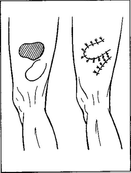
Fig. 137. Local skin plasty with pedicle flap transposition
Direct pedicle flap transplantation can be performed from the upper extremity to the head (the Italian method), from finger to finger, from the trunk to the upper or lower extremity, or from one lower extremity to the other.
In this surgical technique, the flap is harvested from a distant body site that, through specific joint positioning, can be brought into close contact with the defect-bearing body part. Consequently, an appropriate donor site is selected. Depending on the dimensions of the skin defect, a rectangular flap of the required size is outlined along with the subcutaneous tissue. Elevating the flap with fine sharp hooks, it is dissected bluntly and sharply from three sides, followed by meticulous hemostasis. The flap remains connected to the parent base by its fourth (narrow) side. The elevated flap is then mobilized toward the defect and sutured to its margins (Fig. 138). To ensure absolute immobilization of the graft, the approximated areas are secured with a plaster cast. The secondary skin defect created at the donor site is closed by direct suturing. Once the graft has successfully engrafted, the fourth side (the pedicle) is severed from the parent base, and the skin flap is definitively contoured onto the defect.
Direct skin transplantation from distant sites can also be performed using bridge flaps. The method involves outlining a musculocutaneous or adipocutaneous strip on the abdomen, back, or thigh, which is dissected down to the fascia while the underlying defect is closed primarily. The skin flap, remaining attached by two pedicles, is elevated to allow fingers or a hand bearing a skin defect to be slipped underneath it, after which the edges of the "bridge" are sutured to the margins of the skin defect. After the transferred flap has engrafted (in 2–3 weeks), the pedicles are transected to complete the reconstructive procedure.
If direct pedicle flap transplantation is impossible, a delayed, migratory flap plasty according to V. Filatov's method is performed.
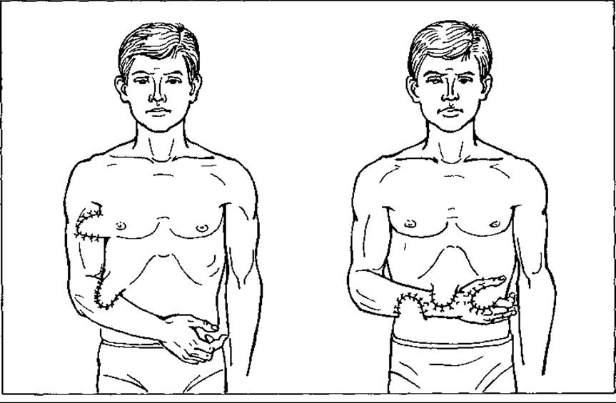
Fig. 138. Direct pedicle flap transplantation
At the selected donor site (most commonly the back, abdomen, or thigh), under local anesthesia, a skin strip 5–6 cm wide is outlined by two parallel incisions 10–12 cm long (the length should not exceed three times the width). The strip, along with the subcutaneous tissue, is dissected and sutured into a tube (Fig. 139). The skin defect created beneath the excised flap is then sutured closed. The newly formed tube remains attached to the donor base by two nourishing pedicles. The excised skin flap should be 10% larger than the skin defect it is intended to cover. After 10–12 days, one end of the pedicle is progressively compressed using a clamp, gradually increasing the duration of compression (5–10–15–20 min). This stage is referred to as "training" or "conditioning" of the flap. Once the graft adapts to the blood supply from the opposite side, the "trained" pedicle is severed. At this point, the skin flap resembles a rounded stem with a single pedicle and begins its "journey." An incision is made in the direction of the skin defect, and the severed end of the flap is sutured into it. After a few days, the flap engrafts, having migrated toward the defect by a distance equal to its length. After some more time, the transplanted end takes ROOT and begins to receive its blood supply from both pedicles. Then, the flap "takes a few more steps" in the same manner and finally reaches its destination—the skin defect. Here, one of its pedicles is divided, and the flap itself is split longitudinally (along the suture line). The newly formed flap is used to close the skin defect by suturing it to the edges of the wound. The flap is nourished by the vessels running through the uncut pedicle. After 10–12 days, the flap engrafts onto the wound bed, and its edges are trimmed along the margins of the skin defect.
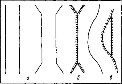
Fig. 139. Diagram of The formation of a migratory flap according to V. P. Filatov's method: a — parallel skin incisions; b — sutured skin edges at the site of the dissected flap; c — Formation of the "suitcase handle"
The skin plasty method described above is effective, but it is time-consuming (sometimes taking 2–3 months). How can this process be accelerated? This can be achieved by providing the flap with a "carrier," which can be the arm. The flap is immediately transplanted to the arm, and then from the arm to its final destination: the leg, cheek, nose, etc.
Free skin grafting involves transplanting skin flaps that completely lose their connection to the donor base for a certain period of time. Available options include transplanting skin epithelium (Mangold's method) as well as grafts of varying thickness: thin, intermediate, and full-thickness.
To gain some understanding of the thickness of skin grafts for free transplantation, one must recall the Anatomical Features of skin structure (Fig. 140).
The skin consists of two layers: the superficially located epidermis and the deep connective tissue layer of the skin proper, known as the dermis. The epidermis is composed of Cytology/practical/35.html">Stratified squamous keratinized epithelium. It comprises five distinct layers: stratum basale, stratum spinosum, stratum granulosum, stratum lucidum, and stratum corneum. It is separated from the dermis by the basement membrane—a network of delicate connective tissue fibers intertwined with the protoplasmic processes of the basal epidermal cells. The stratum basale consists of cylindrical cells responsible for epithelial cell proliferation. It is frequently grouped with the stratum spinosum to form the germinative, or Malpighian, layer.
The germinative layer of the epidermis and the underlying dermis are joined together by a convoluted, undulating line.
The thickness of the dermis is approximately 1–2 mm. It consists of the papillary layer, which lies adjacent to the epithelial covering, and the deeper reticular layer. The papillary layer forms papillae that project into the epidermis. These papillae contain Blood and Lymphatic vessels as well as nerve endings. The reticular layer consists of dense Fibrous connective tissue formed by thick bundles of Collagen fibers and thick elastic fibers interwoven with the elements of the papillary layer.
The orientation of connective tissue bundles within the reticular layer of the skin determines its elasticity and extensibility.
The skin contains sweat and Sebaceous Glands situated within the connective tissue layer of the skin. The former are tubular in structure, while the sebaceous glands predominantly open into hair follicles.
Autologous skin grafting (using the patient's own skin) is most commonly used for skin transplantation, followed less frequently by allogeneic grafting (skin harvested from another human), and even less frequently by brephoplasty (transplantation of skin from a stillborn fetus no older than 6 months). The latter Two Types of dermoplasty require careful consideration of the isosero-logical compatibility between the donor and the recipient.
Skin transplantation can be performed using grafts of varying thickness: thin, intermediate, and full-thickness.
A thin skin graft consists of the epidermis and the papillary layer, with an average thickness ranging from 0.25 to 0.75 mm. The advantage of this technique is that the grafts can be harvested quickly and easily, and the graft maintains a uniform thickness. It is resilient and engrafts reliably even on an infected wound bed. Grafts of unlimited size can be transplanted, and the donor site heals spontaneously and rapidly.
The disadvantage of this method is that the graft tends to contract and may change color (both hyper- and hypopigmentation are possible).
A full-thickness skin graft has a thickness of 0.8–1.1 mm. The advantages of this transplantation method are that the graft undergoes less contraction, retains its original color, develops a subcutaneous layer beneath it, and can be lifted into a fold. However, the graft is highly demanding and will only engraft under aseptic conditions in areas with a robust blood supply. Furthermore, only small grafts can be used, and the donor site must be closed by direct suturing or plastic surgery.
Free skin grafting using small skin islands was pioneered by the French surgeon Reverdin, who in 1869 proposed transplanting small pieces of epidermis onto granulating wounds. The technique of Reverdin plasty is as follows: the skin at the donor site is stretched between two fingers, and small fragments of epidermis (0.5 cm in diameter) are excised using a sharp scalpel or blade and placed onto the granulating surface of the skin defect. Depending on the size of the defect, many such fragments are harvested (sometimes up to 100). These epidermal fragments serve as distinctive islands on the granulation surface from which epithelialization takes place.
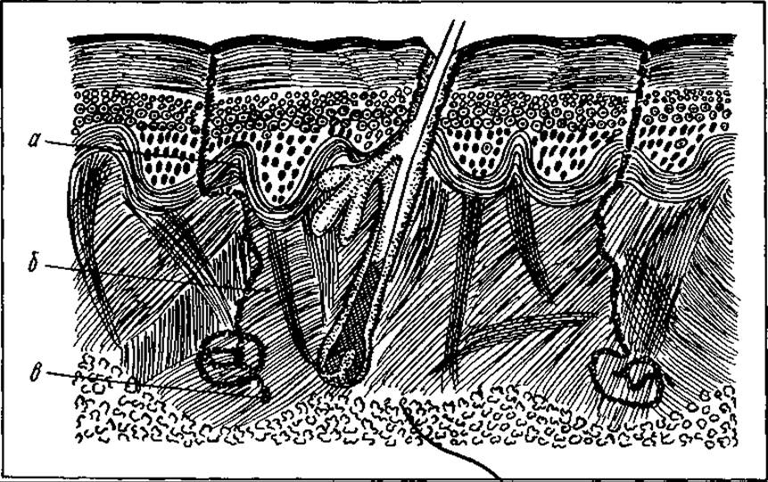
Fig. 140. Diagram of skin structure:
a — incision line for thin superficial skin layer grafts; b — incision line for partial-thickness thick skin grafts; c — incision line for full-thickness grafts
Reverdin's method is meticulous and yields an unsatisfactory cosmetic outcome.
Reverdin's method was superseded by the Janowicz-Chainski technique (1871), which utilizes deeper layers of the skin, though still small in size (up to 0.5 cm in diameter). On the prepared surgical field, a needle is inserted into the skin to elevate it into a conical shape, and the skin at the Base of the cone is excised. These grafts comprise all layers of the skin. Closing large defects requires a significant number of such grafts.
Skin for plastic surgery using Janowicz-Chainski's method is usually harvested from the abdomen or thighs in a checkerboard pattern and applied to undamaged granulation wounds. Following wound healing, small star-shaped scars form at the donor site.
Even Reverdin pointed out the possibility of embedding small pieces of skin deep into granulation tissue. In 1870, this method was applied by P. Pyasetsky. He made incisions On the surface of the granulations and inserted pieces of skin harvested from the donor site into them.
The free-grafting methods described above have the disadvantage that closing large skin defects requires harvesting many skin flaps. Therefore, attempts were made to transplant large skin flaps, notably using Thiersch's method.
This method involves scraping the granulations from the wound surface and stopping bleeding by pressing them with gauze soaked in an isotonic sodium chloride solution. The operative field on the donor surface is prepared, and skin flaps 10 cm long and 2—3 cm wide are excised using a scalpel blade. The epidermis and partially the papillary layer of the skin are sliced off. The flaps are temporarily immersed in a container with isotonic sodium chloride solution and later applied to the granulating surface. To completely cover the surface, Thiersch recommended applying the flaps in a overlapping (shingle-like) manner, with the edge of one flap covering another.
The method of transplanting large partial-thickness skin flaps (so-called split-thickness flaps) involves excising large flaps comprising 1/2 or 3/4 of the skin thickness without reaching the subcutaneous tissue, and transferring them onto skin defects. This method is most commonly used to close fresh skin defects. The edges of the excised flap can be sutured to the edge of the defect. Additionally, interrupted anchoring sutures are placed through the transplanted skin to the underlying tissues. Several holes are made in the flap for the outflow of wound secretion.
Each skin plastic surgery method has its drawbacks. Transplanted small skin fragments (0.5x0.5 cm) create an uneven, bumpy surface, which is undesirable, especially on exposed areas of the body. When using large skin flaps, this drawback is absent, but a new one arises. Wound secretion often accumulates under the transplanted areas, causing them to detach and hindering engraftment. To eliminate this drawback, in 1907 Vogel and Försterling proposed making holes with scissors in the flap obtained by Thiersch's method to allow for the drainage of wound secretion.
To transplant thick skin flaps (full-thickness), they must be harvested without the subcutaneous tissue. This can be done by excising the flap and rolling it onto a rolled gauze bandage (Padret), a special metal roller (Webster), a photographic roller (B. Parin), or a Kocher clamp (Yu. Dzhanelidze).
In 1930, Douglas proposed his own method of free skin grafting using a thick flap. It consists of the following. The outlines of the flap are marked on the donor skin site. Afterwards, circular holes with a diameter of 0.5 cm are made in the skin using a punch. The skin flap is excised without the subcutaneous tissue. The skin fragments punched out remain at the donor site. After harvesting the flap, a wound surface with numerous skin islets remains in its place, which over time stimulate the wound epithelialization processes. The holes in the skin flap scar over shortly after it engrafts. This method makes it possible to harvest thick, large skin flaps for transplantation, after which closing the wound by direct suture would otherwise be difficult (mesh or sieve flap).
In 1937, Dragstedt and Wilson proposed a technique that made it possible to close large tissue defects using skin flaps of a smaller surface area. Having a fusiform shape, they can be significantly stretched due to numerous small through-and-through longitudinal perforations made in a checkerboard pattern.
The authors sutured the transplanted flap to the edges of the wound defect using interrupted sutures. This method can be used to close large-area defects (200x400 cm).
In 1952, Yu. Dzhanelidze proposed a method for transplanting large skin flaps. He outlined a rectangular flap on the donor's skin. After incising one of its long edges, he grasped it with a clamp and rolled the skin onto it, gradually detaching it from the subcutaneous tissue. Stopping just short of the edge, he spread out the flap, stretched it, and perforated it. Only after this did he completely cut it off and transplant it onto the wound defect. The edges of the flap were fixed with sutures to the edge of the skin defect.
The introduction of the operating Microscope and microsurgical techniques into medical practice has made it possible to perform free transplantation of large skin flaps complete with subcutaneous tissue. The method of such transplantation involves connecting the artery and vein supplying and draining the flap to the artery and vein of the defect site. This operation is complex and requires specialized instruments.
During the engraftment of free skin transplants, The Nature of the dressing plays a crucial role. It should not be too tight, compressing cells and intercellular spaces, so as not to hinder the exchange of nutrients and the Development of the vascular network in the graft. Nor should it be too loose, so as to prevent the detachment of the graft by metabolic products accumulating beneath it. It is not recommended to remove the dressing earlier than 8–10 days postoperatively, provided the postoperative course is uncomplicated. To prevent the dressing from sticking to the transplanted skin areas, it is moistened with liquid petrolatum, and the dressing is removed atraumatically. Rough manipulation can cause the graft to detach.
It is very important to precisely determine the thickness of the skin flap. Various instruments (Thiersch, Kolokoltsev, Dьяkonov knives, surgical blades, carefully sharpened amputation knives) and dermatome devices have been proposed for this purpose.
The first such dermatome was designed in 1930 by the surgeon Paget and the engineer Hood. This device resembled a semi-circular drum across which a thin knife blade moved reciprocally. The distance of the blade from the drum could be adjusted depending on the desired thickness of the flap. The skin of the donor site was coated with a special adhesive and fixed to the dermatome drum. By rotating the drum and moving the blade, the adhered skin was sliced off.
In recent years, specialized motor-driven dermatomes and vacuotomes—devices in which the skin from the donor site is fixed and elevated using a special suction head—have been introduced. Such devices can only be used on completely flat and smooth body surfaces. A dermatome powered by compressed air has also been designed.
The successes of reconstructive surgery contribute to the restoration of organs lost by the organism. Modern medicine boldly utilizes the laws of nature, accomplishing what seems impossible. Yet, the main challenge in transplantology remains overcoming the biological incompatibility barrier. Only when this is resolved will organ and tissue transplantation occupy a prominent place in the arsenal of therapeutic and preventive medicine, becoming one of the methods for extending human life expectancy.
Last update: 08/08/2026
Editorial and Educational Adaptation: This material has been compiled based on the primary/original source text. The project team performed an editorial review, corrected technical inaccuracies, structured sections, and adapted the content for an educational format.
What was processed:
- elimination of formatting defects (OCR errors, structural breaks, corrupted characters);
- editorial organization of content;
- standardization of terminology in accordance with academic sources;
- verification of factual statements against the original source text.
All mentions of the author, publication year, and origin of the primary text have been preserved in accordance with the source.