Vertebrate Zoology - V. M. Konstantinov 2011
Chordates
Acrania
The Subphylum Acrania (Cephalochordata) includes a single Class, Leptocardii. This class comprises only one order, Amphioxiformes, which contains a single family, Branchiostomidae, divided into three subfamilies: Branchiostomidae, Epigonichthyidae, and Amphioxidae.
Acrania are a small group of roughly 30 species of primitive, exclusively marine chordate animals. They inhabit temperate and warm seas across the Atlantic, Indian, and Pacific oceans, and can also be found in the Black and Japan seas. They generally prefer Water temperatures between +17 and +30 °C and a salinity of 20-30 ‰.
Branchiostoma (amphioxus) and epigonichthyids dwell in shallow coastal waters, burying the posterior part of their bodies into the sandy sea bottom. When disturbed by Touch or light, the amphioxus darts quickly out of its shelter, swims a short distance, and burrows back into the sediment. Amphioxid lancelets, on the other hand, are found in the plankton of the open ocean.
Lancelets feed on diatoms, detritus, and zooplankton. They reproduce during the warmer months; in the Black Sea, for instance, spawning occurs from late May to early August. Fertilization is external, with females and males simultaneously releasing their Gametes into the water.
The amphioxus is a classic model Organism in zoological research due to its unique Anatomical Features. It retains all the fundamental CHARACTERISTICS OF THE phylum Chordata throughout its life cycle. Studying its anatomy is crucial for understanding the Structural Organization of Chordates, as well as the evolutionary origin and development of vertebrates. It is widely believed that the ancestral stock of vertebrates closely resembled modern lancelets.
Let us examine the organization of Acrania using the European amphioxus (Branchiostoma lanceolatum), which inhabits the Black Sea, as a representative example.
Organization of Acrania (using the amphioxus as an example)
External Morphology. The amphioxus is a semi-transparent, fish-like animal with a laterally compressed body measuring 5–8 cm in length. A fold running along the dorsal side forms the dorsal fin, which continues posteriorly to form a lanceolate caudal fin. Two ventrolateral metapleural folds run along the anterior half of the body; these fuse to enclose the atrial (or peribranchial) cavity and extend backward to the atriopore. The anterior ventral region features a large oral hood surrounded by 10–20 pairs of cirri. This depression (the oral hood) marks the beginning of the digestive tract (Fig. 1). The amphioxus was first described by P. S. Pallas in 1774.
Integument. The Skin of the amphioxus consists of two layers: an outer epidermis and an inner dermis (cutis). The epidermis is single-layered and covered externally by a thin cuticle. Its Cells are cylindrical and interspersed with goblet-shaped glandular cells, while some cells bear sensory hairs. The dermis is poorly developed, consisting of a gelatinous, loose Connective Tissue.
Musculature. The musculature is metameric and poorly differentiated, serving only for simple swimming and burrowing movements. It is distributed unevenly across the body, with the bulk concentrated dorsally and in the caudal region. The Muscle layer is composed of longitudinal bands divided into segments called myomeres. Adjacent myomeres are separated by connective tissue partitions known as myosepta, which provide structural support for the muscle fibers. These fibers are bent at acute angles pointing toward the anterior end of the body, meaning the myomeres are essentially nested cones. The myomeres on the left and right sides of the body are arranged asynchronously: a single myomere on one side lies opposite the halves of two adjacent myomeres on the opposite side. This staggered arrangement facilitates lateral body bending during swimming. The muscle layer on the ventral side of the body (specifically, its anterior two-thirds) is flat and thin.

Fig. 1. Diagram of a longitudinal section through the body of an amphioxus:
1 — oral aperture surrounded by cirri; 2 — caudal fin; 3 — dorsal fin; 4 — metapleural fold; 5 — atriopore; 6 — notochord; 7 — myomere (shown only in the caudal region); 8 — myoseptum; 9 — nerve cord; 10 — velum; 11 — gill slits; 12 — intestine; 13 — hepatic caecum; 14 — peribranchial cavity; 15 — endostyle; 16 — Gonads; 17 — anus
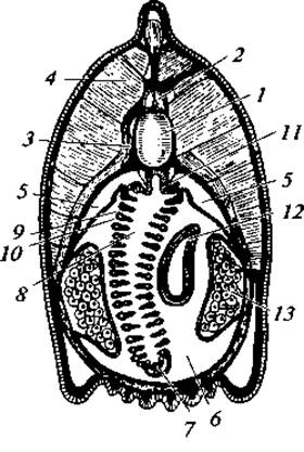
Fig. 2. Cytology/practical/72.html">Cross section of an amphioxus through the pharyngeal region:
1 — notochord; 2 — nerve cord; 3 — connective tissue sheath; 4 — myomere; 5 — coelom; 6 — peribranchial cavity; 7 — endostyle; 8 — pharyngeal cavity; 9 — gill slit; 10 — interbranchial septum; 11 — nephridium; 12 — hepatic caecum; 13 — gonad
Skeleton. The Axial Skeleton is represented by a notochord running along the longitudinal axis of the body. Enclosing the notochord is a thick connective tissue sheath that also surrounds the overlying nerve cord (Fig. 2). Extensions of this sheath connect with the myosepta and the subcutaneous connective tissue.
In the region of the gill slits, There is a complex supporting lattice of fibrous, acellular bars. Unpaired fins are supported by rods of dense gelatinous tissue, and similar structures form the Skeleton of the oral hood and cirri.
Nervous system. The Central Nervous System consists of a dorsal longitudinal nerve cord containing an internal cavity called the neurocoel. The dorsal edges of the tube do not fuse completely, leaving a narrow longitudinal fissure that opens into the neurocoel. At the anterior end of the nerve cord, the neurocoel expands slightly—a region thought to correspond to the third Brain ventricle of vertebrates. Damage to this anterior section of the nerve cord leads to a loss of motor coordination.
During early development, the cavity of the nerve cord communicates with the exterior via an opening called the neuropore. In adults, a depression known as the olfactory pit remains at the site of the neuropore on the anterodorsal surface of the HEAD.
Scattered along the entire length of the nerve cord, lining the margins of the neurocoel, are photoreceptive Organs known as Hesse ocelli. Each ocellus consists of two cells: a light-sensitive clear Cell and a pigmented dark cell. Consequently, the entire nerve cord of the amphioxus is light-sensitive.
The Peripheral Nervous System is formed by nerves branching out from the nerve cord, with two pairs of nerves (dorsal and ventral) corresponding to each muscle segment. Functionally, the dorsal nerves are mixed (sensory and motor), whereas the ventral nerves are exclusively motor. Unlike the vast majority of vertebrates, the dorsal and ventral nerve branches in the amphioxus are not interconnected, suggesting an absence of the reflex arcs characteristic of vertebrate nervous systems.
Sense Organs. The amphioxus possesses primitive sense organs. Light stimuli are detected by Hesse ocelli. The olfactory pit presumably perceives Chemical properties of the water, while scattered sensory cells throughout the epidermal layer respond to mechanical wave stimuli.
Feeding and Respiration. Feeding and respiration are largely passive processes. Lancelets feed on microscopic plants and animals suspended in the water Column. The cirri along the edge of the oral hood stir up detritus (see Fig. 1), while the ciliated epithelium lining the oral vestibule directs the food-laden water current toward the Mouth. Located deep within the hood is the mouth, which is bordered by guiding folds known as the velum. The voluminous Pharynx is pierced by numerous (roughly 100 pairs) obliquely arranged gill slits. These slits do not open directly to the exterior, but rather into the atrial (or peribranchial) cavity. The atrial cavity is bounded by fused metapleural folds; only at their posterior end do the folds remain unfused, forming an opening called the atriopore. Thus, water passes from the pharynx through the gill slits into the atrial cavity and exits to the exterior via the atriopore. The peribranchial cavity protects the respiratory apparatus from clogging with sediment particles—a vital adaptation for organisms that spend most of their time buried in sand. Some evidence also suggests that cutaneous respiration takes place.
Along the mid-ventral line of the pharynx runs a longitudinal groove known as the endostyle, which is lined with glandular and ciliated cells. Near the mouth, this groove bifurcates and extends up the sides of the pharynx as two ciliated bands, merging dorsally into an epibranchial groove that runs posteriorly toward the intestine. The endostyle Functions as follows: its glandular cells secrete mucus that traps food particles precipitating out of the water onto the endostyle. The beating of the endostyle's cilia generates a current that propels the mucus and entrapped food parcels forward toward the mouth. Near the mouth, the food mass is directed upward and transported along the epibranchial groove into the intestine. Additionally, folds associated with the gill slits are believed to assist in food particle transport.
The intestine is straight and undivided into distinct sections. A hollow blind pouch, known as the hepatic cecum, extends from the ventral side of the anterior gut; it is homologous to the vertebrate Liver.
Circulatory system. The CIRCULATORY SYSTEM OF amphioxus is closed, primitive, and represented by arterial and venous vessels (Fig. 3). Unlike vertebrates, amphioxus lacks a Heart.
Arterial System. A contractile ventral aorta runs beneath the pharynx. Paired branchial Arteries diverge from it on both sides, passing through the partitions between the gill slits. The branchial arteries are thin-walled; dorsal to the pharynx, they empty into paired supra-branchial vessels, or roots of the dorsal aorta, delivering oxygenated Blood. At the level of the posterior end of the pharynx, these fuse to form the main arterial trunk—the dorsal aorta—which runs beneath the notochord along the body to its posterior tip. Along its course, the dorsal aorta branches into vessels supplying the Internal Organs. Carotid arteries extend anteriorly from the aortic roots, supplying blood to the head region. Venous system. Blood from the anterior end of the body collects into paired jugular (or anterior cardinal) Veins, which carry blood posteriorly. Blood from the posterior end of the body is drained by paired posterior cardinal veins, carrying blood forward. Posterior to the pharynx, the anterior and secondary cardinal veins of their respective sides fuse to form two (left and right) wide, thin-walled Cuvierian ducts. From the Cuvierian ducts, blood empties into the venous sinus, from which the ventral aorta originates. Venous blood from the internal organs collects into the sub-intestinal vein, which, upon reaching the hepatic cecum, breaks up into a capillary network within it, forming the hepatic portal system. From there, blood flows through the hepatic vein into the venous sinus.
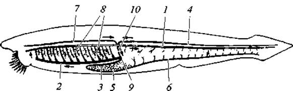
Fig. 3. Diagram of the circulatory system of amphioxus:
1 — dorsal aorta; 2 — ventral aorta; 3 — hepatic vein; 4 — posterior cardinal vein; 5 — HEPATIC PORTAL VEIN; 6 — sub-intestinal vein; 7 — anterior cardinal vein; 8 — branchial arteries; 9 — venous sinus; 10 — Cuvierian duct
Due to the absence of a heart in amphioxus, blood flow is driven by the pulsation of the ventral aorta and the lower PARTS OF THE branchial vessels ("branchial hearts"). The blood is colorless: it contains neither formed elements nor pigments.
Excretory system. The excretory system is represented by numerous (up to 90 pairs) nephridia located in the pharyngeal region. One end of the nephridial tubule features a series of openings connecting it to the body cavity, or coelom, which in amphioxus extends above the pharyngeal region as a pair of longitudinal canals. These openings—nephrostomes—are lined with club-shaped cells called solenocytes. A solenocyte contains a fine tubule with an incorporated cilia flagellum. Within the body wall thickness, in close proximity to the nephridial tubules, lies a dense network of blood capillaries through which fluids containing Metabolic waste products are presumably filtered into the coelom. The other end of the nephridial tubule opens into the atrium, where excretory products are collected and expelled outward through the atriopore.
The excretory system of amphioxus is very similar to that of certain segmented worms.
Reproductive System. Amphioxuses are dioecious animals. Their gonads are numerous and metamerically arranged. They lie within the coelom and protrude into the atrial cavity as two rows of rounded (Ovaries) or slightly elongated (Testes) bodies—about 25 pairs. Mature Germ Cells are released into the peribranchial cavity via temporarily formed genital ducts, and are subsequently expelled through the atriopore. Fertilization in amphioxus is external; the eggs are small (0.1 mm in diameter) and oligolecithal.
Let us examine The Development of amphioxus in greater detail.
Development. The development of amphioxus was first studied by A. O. Kovalevsky (1867). This topic is of great interest, as analyzing the Developmental Stages of the most primitive living chordate provides certain insights into the Early stages of chordate phylogeny and offers a simplified schematic picture of their embryonic development.
Figures 4 and 5 illustrate successive stages of embryonic development in amphioxus up to the larval stage.
Cleavage of the fertilized egg is total and nearly equal: during blastula formation, it is evident that cells on its lower side, corresponding to the vegetative (“nutritive”) pole of the egg, are larger than those on the upper side. Consequently, the inner layer of the subsequent gastrula stage is formed by larger cells. Cleavage occurs very rapidly. In the ectoderm on the upper side of the embryo, the neural plate differentiates; its edges fold and subsequently fuse. The resulting neural tube temporarily maintains communication with the external environment at its anterior end (via the neuropore) and with the gastrula cavity (i.e., the archenteron) at its posterior end (via the neurenteric canal). Later, the neurenteric canal disappears entirely, while an olfactory pit remains at the site of the neuropore.
Simultaneously, the endoderm differentiates. A longitudinal outpocketing appears on the DORSAL SIDE OF the archenteron. Subsequently, it detaches and transforms into a solid cord—the notochord.
Around the same time, two rows of symmetrically arranged outpocketings of the archenteron appear on either side of the notochord anlage. As they grow, they detach from it to form a paired series of mesoderm anlagen—metamerically arranged coelomic pouches. During further development, each coelomic pouch divides into two sections: an upper somite and a lower lateral plate. Subsequently, the coelomic cavities do not fuse with one another, but instead disappear; the cavities of the lateral plates, however, fuse together to form the secondary body cavity, or coelom.
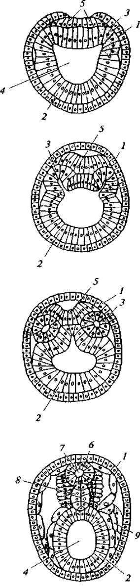
Fig. 4. Embryonic development of amphioxus (cross-sections through four successive stages of embryonic development):
1 — ectoderm; 2 — endoderm; 3 — mesoderm; 4 — enteric cavity; 5 — neural plate; 6 — neural tube; 7 — neurocoel; 8 — notochord; 9 — secondary body cavity
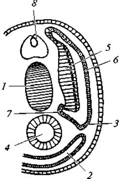
Fig. 5. Cross-section through an amphioxus larva:
1 — notochord; 2 — secondary body cavity; 3 — gonadotome; 4 — intestine; 5 — myotome; 6 — cutaneous layer; 7 — sclerotome; 8 — neural tube
Further Differentiation of the somite gives rise to the following anlagen: 1) the sclerotome (the lower inner part of the somite), which gives rise to cells forming the connective tissue Sheath of the notochord and neural tube, skeletal fin rays, and presumably myosepta; 2) the myotome (the part of the somite adjacent to the notochord), which forms the trunk musculature; 3) the cutaneous layer (the upper and outer part of the somite), which forms the connective tissue component of the skin, i.e., the cutis (see Fig. 5).
The Peritoneum, mesenteries (within which the main Blood Vessels develop as longitudinal channels), and intestinal musculature all develop from the lateral plate. Nephridial tubules develop as finger-like outgrowths of the walls of the secondary body cavity. Gonads develop as outgrowths of the body wall region corresponding to the junction between the somite and the lateral plate—the gonadotome.
The mouth forms through the outpocketing of the archenteron at the end opposite to the blastopore (primitive mouth), coupled with an opposing invagination of the ectoderm. A perforation occurs where these structures meet. The formation of the mouth and gill slits is asymmetrical. The oral opening is established on the lower left side of the embryo. The left gill slits (14 in total) initially appear on the ventral side and subsequently migrate to the right side of the embryo. Following this, another row of slits (8 in total) appears dorsal to the previously mentioned 14 slits. Later, the lower row of slits shifts to the ventral side and only then to the left side of the body, during which their number decreases from 14 to 8. The number of gill slits on both sides then increases sharply. Eventually, the mouth shifts to the ventral side.
The atrial cavity initially forms as a groove on the ventral surface of the body. The metapleural folds that shape this groove grow toward each other and fuse to enclose a cavity, opening to the outside only at its posterior end where the folds remain unfused. Overall, the larval development of amphioxus lasts about three months.
Ancestors and Systematics of Acrania
Paleontology provides no fossil evidence regarding the ancestors of modern acranians. Therefore, THE ORIGIN OF this group must be inferred from comparative anatomical and embryological data. According to A. N. Severtsov, the ancestors of acranians were free-swimming, bilaterally symmetrical animals. They lacked an atrial cavity, and the notochord did not extend to the anterior end of the body. There were fewer gill slits (17–20), which appear to have been arranged symmetrically. This group gave rise to two branches. One retained a free-swimming lifestyle and led to the vertebrates. The other adapted to a sluggish, benthic, or burrowing lifestyle. The Asymmetry in the arrangement of the gill slits observed during certain stages of modern acranian ontogeny is likely a remnant of a phylogenetic phase when the ancestors of Acrania were benthic animals resting on the sea floor on one side, with their gill slits shifted toward the upper surface.
Subsequently, some acranians transitioned to an infaunal lifestyle within the bottom sediment. As an adaptive mechanism to protect their gill slits from clogging by solid particles, they developed metapleural folds and a peribranchial (atrial) cavity. This group includes modern lancelets and epigonichthyids. Conversely, another Lineage remained in the water column, becoming part of the plankton (amphioxids).
Currently, scientists have proposed an alternative perspective on the origin of acranians. It is hypothesized that lancelets are neotenic forms of once sessile bottom-dwelling animals (possibly related to tunicates) that acquired The ability to reproduce at the larval stage.
As previously mentioned, the subphylum Acrania comprises a single class — Cephalochordata, a single order — Amphioxiformes, and, according to modern systematists, a single family — Branchiostomidae, encompassing approximately 30 species. Some zoologists divide them into three subfamilies, discussed below.
Typical lancelets (Branchiostoma), which serve as the representative model for the subphylum, are characterized by a symmetrical body plan. Their gonads are paired, and the metapleural folds are of equal length. Their body length reaches up to 8 cm, with about 20 species known.
Epigonichthyid lancelets (Epigonichtys) are smaller, with a body length of up to 5 cm. They exhibit certain asymmetrical features: the gonads are present only on the right side of the body, and the right metapleural fold is longer than the left. There are 6 known species.
Amphioxid lancelets (Amphioxidae) retain certain larval characteristics: they lack an atrial cavity, and the mouth is shifted to the left side and almost devoid of tentacles. Their body length reaches up to 16 mm. Unlike other lancelets, they lead a pelagic rather than a benthic lifestyle. It is hypothesized that they may represent the larvae of epigonichthyids.
In certain regions, lancelets are abundant. Along the coasts of Southeast Asia, lancelets are locally harvested by sifting surface sand from the sea floor. Overall, they play a significant role in marine biocenoses, serving as a key link in the food webs of both benthic and planktonic animals.
Last update: 13/08/2026
Editorial and Educational Adaptation: This material has been compiled based on the primary/original source text. The project team performed an editorial review, corrected technical inaccuracies, structured sections, and adapted the content for an educational format.
What was processed:
- elimination of formatting defects (OCR errors, structural breaks, corrupted characters);
- editorial organization of content;
- standardization of terminology in accordance with academic sources;
- verification of factual statements against the original source text.
All mentions of the author, publication year, and origin of the primary text have been preserved in accordance with the source.